

Recommandé
Contenu connexe
Tendances
Tendances (20)
En vedette
En vedette (11)
Similaire à Burn
Similaire à Burn (20)
Dernier
Dernier (20)
Burn
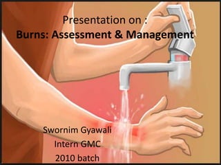
- 1. Presentation on : Burns: Assessment & Management Swornim Gyawali Intern GMC 2010 batch
- 2. Outline • Objective • Introduction • Type of Burn injury • Classification of Burns • Pathophysiology of Burns • Assessment of the Burn wound • Management of Burns – Primary – Secondary • Complications of Burn Injuries
- 3. Objectives • At end of this presentation we be able to know 1. definition and causes of Burn injuries 2. Types and classification of burns 3. pathophysiology of burns 4. Management of a patient who sustained burn injury 5.Complications of burns
- 4. Introduction Definition • A burn is a coagulative destruction of the surface layers of the body. • It occur when some or all of the cells in the skin or other tissues are destroyed by heat cold electricity Radiation Lightening caustic chemicals
- 5. Types of Burn Injury • Thermal Flame : fire injury Scald : moist heat/steam Flash : explosion Contact : to hot surfaces
- 6. • Cold exposure (frostbite) Usually occurs in distal parts of the body Common sites: Fingers, Toes, Nose and Ears Severe Vasoconstriction & Decreased Blood flow Ischemia • Chemical burns Cause progressive damage Acid produces tissue coagulative Necrosis. Alkaline burns generate colliquation Necrosis. Systemic absorption of some chemicals is life threatening
- 7. • Electrical mechanisms of injury : i. Electrical current injury ii. Electrothermal burns from arcing current iii. Flame burn caused by ignition of clothes Deep destruction of muscles rhabdomyolysis myoglobinuria ATN ARF • Inhalation Hot smoke • Radiation sunburn
- 8. Pathophysiology of Burn Local Changes 1. Burn causes coagulative necrosis of the epidermis and underlying tissues 2. depth of injury: temperature & duration of exposure area of cutaneous injury
- 10. Assessment of The Burn Wound • Burn Depth Cutaneous burns are classified according to the depth of tissue injury: 1. superficial or epidermal (first-degree), 2. partial-thickness (second degree), or 3. full thickness (third degree). 4. Burns extending beneath the subcutaneous tissues and involving fascia, muscle and/or bone are considered fourth degree
- 12. First degree (Superficial) • Red, erythematous • Very sensitive to touch • Very painful • Usually moist • No blisters Second degree (partial- thickness) • Erythematous or whitish with a fibrinous exudate • Wound base is sensitive to touch and Painful • Commonly have blisters • Surface may blanch to pressure Third degree (Full thickness) • Surface may be: White, Black, leathery, Pale or Bright red • Generally anesthetic or hypoesthetic • Subdermal vessels do not blanch • No blisters • Hair easily pulled from its follicle Fourth degree • Involves deep tissues including fascia,
- 14. Assessment of The Burn Wound (cont’d) • Total percentage of body surface area (TBSA) 1. Lund-Browder chart
- 15. • Rule of Nines
- 18. Management; Primary Survey Initial Intervention Airway maintenance with cervical spine control Breathing and Ventilation Circulation with Haemorrhage Control Disability: Neurological Status Exposure with Environmental Control
- 19. Diagnostic tests and monitoring • Arterial blood gas • Chest x-ray • Serial peak expiratory flow rates (PEFR) • Pulse oximetry • Capnography • fiberoptic laryngoscopy and bronchoscopy
- 20. Treatment • Supplemental oxygen and airway protection • Close monitoring of fluid resuscitation • Mechanical ventilation • Inhaled nitric oxide • aerosolized heparin and N- acetylcysteine (NAC)
- 21. Fluid resuscitation American Burn Association's practice guidelines, patient with greater than 15 percent total body surface area (TBSA) non-superficial burns should receive formal fluid resuscitation. Fluid selection Formulae 1. Parkland : 4ml x wt (Kg) x % TBSA burn -Ringer’s lactate or Hartman solution 2. Evans :1ml x wt x %TBSA 3. Brooke :1.5ml x wt x %TBSA 4. Modified Brook:2ml x wt x % TBSA
- 22. Management; secondary Survey (cont’d) • History • Thorough physical examination • Lab studies and monitoring CBC Electrolytes RFT Glucose Venous blood gas Caboxyhemoglobin Arterial blood gas Chest x-ray ECG
- 23. Management; Secondary Survey (cont’d) Chemoprophylaxis Tetanus immunization Antibiotic Wound management Wound dressing and care Escharotomy Chest - at the anterior axillary line Extremity - can be done at a bedside without local anesthesia
- 25. Nutrition • Hypermetabolism develops as a response to injury • If TBSA >40%, lean body weight ↓ by 25% over the first 3 weeks • Patient with major burn needs high calorie in the form of: CHO (50%), protein (20%) , fat (30%) and some vitamins & minerals
- 26. Nutritional Requirement Calculations Curreri formula • Age 16–59 years: (25)W + (40)TBSA • Age 60+ years: (20)W + (65)TBSA Sutherland formula • Children: 60 kcal /kg + 35 kcal%TBSA • Adults: 20 kcal /kg + 70 kcal%TBSA Protein needs • Greatest nitrogen losses between days 5 and 10 • 20% of kilocalories should be provided by proteins
- 27. Burn Complications 1. INFECTION 2. Curling ulcer- stress ulcers 3. Contracture 4. Marjolin’s ulcer, Hypertrophic scar, keloid Pschological aspect • PTSD • Flash backs • Avoidance behavior • Sleep disturbance
- 28. Minimizing complications 1. Hand washing before & after touching each patient. 2. Aseptic techniques for dressing & procedures 3. Early nutritional support 4. Early excision of deep burns 5. Use of topical antimicrobials 6. Early excision and grafting
- 29. Thank you !!! • Queries ????
- 30. Refrences 1. SCHWARTZ :Principles of surgery ,9th edi.2008 2. BAILEY & LOVE : Short practice of surgery ,25th edi,2008 3. American Burn Association's practice guidelines, 2012 4. Internet (pictures) 5. Medscape.com