Best Rate (Patna ) Call Girls Patna ⟟ 8617370543 ⟟ High Class Call Girl In 5 ...
shock.pdf
1. M E D I C I N E
Continuing Medical Education
The Nomenclature, Definition
and Distinction of Types of Shock
Thomas Standl, Thorsten Annecke, Ingolf Cascorbi, Axel R. Heller,
Anton Sabashnikov, Wolfram Teske
Summary
Background: A severe mismatch between the supply and demand of oxygen is the
common feature of all types of shock. We present a newly developed, clinically
oriented classification of the various types of shock and their therapeutic impli-
cations.
Methods: This review is based on pertinent publications (1990–2018) retrieved by a
selective search in PubMed, and on the relevant guidelines and meta-analyses.
Results: There are only four major categories of shock, each of which is mainly
related to one of four organ systems. Hypovolemic shock relates to the blood and
fluids compartment while distributive shock relates to the vascular system; cardio-
genic shock arises from primary cardiac dysfunction; and obstructive shock arises
from a blockage of the circulation. Hypovolemic shock is due to intravascular
volume loss and is treated by fluid replacement with balanced crystalloids.
Distributive shock, on the other hand, is a state of relative hypovolemia resulting
from pathological redistribution of the absolute intravascular volume and is treated
with a combination of vasoconstrictors and fluid replacement. Cardiogenic shock is
due to inadequate function of the heart, which shall be treated, depending on the
situation, with drugs, surgery, or other interventional procedures. In obstructive
shock, hypoperfusion due to elevated resistance shall be treated with an immediate
life-saving intervention.
Conclusion: The new classification is intended to facilitate the goal-driven treatment
of shock in both the pre-hospital and the inpatient setting. A uniform treatment strat-
egy should be established for each of the four types of shock.
Cite this as:
Standl T, Annecke T, Cascorbi I, Heller AR, Sabashnikov A, Teske W:
The nomenclature, definition and distinction of types of shock.
Dtsch Arztebl Int 2018; 115: 757–68. DOI: 10.3238/arztebl.2018.0757
Department of Anesthesiology, Intensive and Palliative Care Medicine, Städtisches Klinikum
Solingen gGmbH: Prof. Dr. med. Thomas Standl, MHBA
Department of Anesthesiology and Intensive Care Medicine, University Hospital of Cologne:
Prof. Dr. med. Thorsten Annecke, DESA
Institute of Clinical and Experimental Pharmacology at the University Medical Center Schleswig-
Holstein, Campus Kiel: Prof. Dr. med. Dr. rer. nat. Ingolf Cascorbi
Surgical Center/Emergency Department, Department of Anesthesiology and Intensive Care, Univer-
sity Hospital Carl Gustav Carus, Technische Universität Dresden: Prof. Dr. med. Axel R. Heller, MBA,
DEAA
Department of Cardiothoracic Surgery, Cardiac Center, University Hospital of Cologne:
PD Dr. med. Anton Sabashnikov
Department of Orthopedics and Trauma Surgery, Kath. Krankenhaus Hagen gGmbH:
PD Dr. med. Wolfram Teske
I
n the first descriptions of shock the focus was
exclusively on traumatic hemorrhagic shock, but later
this changed and five different types of shock came to
be distinguished (1). Although it is true that all types of
shock can lead to the same final stage of multiorgan
failure as a result of the imbalance between oxygen de-
mand and supply, the differences in their pathogenesis
and pathophysiology make it desirable to change their
classification, partly for teaching purposes, but also,
especially, because different therapeutic measures are
needed for the different types of shock. The new classifi-
cation makes no claim to be binding, and the therapeutic
effects are as a rule limited primarily to restoration of
vital functions, in particular cardiovascular function con-
sistent with survival.
For the reasons given above, the new classification
comprises just four main categories:
● Hypovolemic shock
● Distributive shock
● Cardiogenic shock
● Obstructive shock.
Of these, hypovolemic shock is divided into four
subcategories and distributive shock into three. Ob-
structive shock has been given a category of its own.
Although this nomenclature and classification is
schematic and there is some overlapping between the
main groups, these four main groups can be basically
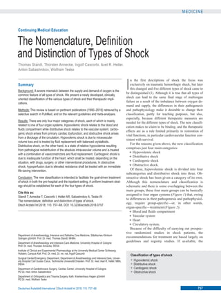
assigned to four organ systems (Figure 1) that, owing
to differences in their pathogenesis and pathophysiol-
ogy, require group-specific—or, in other words,
organ-specific—treatment (Figure 2):
● Blood and fluids compartment
● Vascular system
● Heart
● Circulatory system.
Because of the difficulty of carrying out prospec-
tive randomized studies in shock patients, the
recommendations for treatment are based largely on
guidelines and registry studies. If available, the
Classification of types of shock
• Hypovolemic shock
• Distributive shock
• Cardiogenic shock
• Obstructive shock
Deutsches Ärzteblatt International | Dtsch Arztebl Int 2018; 115: 757–68 757
2. M E D I C I N E
recommendation grade (RG) from the guidelines is
given. Where no recommendation grade is available,
the recommendation is that of the present authors
(eTable 1). The effects of the interventions presented
on survival and disability-free survival are in some
cases not strong.
Learning goals
After reading this article, the reader should:
● Be familiar with the new classification of types of
shock
● Understand the different pathogenesis and patho-
physiology of the four main categories of shock
● Know the different therapeutic approaches to the
various types of shock.
Hypovolemic shock
Hypovolemic shock is a condition of inadequate organ
perfusion caused by loss of intravascular volume,
usually acute. The result is a drop in cardiac preload to
a critical level and reduced macro- and microcircu-
lation, with negative consequences for tissue
metabolism and the triggering of an inflammatory
reaction.
Hypovolemic shock is divided into four subtypes
(2):
● Hemorrhagic shock, resulting from acute hemor-
rhage without major soft tissue injury
● Traumatic hemorrhagic shock, resulting from
acute hemorrhage with soft tissue injury and, in
addition, release of immune system activators
● Hypovolemic shock in the narrower sense, result-
ing from a critical reduction in circulating plasma
volume without acute hemorrhage
● Traumatic hypovolemic shock, resulting from a
critical reduction in circulating plasma volume
without acute hemorrhage, due to soft tissue injury
and the release of immune system mediators.
Pathogenesis and pathophysiology
The characteristic feature of both, hemorrhagic and
traumatic hemorrhagic shock is bleeding. However,
differences exist between the two subcategories in
terms of the extent of soft tissue damage. Clinically the
most significant cause of hemorrhagic shock is acute
bleeding from an isolated injury to a large blood vessel,
gastrointestinal bleeding, nontraumatic vascular
rupture (e.g., aortic aneurysm), obstetric hemorrhage
(e.g., uterine atony), and hemorrhage in the region of
the ear, nose, and throat (vascular erosion). The shock
is triggered by the critical drop in circulating blood
volume; massive loss of red blood cells intensifies the
tissue hypoxia.
Traumatic hemorrhagic shock is distinguished
from hemorrhagic shock by the additional presence of
major soft tissue injury which aggravates the shock. A
typical example of this type of shock is polytrauma,
most usually caused by road traffic accidents and falls
from a great height. Diffuse bleeding, hypothermia
(especially ≤ 34 °C), and acidosis lead to life-
threatening coagulopathy (3, 4). The soft tissue injury
leads to postacute inflammation, further reinforcing
this process. At the microcirculatory level, leuko-
cyte–endothelium interactions (5) and destruction of
endothelial membrane-bound proteoglycans and gly-
cosaminoglycans cause microvascular dysfunction
with capillary leak syndrome. At the intracellular
level a metabolic imbalance arises (6) with possible
mitochondrial damage (7) and a negative influence on
the vasomotor system (8).
Hypovolemic shock in the narrower sense and trau-
matic hypovolemic shock show significant fluid loss
without hemorrhage.
Hypovolemic shock in the narrower sense arises
from external or internal fluid loss coupled with
inadequate fluid intake. It can be caused by hyperther-
mia, persistent vomiting and diarrhea (e.g., cholera),
or uncompensated renal losses (e.g., diabetes insipid-
us, hyperosmolar diabetic coma). Sequestration of
large quantities of fluid in the abdomen, e.g., in ileus
or liver cirrhosis, also leads to a reduction of
circulating plasma volume. The pathologically raised
hematocrit as well as the increased leukocyte and
platelet interactions additionally impair the rheologic
properties of the blood and can lead to persistent
organ damage even after the patient has been treated
for shock (“no-reflow phenomenon”).
Typical causes of traumatic hypovolemic shock are
large surface burns, chemical burns, and deep skin
lesions. The trauma also activates the coagulation
cascade and the immune system, potentiating the
impairment of the macro- and microcirculation. The
inflammatory reaction results in damage to the en-
dothelium, increases capillary leak syndrome, and
causes severe coagulopathy (9, 10).
It may be possible to draw some cautious
conclusions about the incidence of traumatic hypo-
volemic and traumatic hemorrhagic shock from the
Trauma Registry of the German Trauma Society
(Deutsche Gesellschaft für Unfallchirurgie). In the
2017 annual report, out of 40 836 patients, 27 147
(66%) had a maximum severity of injury of AIS 3
Physiology of hypovolemic shock
The result is a drop in cardiac preload to a critical level and
reduced macro- and microcirculation, with negative con-
sequences for tissue metabolism and the triggering of an
inflammatory reaction.
Hypovolemic shock
Hypovolemic shock is a condition of inadequate organ per-
fusion caused by loss of intravascular volume, usually acute.
758 Deutsches Ärzteblatt International | Dtsch Arztebl Int 2018; 115: 757–68
3. M E D I C I N E
(Abbreviated Injury Score) or more, and 10 639
(26%) had life-threatening injuries (ISS, Injury Se-
verity Score ≥ 11), on the basis of which the number
of patients can be calculated to be around 30 000 per
year. The incidence of gastrointestinal hemorrhage in
Germany is around 100 000 patients per year, of
whom roughly 10 000 suffer hypovolemic shock.
These figures, together with those for the remaining
subtypes of hypovolemic shock, lead to a total of
about 50 000 patients per year (Table 1).
Treatment
The preclinical and clinical treatment of hypovolemic
shock consists of immediate intravascular volume
replacement (fluid resuscitation) with balanced crystal-
loids (recommendation grade: B) using wide-bore
Causes
Typical causes of traumatic hypovolemic shock are large
surface burns, chemical burns, and deep skin lesions.
Hypovolemic shock in the narrower sense and traumatic
hypovolemic shock
Hypovolemic shock in the narrower sense and traumatic hypo-
volemic shock show significant fluid loss without hemorrhage.
FIGURE 1
Synoptic view of the four types of shock (inner, white field) with the organ systems primarily associated with them (outer corners), sites
and mechanisms of manifestation (outside the circle), and pathogenetic and pathophysiologic features (outer and middle sectors of the circle).
To maintain clarity, mixed types of shock are not depicted.
Blood and fluid
compartment
Vascular system
Heart Circulatory system
Volume
Permeability
T
o
n
e
r
e
g
u
l
a
t
i
o
n
L
e
s
s
e
r
c
i
r
c
u
l
a
t
i
o
n
Greater circulation
Various locations
Heart valves
C
a
r
d
i
a
c
c
o
n
d
u
c
t
i
o
n
s
y
s
t
e
m
M
y
o
c
a
r
d
iu
m
B
lo
o
d
(
w
h
o
le
)
Body fluids
Plasma
S
e
p
t
i
c
A
n
a
p
h
y
l
a
c
t
i
c
Anaphylactoid
Neurogenic
Determined
by right heart
D
e
t
e
r
m
i
n
e
d
b
y
a
f
t
e
r
l
o
a
d
D
e
te
r
m
in
e
d
b
y
p
r
e
lo
a
d
De
co
m
pe
ns
at
ed
st
en
os
is
A
c
u
t
e
i
n
s
u
f
f
i
c
i
e
n
c
y
Brady-
and tachy-
arrhythmias
Myocardial
pump failure
Hemor-
rhagic
Traumatic-
hemorrhagic
Hypovolemic
(narrower
sense)
T
ra
u
m
a
ti
c
-
h
y
p
o
v
o
le
m
ic
D
i
s
t
r
i
b
u
t
i
v
e
S
h
i
f
t
E
x
t
r
a
c
a
r
d
i
a
c
Output
C
a
r
d
i
a
c
L
o
s
s
Imbalance
Shock
O2 supply
O2 demand
O
b
s
t
r
u
c
t
i
v
e
C
a
r
d
i
o
g
e
n
i
c
H
y
p
o
-
v
o
l
e
m
i
c
Deutsches Ärzteblatt International | Dtsch Arztebl Int 2018; 115: 757–68 759
4. M E D I C I N E
peripheral venous access and, in a patient who is hem-
orrhaging, rapid bleeding control (Table 2). To prevent
or alleviate hypoxia, endotracheal intubation with nor-
moventilation usually follows (recommendation grade:
A). The extent of blood loss can be roughly estimated
using the ATLS (Advanced Trauma Life Support) score
(11). Trauma patients with shock should be transferred
directly to a trauma center (recommendation grade: B).
Surgical management should be undertaken as
soon as possible using the damage control surgery
(DCS) approach (12). Persisting hypotension,
especially in patients with head trauma, should
prompt administration of a vasconstrictor (e.g.,
norepinephrine) to achieve a systolic arterial pressure
(SAP) ≥ 90 mmHg (recommendation grade: B) (13).
In patients with controllable bleeding up to
age-specific and comorbidity-specific hemoglobin
threshold values, red cell concentrate (RCC) trans-
fusions are given. Those with uncontrolled bleeding,
irrespective of the current hemoglobin value, should
receive transfusions of RCC, fresh frozen plasma
(FFP), and platelet concentrates (PC). Patients with
traumatic or peripartum bleeding should also be given
1 to 2 g tranexamic acid at an early stage (recommen-
dation grade: A) (14–16). Multidisciplinary treatment
includes early stabilization of coagulation by means
of coagulation factors, either as individual factors or
as FFP, together with surgical prevention of further
blood loss (17).
In patients with gunshot or stab wounds to the
body cavities or a ruptured aortic aneurysm, blood
pressure shall be stabilized at a permissive hypo-
tension (SAP = 70 to 80 mmHg) by norepinephrine
infusion and moderate volume replacement until
Distributive shock
Distributive shock is a state of relative hypovolemia resulting
from pathological redistribution of the absolute intravascular
volume and is the most frequent form of shock.
Multidiscipinary treatment
Multidisciplinary treatment includes early stabilization of co-
agulation by means of coagulation factors, either as individual
factors or as fresh frozen plasma (FFP), together with surgical
prevention of further blood loss.
FIGURE 2
System/priority Indicative findings Shock type Pathophysiology
A
B
C
D
E
Obstruction, „seesaw“ breathing,
muffled speech, cyanosis
Tachypnea, rhonchus, SaO2↓,
hyperresonant sound on per-
cussion, breathing barely audible,
(tension) pneumothorax
Arterial hypotension,
volume loss,
tachy- (brady-)cardia,
capillary refill time >2 s, lactate↑
ECG changes, oliguria
Altered consciousness,
restlessness,
loss of consciousness
Cool pale (warm) skin,
cold sweat, flush, fever,
localized complaints/pain
Pathophysiology strongly
influences treatment
Airway
Breathing
Circulation
Disability
(neurology)
Exposure
Cardiac output
or SvO2
Volume-related
● Shift
→ distributive
● Loss
→ hypovolemic
● Extracardiac
→ obstructive
Output-related
● Cardiac
→ cardiogenic
Echo: no abnormality
detected
Echo:
ventricular filling↓
Echo: variable
depending on
cause
Echo: contractility↓
ventricular filling ↑
Low
Cardiac
preload
The history strongly influences the suspected diagnosis
High
Variable
depending on
cause
Low
Normal
or high
Algorithm for
differential
diagnosis
as the basis for
treatment of the
different types of
shock
SvO
2
, central
venous oxygen
blood saturation
760 Deutsches Ärzteblatt International | Dtsch Arztebl Int 2018; 115: 757–68
5. M E D I C I N E
bleeding control is achieved (recommendation
grade: B) (13).
For patients with large burns, the modified Brooke
formula can give an indication of the volume replace-
ment required in the first 24 h (18).
Distributive shock
Distributive shock is a state of relative hypovolemia re-
sulting from pathological redistribution of the absolute
intravascular volume and is the most frequent form of
shock (Table 1). The cause is either a loss of regulation
of vascular tone, with volume being shifted within the
vascular system, and/or disordered permeability of the
vascular system with shifting of intravascular volume
into the interstitium. The three subtypes are septic,
anaphylactic/anaphylactoid, and neurogenic shock.
Septic shock
Sepsis is defined according to the current Sepsis-3
criteria as a dysregulated response by the body to an in-
fection resulting in life-threatening organ dysfunctions.
These are characterized and quantified by an increase
in SOFA (Sequential Organ Failure Assessment) score
by ≥ 2 points (eTable 2) (19). In the emergency care
setting, the “Quick SOFA” (qSOFA) score can be used
for screening, requiring only a preliminary examination
of state of consciousness, respiration rate, and blood
pressure. If there are pathological alterations of these
parameters (obtunded consciousness, respiration rate
≥ 22/min, systolic blood pressure ≤ 90 mmHg), and if
infection is suspected, the presence of sepsis may be
assumed (20).
A lactate value above 2 mmol/L and persistent
hypotension requiring the administration of vaso-
pressors to keep mean arterial blood pressure (MAP)
above 65 mmHg define septic shock (21). Hypo-
volemia as the sole cause of circulatory failure must
be ruled out, for example by echocardiography (19,
21).
Pathogenesis and pathophysiology
Patients over the age of 65 years with immunosuppres-
sion or underlying malignant disease are dispropor-
tionately affected. In some patients the inflammatory
response is small or nonexistent (19, 22, 23). In Ger-
many about 280 000 patients annually are affected by
sepsis; the incidence is rising every year by about 5.7%,
and between 2007 and 2013 the mortality fell from
27.0% to 24.3% (20). About 35% of these patients
suffer from septic shock, representing a total of about
100 000 patients per year (Table 1).
The core of the pathophysiology is the endothelial
dysfunction, which leads to dysregulation of vascular
tone resulting in vasodilation, impaired distribution,
and volume shifting in the macro- and microcircu-
lation, and to a rise in vascular permeability (capillary
leak syndrome) (22–25). Frequently, biventricular im-
paired myocardial function is also present in the form
of septic cardiomyopathy (26), which contributes to
patient mortality (26, 27). Septic shock is a mixed
form of a variety of pathologies (hypovolemia,
vasodilation, impaired cardiac function, and
mitochondrial dysfunction) and is usually associated
with complex coagulopathies (22–25).
Treatment
Apart from an increased level of alertness and rapid
diagnosis, septic shock requires treatment to support
the circulation by the infusion of balanced crystalloid
solutions (recommendation grade: A), administration of
vasopressors (norepinephrine, vasopressin if needed),
in some cases also inotropic drugs (e.g., dobutamine),
and organ replacement therapy (recommendation
grade: B) (Table 2). Advanced invasive monitoring is
indicated to allow tailored therapy for the impaired
hemodynamics. Echocardiography has a central part to
play here (22, 24, 28). In all sepsis patients, as soon as
samples have been obtained for microbiological study,
calculated broad-spectrum antibiotic therapy and (if
possible) source control (causal treatment) should be
started as soon as possible (recommendation grade: A)
(29). Noninfectious disease involving extensive medi-
ator activation (e.g., acute pancreatitis) may lead to a
clinical presentation similar to that of septic shock. This
TABLE 1
Relative incidences of the various types of shock
Type of shock
Hypovolemic
Distributive
Cardiogenic
Obstructive
Relative incidence
(authors’ own
calculations)
27%
59%
Made up of: septic 55%,
anaphylactic and
neurogenic 4%
13%
1%
Relative incidence
(representative published
figures [25])
16%
66%
Made up of: septic 62%,
anaphylactic and
neurogenic 4%
16%
2%
Prevalence
In Germany about 280 000 patients are affected by sepsis
every year; the incidence is rising every year by about 5.7%,
and between 2007 and 2013 the mortality fell from 27.0% to
24.3%. About 35% of these patients suffer from septic shock.
Septic shock
Sepsis is defined according to the current Sepsis-3 criteria as
a dysregulated response by the body to an infection resulting
in life-threatening organ dysfunctions.
Deutsches Ärzteblatt International | Dtsch Arztebl Int 2018; 115: 757–68 761
6. M E D I C I N E
TABLE 2
Typical drugs for treatment of the various types of shock
Drug
Blood and coagulation products
Red cell concen-
trates (RCC)
Fresh frozen plasma
(FFP)
Coagulation factors
(fibrinogen, PPSB =
F II, VII, IX and X)
Platelet concen-
trates (PC)
Tranexamic acid
Solutions for infusion
Isotonic balanced
full electrolyte
solutions
Vasoconstrictors, positive inotropic agents, and vasodilators
Epinephrine*1,* 2
Dobutamine*2
Norepinephrine*2
Milrinone*2
Levosimendan*2
Vasopressin*3
Indication
Hemorrhagic shock, traumatic
hemorrhagic shock, all other
types of shock in patients with
signs of anemic hypoxia
Hemorrhagic shock, traumatic
hemorrhagic shock, all other
types of shock in patients with
acquired coagulopathy and
bleeding
Hemorrhagic shock, traumatic
hemorrhagic shock, all other
types of shock in patients with
acquired coagulopathy and
bleeding
Trauma and hemorrhage-
induced coagulopathy with
thrombocytopenia
Hemorrhagic shock, traumatic
hemorrhagic shock, peripartum
hemorrhage
All types of shock, when cardiac
preload is concomitantly
reduced due to intravascular
volume depletion or obstruction
All types of shock, when use of
other catecholamines fails to
achieve adequate vasoconstric-
tion and increased inotropy:
cardiopulmonary resuscitation,
anaphylactic shock
Cardiogenic shock, all types of
shock with insufficient ventricu-
lar pump function
All types of shock with reduced
peripheral resistance
Cardiogenic shock
Cardiogenic shock
Shock states, especially septic
shock, when norepinephrine
alone does not achieve the
required vasoconstriction and
lost volume has been replaced
Main effect
Replace lost red blood cells,
increase blood oxygen con-
centration, increase blood
coagulability
Replaces coagulation factors
and volume
Selectively replace individual
factors after loss/use of vitamin
K inhibitor and NOAC-induced
hemorrhage
Replaces platelets
Inhibits plasmin activation,
reduces hyperfibrinolysis
Replaces fluids lost due to
electrolyte imbalance or volume
shift, increases stroke volume
by raising cardiac preload
α1-Receptor-mediated vaso-
constriction
β1-Receptor-mediated positive
inotropia
β2-Receptor-mediated
bronchodilation
Predominantly β1-receptor-
mediated positive inotropic
effect
Predominantly α1-receptor-
mediated vasoconstriction, (low)
positive inotropic effects
PDE-3 inhibitor: positive
inotropic and vasodilatory effect
Calcium sensitizer
V1-mediated (catecholamine-
independent) vasoconstriction
Important adverse effects
Hyperkalemia (check length of
storage of RCC), acute trans-
fusion reaction, sensitization in
case of non-identical subgroup
infection (cytomegaly, HIV,
hepatitis A, B, C, E)
Anaphylaxis, acute transfusion
reaction, sensitization in case of
non-identical subgroup infection,
volume overload, TRALI, infec-
tion (cytomegaly, HIV, hepatitis
A, B, C, E)
Risk of thromboembolism,
contraindication: HIT2
Acute transfusion reaction, sen-
sitization in case of non-identical
subgroup infection, anaphylaxis
Diarrhea, vomiting, nausea,
allergic dermatitis; adminis-
tration later than 3 h after
trauma may be harmful
Volume overload, pulmonary
edema, peripheral edema
Myocardial ischemia, stress
cardiomyopathy, tachyaryth-
mias, oliguria/anuria
Rise in heart rate ≥ 30/min, rise
in BP ≥ 50 mmHg, headache,
cardiac arrhythmias, possible
drop in BP due to β2-
receptor-mediated vasodilation
Peripheral ischemia, rise in BP,
reflex bradycardia, cardiac
arrhythmias
Drop in BP due to vasodilation,
ventricular ectopic beats and
tachycardia, ventricular
fibrillation, headache
Drop in BP due to vasodilation,
ventricular tachycardia, head-
ache, extrasystoles, atrial
fibrillation, heart failure,
myocardial ischemia, dizziness,
gastrointestinal disorders
Ischemia, reduced cardiac
output, bradycardia,
tachyarrhythmia, hyponatremia,
ischemia
Dosage
According to effect, need, and
transfusion trigger in the individ-
ual case, 1 RCC raises Hb
value by approx. 1 g/dL. In
patients with massive
hemorrhage: RCC:FFP:PC =
4:4:1
Initially 20 mL/kg, then accord-
ing to effect and individual need.
1 mL/kg raises the coagulation
factor(s) concerned by approx.
1%.. In patients with massive
hemorrhage: RCC:FFP:PC =
4:4:1
1 IU/kg causes the relevant
factor to rise by approx. 0.5–1%
1 apheresis PC raises the pla-
telet count by approx. 20 G/dL.
In patients with massive hemor-
rhage: RCC:FFP:PC = 4:4:1
Early (<3 h) in patients with
hemorrhage, especially when
peripartum or due to trauma:
1–2 g i. v.
Initially 10–20 mL/kg i. v.
repeatedly according to effect
and volume response
0.3–0.6 mg i.m. (autoinjector in
anaphylaxis cases), continu-
ously according to effect and
need: 0.05 to 1.0 (up to a maxi-
mum of 5.0) µg/kg per min i. v.
Bolus doses: 5–10 µg i. v.; with
CPR: 1 mg i. v. every 3–5 min
Continuously according to effect
and need: 2.5 to 5 (up to a
maximum of 10) µg/kg per min
i. v.
Continuously according to effect
and need: 0.1–1.0 µg/kg per
min i. v.
Bolus administration: 5–10 µg
i. v.
Continuously according to effect
and need: 0.375–0.75 µg/kg per
min i. v.
Single use only: 0.05–0.2 µg/kg
per min/24 h i. v.
Continuously according to effect
and need: 0.01 up to max. 0.03
U/min i. v.
762 Deutsches Ärzteblatt International | Dtsch Arztebl Int 2018; 115: 757–68
7. M E D I C I N E
is due to activation of the same mediator cascade by
noninfectious molecular signals of soft tissue damage (22).
The pathophysiology and pathogenesis of toxic
shock syndrome (TSS) are related to those of septic
shock. TSS is characterized by fever, severe hypoten-
sion, and skin rash as the main symptoms. It is usually
triggered by toxins from certain staphylococci. The
incidence is 0.5 / 100 000, and mortality is between
2% and 11%. Treatment is the same as that recom-
mended for septic shock.
Anaphylactic and anaphylactoid shock
Anaphylactic shock is characterized by massive
histamine-mediated vasodilation and maldistribution
with a shift of fluid from the intravascular to the
extravascular space.
Pathogenesis and pathophysiology
Anaphylaxis is an acute systemic reaction usually
mediated by IgE-dependent hypersensitivity reactions.
The central role is played by mast cells and the
histamine they release. In Germany, the incidence of
anaphylactic reactions is 50 per 100 000 / year; they are
the reason for about 1% of emergency admissions.
Lifetime prevalence is reported at 0.5% to 2% and
mortality at 2% to 20%. On a conservative assumption
that 10% of these patients suffer shock, this results in a
total of 8000 shock patients a year. The most frequent
trigger in children is food products (58%), whereas in
adults it is insect venom (55%, of which 70% are wasp
stings and 20% bee stings), followed by drugs (21%,
two-thirds of these being diclofenac, acetylsalicylic
acid, and antibiotics, and 1% being ACE inhibitors or
Clinical presentation of anaphylactic shock
The clinical presentation varies greatly from one individual to
another according to the dose and site of entry of the antigen
and the degree of sensitization. Initially, skin manifestations,
abdominal symptoms, or respiratory symptoms may be
prominent.
Anaphylactic and anaphylactoid shock
Anaphylactic shock is characterized by massive histamine-
mediated vasodilation and maldistribution with a shift of fluid
from the intravascular to the extravascular space.
Sources of dosage recommendations:
*1
Guideline for acute therapy and management of anaphylaxis. S2 guideline (31), *2
German–Austrian S3 guideline “Infarction-related cardiogenic shock—diagnosis, monitoring, and therapy”
(37), *3
drug information for Empressin® February 2015, *4
drug information for Akrinor® September 2016, *5
Angus and van der Poll 2013 (24), *6
drug information for Hydrocortison® March
2018, *7
drug information for Astonin-H® June 2014.
DIC, disseminated intravascular coagulation; RCC, red cell concentrates; FFP, fresh frozen plasma; HIT2, heparin-induced thrombocytopenia type 2; i. m., intramuscular;
i. v., intravenous; PC, platelet concentrates; TRALI, transfusion-related acute lung injury; PPSB, prothrombin, proconvertin, Stuart factor, and antihemophilic B factor; CPR, cardiopulmonary
resuscitation; BP, blood pressure; PDE-3, phosphodiesterase 3
Drug
Cafedrine hydro-
chloride 200 mg
Theodrenaline-
hydrochloride
10 mg*4
Glyceryl trinitrate*2
Sodium
nitroprusside*2
Anti-inflammatory and antiallergic drugs
Dimetindene
maleate*1
Methylpred-
nisolone*1
Hydrocortisone*5, *6
Fludrocortisone*7
Indication
Neurogenic shock
Cardiogenic shock
Cardiogenic shock
Anaphylaxis/
anaphylactic shock
Anaphylaxis/
anaphylactic shock
Septic shock with persistent
instability after fluid and
vasopressor therapy
Adrenal insufficiency
Neurogenic shock
Septic shock?
Main effect
β1-Receptor-mediated inotropy
and α1-receptor-mediated
vasoconstriction
Rise in BP with peripheral
resistance unchanged and
moderately reduced heart rate
Vasodilation to reduce preload
in particular
Vasodilation to reduce afterload
Blocks H1-receptor-mediated
action of histamine
Synthetic glucocorticoid, potent
anti-inflammatory effect
Endogenous glucocorticoid,
substituted in patients with re-
duced or no cortisol production
Mineralocorticoid
Important adverse effects
Palpitations, symptoms of
angina pectoris, cardiac
arrhythmias
Development of tolerance
Risk of cyanide toxicity
Drowsiness, fatigue, dizziness,
nausea, dry mouth
Glucocorticoid-associated
adverse effects only when given
long-term
See Methylprednisolone
If given long-term: edema,
hypertension, hypokalemia
Dosage
¼–1 ampoule (2 mL) usually
diluted with NaCL 0.9%
to a total of 10 mL
i. v. Maximum: 3 ampoules/24 h
Continuously according to effect
and need: 0.3–4 µg/kg per min
i. v.
Initially: 0.1 µg/kg per min i. v.,
then: double the dose every 3–5
min up to 10 µg/kg per min i. v.
4–8 mg over 30 s/24 h i. v.
0.5–1 g/24 h i. v.
Initially: 100 mg over 10 min
then: 200–500 mg/24 h i. v.
0.1–0.2 mg/24 h p. o.
Deutsches Ärzteblatt International | Dtsch Arztebl Int 2018; 115: 757–68 763
8. M E D I C I N E
beta-blockers). Intensifying factors include physical
effort, stress, and acute infection.
Anaphylactoid shock is caused by physical,
chemical, or osmotic hypersensitivity reactions that
are IgE-independent. Mediators are released from
mast cells and basophilic granulocytes independently
of any antigen–antibody reaction or presensitization.
Typical triggers are X-ray contrast media.
The clinical presentation varies greatly from one
individual to another according to the dose and site of
entry of the antigen and the degree of sensitization.
Initially, skin manifestations, abdominal symptoms,
or respiratory symptoms may be prominent. Anaphy-
lactic reactions may resolve spontaneously or may
progress despite appropriate therapy. In anaphylaxis
with fatal outcome, thromboembolic events are seen
as often as arrhythmias and ventricular dysfunction (30).
Treatment
Patients with severe anaphylactic reactions require
constant monitoring, as late reactions including
arrhythmias, myocardial ischemia, and respiratory fail-
ure may manifest as late as 12 hours after the initial
event. In terms of drug treatment, for anaphylactic
shock especially the administration of epinephrine
(plus norepinephrine, if necessary) and forced fluid
replacement are required (31). In patients with
bronchospasm, β-sympathomimetics and, as second-
line treatment, glucocorticoids are indicated (as they
are in patients with delayed progressive symptoms)
(31). Histamine antagonists suppress the histaminergic
effects (Table 2). Treatment for anaphylactoid shock is
the same as for anaphylactic shock.
Neurogenic shock
Neurogenic shock is a state of imbalance between
sympathetic and parasympathetic regulation of cardiac
action and vascular smooth muscle. The dominant signs
are profound vasodilation with relative hypovolemia
while blood volume remains unchanged, at least initially.
Pathogenesis and pathophysiology
The pathomechanisms of neurogenic shock can be
divided into three groups (eFigure):
● Direct injury to the centers for circulatory regu-
lation due to compression (brainstem trauma),
ischemia (e.g., basilar artery thrombosis), or the
influence of drugs
● Altered afferents to the circulatory center in the
medulla oblongata due to fear, stress, or pain or
dysregulated vagal reflexes
● Interruption of the descending connection from the
bulbar regulatory centers to the spinal cord,
especially in patients who have sustained trauma
above the middle of the thoracic spine (paraple-
gia).
At 15% to 20%, spinal cord injuries are the most
common cause of neurogenic shock (32), followed by
surgical intervention in the lumbar region (33). Neu-
rogenic shock can occur due to cerebral ischemia,
subarachnoid hemorrhage, meningitis, or, more
rarely, during or after epileptic seizures, rapid onset of
Guillain–Barré syndrome, pandysautonomia, or
cerebral herniation. Occasionally, neurogenic shock
can be triggered by stress or severe pain, or even after
a karate kick.
Neurogenic shock is characterized by the sudden
drop of SAP to <100 mmHg and heart rate to <60/min
with obtunded consciousness (rapid onset in bulbar
injury) and, in patients with high spinal cord injury,
loss of spinal reflexes (34). The capacity of the
splanchnic venous system and skeletal musculature
rises while systemic venous pressure drops markedly.
Mortality is around 20%.
Treatment
The critical element in treating neurogenic shock is the
treatment of the cause. In addition to rapid fluid
replacement, norepinephrine is given at increasing
dosages until peripheral vascular resistance rises (Table
1). To restore vascular tone, direct- or indirect-acting
sympathomimetics can also be given (35). Miner-
alocorticoids to increase plasma volume are also a
therapeutic option.
Cardiogenic shock
Cardiogenic shock is primarily a disorder of cardiac
function in the form of a critical reduction of the
heart’s pumping capacity, caused by systolic or
diastolic dysfunction leading to a reduced ejection
fraction or impaired ventricular filling. It is defined by
SAP <90 mmHg or mean arterial blood pressure of 30
mmHg below the baseline value and cardiac index
(CI) <1.8 L/min/m2
without pharmacologic or mech-
anical support or <2.0 L/min/m2
with support (36).
According to the German–Austrian S3 guideline,
cardiac index determination is not required for a clinical
diagnosis of cardiogenic shock (37). In addition to
these hemodynamic and clinical criteria, evidence
of cardiac dysfunction is required, together with
the exclusion of other types of shock (differential
diagnosis).
Cardiogenic shock
Cardiogenic shock is primarily a disorder of cardiac function in
the form of a critical reduction of the heart’s pumping capacity,
caused by systolic or diastolic dysfunction leading to a re-
duced ejection fraction or impaired ventricular filling.
Neurogenic shock
Neurogenic shock is a state of imbalance between sympathetic
and parasympathetic regulation of cardiac action and vascular
smooth muscle. The dominant signs are profound vasodilation
with relative hypovolemia while blood volume remains un-
changed, at least initially.
764 Deutsches Ärzteblatt International | Dtsch Arztebl Int 2018; 115: 757–68
9. M E D I C I N E
Pathogenesis and pathophysiology
The cardiac dysfunction may be due to myocardial,
rhythmologic, or mechanical causes (Figure 1). With
the myogenic form, reduction of pump function due to
acute coronary syndrome (ACS) is the preeminent
cause. Other causes include various cardiomyopathies,
myocarditis, pharmacotoxicity, and blunt trauma to the
heart. Mechanical causes include advanced acute and
chronic valvular disease and mechanical complications
after myocardial infarction or caused by intracavitary
structures impeding flow (thrombi or tumors). Tachy-
cardia and bradycardia may also result in the clinical
picture of cardiogenic shock. Based on an average of
280 000 myocardial infarctions in Germany and an 8%
incidence of cardiogenic shock among these cases, it
can be estimated that 23 000 patients suffer cardiogenic
shock every year (Table 1). The main symptoms of car-
diogenic shock are agitation, disturbed consciousness,
cool extremities, and oliguria. Death in patients in
cardiogenic shock is usually caused by hemodynamic
instability, multiorgan failure, and systemic inflam-
mation.
To maintain adequate cardiac output and hence suf-
ficient organ perfusion, systemic counter-regulation
mechanisms such as the sympathetic nervous system
and neurohumoral, renal, and local vasoregulation are
activated.
Treatment
Echocardiography and invasive monitoring are the
pillars of diagnosis. The primary goal of treatment is re-
moving the cardiac causes of the shock. This includes
the earliest possible coronary reperfusion in ACS by
means of percutaneous coronary intervention (PCI)
with the insertion of stents (bare metal stent, BMS;
drug-eluting stent, DES) (recommendation grade: A),
surgical or other interventional treatment of mechanical
causes and structural heart disease, and surgical or in-
terventional ablation, and pacemaker therapy (36, 38).
In addition to this, symptomatic treatment is under-
taken with the aim of improving end organ perfusion,
microcirculation, and cellular oxygen utilization. This
includes not just catecholamines such as dobutamine
(recommendation grade: B), norepinephrine
(recommendation grade: B), and epinephrine (recom-
mendation grade: 0), vasodilators (recommendation
grade: 0), calcium sensitizers (recommendation grade:
0), PDE3 inhibitors (recommendation grade: 0),
antiarrhythmic drugs, and more (Table 2), but also
mechanical circulatory support such as intra-aortic
balloon counterpulsation (recommendation grade: B),
surgical and percutaneous interventional implantable
ventricular support systems, and extracorporeal
membrane oxygenation (ECMO) (37, 38).
Obstructive shock
Obstructive shock is a condition caused by the obstruc-
tion of the great vessels or the heart itself. Although the
symptoms resemble those of cardiogenic shock, ob-
structive shock needs to be clearly distinguished from
the latter because it is treated quite differently (39).
Pathogenesis and pathophysiology
Disorders involving impaired diastolic filling and re-
duced cardiac preload include vena cava compression
syndrome, tension pneumothorax, pericardial tampon-
ade, and high-PEEP ventilation. A pulmonary artery
embolism or mediastinal space-occupying mass in-
creases right-ventricular afterload, while at the same
time left ventricular preload is reduced by obstructions
in the pulmonary flow. The same mechanisms occur
with an intracardial mass. Obstruction of the aortic
flow can be distinguished from this, as it leads to a rise
in left ventricular afterload (e.g., Leriche syndrome
[aortoiliac occlusive disease], aortic dissection, and
high-grade aortic valve stenosis). After trauma,
especially, combined shock forms are seen, e.g., with
tension pneumothorax and hemorrhage. No figures
exist for the incidence of obstructive shock, but it is
likely to be the rarest form of shock.
The pathophysiology of obstructive shock can be
classified according to the location of the obstruction
in the vascular system in relation to the heart (Figure
1). Mechanical intra- or extravascular or luminal
factors reduce blood flow in the great vessels or car-
diac outflow with a critical drop in cardiac output and
global oxygen supply. The result is a state of shock
with tissue hypoxia in all organ systems. Common to
all these obstructive states is the often rapid, massive
drop in cardiac output and blood pressure.
The symptoms of obstructive shock are nonspecific
and the condition is characterized by the compensa-
tory autonomic response in the form of tachycardia,
tachypnea, oliguria, and altered consciousness. Hypo-
tension may be quite modest initially and this can lead
to underestimation of the clinical situation (39). For
the differential diagnosis, careful clinical examination
is essential (auscultation, percussion, ultrasonography
including echocardiography), but it must be accurate
and prompt, because of the speed with which the state
of shock progresses. Obstruction of intrathoracic
blood flow can lead to cervical venous congestion or
Obstructive shock
Obstructive shock is a condition caused by the obstruction of
the great vessels or the heart itself. Although the symptoms
resemble those of cardiogenic shock, obstructive shock needs
to be clearly distinguished from the latter because it is treated
quite differently.
Main symptoms of cardiogenic shock
The main symptoms of cardiogenic shock are agitation, dis-
turbed consciousness, cool extremities, and oliguria. Death in
patients in cardiogenic shock is usually caused by hemody-
namic instability, multiorgan failure, and systemic inflam-
mation.
Deutsches Ärzteblatt International | Dtsch Arztebl Int 2018; 115: 757–68 765
10. M E D I C I N E
to atypical peripheral pulses. Tension pneumothorax
may be associated with subcutaneous emphysema and
deviation of the trachea visible in the neck, while
aortic dissection or Leriche syndrome may cause pain
in the chest or abdomen. The “4 H’s and 4 T’s” rule of
reversible causes of cardiocirculatory arrest (40)
involve three obstructive causes: pericardial tampon-
ade, tension pneumothorax, and thromboembolism.
Treatment
Obstructive shock needs immediate causal treatment.
Simple measures may suffice, such as changing the
position of a patient with caval compression syndrome
or adjusting the ventilation of the patient where the
level of PEEP is too high. According to the underlying
cause of the obstruction, a pulmonary embolism is
treated with thrombolysis; tension pneumothorax or
pericardial tamponade are relieved immediately by
thoracic or pericardial drainage (recommendation
grade: A); and Leriche syndrome is treated by surgical
embolectomy.
Conflict of interest statement
Professor Annecke has received third-party funding or equipment for
research projects or for carrying out clinical studies from CytoSorbents,
Pulsion/Maquet, Corpuls, Köhler Chemie, Aerogen, and Medtronic.
Professor Standl has received lecture fees and reimbursement of confer-
ence fees and travel expenses from B. Braun, MSD, Pajunk, Grünenthal,
and Fresenius.
The other authors declare that no conflict of interest exists.
Manuscript received on 28 September 2017, revised version accepted on
27 August 2018.
Translated from the original German by Kersti Wagstaff, MA.
References
1. Adams HA, Baumann G, Gänsslen A, et al.: Die Definitionen der
Schockformen. Intensivmed 2001; 38: 541–53.
2. Adams HA, Baumann G, Cascorbi I, et al.: Interdisziplinäre Behand-
lungspfade: Hypovolämischer Schock. Eine Empfehlung der IAG
Schock der DIVI. Monographie Deutscher Ärzteverlag, Köln, 2010.
3. Gänsslen A, Adams HA, Baumann G, et al.: Hämostase im Schock.
Teil 4: Spezielle pathophysiologische Aspekte. Anästh Intensivmed
2016; 57: 58–67.
4. Mutschler M, Nienaber U, Brockamp T, et al.: Renaissance of base
deficit for the initial assessment of trauma patients: a base deficit
based classification for hypovolemic shock developed on data from
16,305 patients derived from the Trauma Register DGU. Crit Care
2013, 17: R42.
5. Deitch E, Condon M, Feketeova E, et al.: Trauma-hemorrhagic shock
induces a CD36-dependent RBC endothelial-adhesive phenotype. Crit
Care Med 2014; 42: e200–10.
6. Slaughter A, Nunns G, Alessandro A, et al.: The metabolopathy of
tissue injury, hemorrhagic shock and resuscitation in a rat model.
Shock 2018; 49: 580–90.
7. Karamercan M, Weiss S, Villarroel J, et al.: Can peripheral blood
mononuclear cells be used as a proxy for mitochondrial dysfunction in vital
organs during hemorrhagic shock and resuscitation? Shock 2013; 40:
476–84.
8. Song R, Bian H, Wang X, Huang X, Zhao K: Mitochondrial injury
underlies hyporeactivity of arterial smooth muscle in severe shock. Am
J Hypertension 2011; 24: 45–51.
9. Sherren PB, Hussey J, Martin R, Kundishora T, Parker M and
Emerson B: Acute burn induced coagulopathy. Burns 2013; 39:
1157–61.
10. Mitra B, Wasiak J, Cameron PA, O’Reilly G, Dobson H and Cleland H:
Early coagulopathy of major burns. Injury 2013; 44: 40–3.
11. Lawton LD, Roncal S, Leonard E, et al.: The utility of advanced
trauma life support (ATLS) clinical shock grading in assessment of
trauma. Emerg Med J 2014; 31: 384–9.
12. Khan S, Davenport R, Raza I, et al.: Damage control resuscitation
using blood component therapy in standard doses has a limited effect
on coagulopathy during trauma hemorrhage. Intensive Care Med
2015; 41: 239–47.
13. S3-Leitlinie Polytrauma/Schwerverletzten-Behandlung, AWMF Reg-
ister-Nr. 012/019, Stand 7/2016.
14. Shakur H, Roberts I, Bautista R, Caballero J, et al.: Effects of
tranexamic acid on death, vascular occlusive events, and blood trans-
fusion in trauma patients with significant haemorrhage (CRASH-2):
a randomised, placebo-controlled trial. Lancet 2010; 376: 23–32.
15. Roberts I, Shakur H, Afolabi A, et al.: The importance of early treat-
ment with tranexamic acid in bleeding trauma patients: an exploratory
analysis of the CRASH-2 randomised controlled trial. Lancet 2011;
377: 1096–101.
16. Shakur H, Roberts I, Fawole B, et al.: Effect of early tranexamic acid
administration on mortality, hysterectomy, and other morbidities in
women with post- partum haemorrhage (WOMAN): an international,
randomised, double-blind, placebo-controlled trial. Lancet 2017; 389:
2105–16.
17. Rossaint R, Bouillon B, Vladimir Cerny V, et al.: The European guide-
line on management of major bleeding and coagulopathy following
trauma: fourth edition. Critical Care 2016; 20: 100.
18. Aoki K, Yoshino A, Yoh K, et al.: A comparison of Ringer’s lactate and
acetate solutions and resuscitative effects on splanchnic dysoxia in
patients with extensive burns. Burns 2010; 36: 1080–5.
19. Singer M, Deutschmann CS, Seymour CW, et al.: The third
international consensus definitions for sepsis and septic shock
(Sepsis-3). JAMA 2016; 315: 801–10.
20. Fleischmann C, Thomas-Rueddel DO, Hartmann M, et al.: Hospital
incidence and mortality rates of sepsis. Dtsch Arztebl Int 2016; 113:
159–66.
21. Shankar-Hari M, Phillips GS, Levy ML, et al.: Developing a new defini-
tion and assessing new clinical criteria for septic shock. JAMA 2016;
315: 775–87.
22. Gotts EJ, Matthay MA: Sepsis: pathophysiology and clinical manage-
ment. BMJ 2016; 353: i1585.
23. Delano MJ, Ward PA: The immune system`s role in sepsis progres-
sion, resolution, and long term outcome. Immunological Reviews
2016; 274: 330–53.
24. Angus DC, van der Poll T: Severe sepsis and septic shock. N Engl J
Med 2013; 369: 840–51.
25. Vincent JL, De Backer D: Circulatory Shock. N Engl J Med 2013; 369:
1726–34.
26. Antonucci A, Fiaccadori E, Donadello K, et al.: Myocardial depression
in sepsis: From pathogenesis to clinical manifestations and treatment.
J Crit Care 2014; 29: 500–11.
27. Werdan K, Oelke A, Hettwer S, et al.: Septic cardiomyopathy: he-
modynamic quantification, occurence, and prognostic implications.
Clin Res Cardiol 2013; 100: 661–68.
28. Cecconi M, De Backer D, Antonelli M, et al.: Consensus on circulatory
shock and hemodynamic monitoring. Task force of the European
Society of Intensive Care Medicine. Intensive Care Med 2014; 40:
1795–1815.
29. Rhodes A, Evans LE, Alhazzani W, et al.: Surviving Sepsis Campain:
International Guidelines for Management of Sepsis and Septic Shock:
2016. Intensive Care Med 2017; 43: 302–77.
Pathophysiology of obstructive shock
The pathophysiology of obstructive shock can be classified
according to the location of the obstruction in the vascular
system in relation to the heart.
Treatment of obstructive shock
Obstructive shock needs immediate causal treatment. Simple
measures may suffice, such as changing the position of a
patient with caval compression syndrome or adjusting the
ventilation of the patient when the level of PEEP is too high.
766 Deutsches Ärzteblatt International | Dtsch Arztebl Int 2018; 115: 757–68
11. M E D I C I N E
30. Triggiani M, Montagni M, Parente R, Ridolo E: Anaphylaxis and
cardiovascular diseases: a dangerous liaison. Curr Opin Allergy Clin
Immunol 2014;14: 309–15.
31. Ring J, Beyer K, Biedermann T, et al.: Guideline for acute therapy und
management of anaphylaxis. S2 guideline. Allergo J Int 2014; 23:
96–112.
32. Pastrana EA, Saavedra FM, Murray G, et al.: Acute adrenal insuffi-
ciency in cervical spinal cord injury. World Neurosurg 2012; 77: 561–3.
33. Matsumoto T, Okuda S, Haku T, et al.: Neurogenic shock immediately
following posterior lumbar interbody fusion. Global Spine J 2015; 5:
e13–e6.
34. Summers RL, Baker SD, Sterling SA, et al.: Characterization of the
spectrum of hemodynamic profiles in trauma patients with neurogenic
shock. J Critical Care 2013; 28: 531.e1–531.e5.
35. Wood GC, Boucher AB, Johnson JL, et al.: Effectiveness of pseudo-
ephedrine as adjunctive therapy for neurogenic shock after acute
spinal cord injury: a case series. Pharmacotherapy 2014; 34: 89–93.
36. Furer A, Wessler J, Burkhoff D: Hemodynamics of cardiogenic shock.
Interv Cardiol Clin 2017; 6: 359–71.
37. Werdan K, Russ M, Buerke M et al.: Deutsch-österreichische S3-
Leitlinie Infarktbedingter kardiogener Schock – Diagnose, Monitoring
und Therapie. Kardiologe 2011; 5: 166–224.
38. Nuding S, Werdan K, Prondzinsky R: Optimal course of treatment in
acute cardiogenic shock complicating myocardial infarction. Expert
Rev Cardiovasc Ther 2018; 16: 99–112.
39. Pich H, Heller AR: Obstruktiver Schock. Anaesthesist 2015; 64:
403–19.
40. Soar J, Nolan JP, Bottiger BW, et al.: European Resuscitation Council
Guidelines for Resuscitation 2015: Section 3. Adult advanced life
support. Resuscitation 2015; 95: 100–47.
Corresponding author:
Prof. Dr. med. Thomas Standl, MHBA
Klinik für Anästhesie, Operative Intensiv- und Palliativmedizin
Städtisches Klinikum Solingen gGmbH
Gotenstr. 1,
42653 Solingen, Germany
standl@klinikumsolingen.de
►Supplementary material
eTables, eFigure:
www.aerzteblatt-international.de/18m0757
Further information on CME
● Participation in the CME certification program is possible
only over the Internet: cme.aerzteblatt.de. This unit can be
accessed until 3 February 2019. Submissions by letter,
e-mail or fax cannot be considered.
● The following CME units can still be accessed for credit:
– ”The neurophysiology and treatment of motion sickness”
(issue 41/2018) until 6 January 2019
– ”The diagnosis and treatment of anxiety disorders”
(issue 37/2018) until 9 December 2018
– ”Arterial Hypertension”
(issue 33–34) until 11 November 2018
● This article has been certified by the North Rhine Academy
for Continuing Medical Education. Participants in the CME
program can manage their CME points with their “uniform
CME number” (einheitliche Fortbildungsnummer, EFN).
The EFN must be stated during registration on
www.aerzteblatt.de (“Mein DÄ”) or else entered in “Meine
Daten,” and the participant must agree to communication of
the results. The 15-digit EFN is found on the CME card
(8027XXXXXXXXXXX).
Deutsches Ärzteblatt International | Dtsch Arztebl Int 2018; 115: 757–68 767
12. M E D I C I N E
CME credit for this unit can be obtained via cme.aerzteblatt.de until 3 February 2019
Only one answer is possible per question. Please select the answer that is most appropriate.
Question 1
What is the cause of hypovolemic shock?
a) Increased vasoregulation with volume shift
b) Inadequate organ perfusion caused by loss of intravascular
volume, usually acute
c) Cardiac output and myocardial pump failure
d) Right heart–related circulatory failure due to obstruction
e) Decompensated valve stenosis
Question 2
What is a typical feature of hemorrhagic shock?
a) Acute hemorrhage
b) Pallor of the lower extremities
c) Raised body temperature
d) Microvascular dysfunction
e) Bradycardia
Question 3
Which of the following is often accompanied by traumatic
hemorrhagic shock?
a) Persistent diarrhea
b) Acute cholera
c) Diabetic coma
d) Polytrauma sustained in a road traffic accident
e) Cirrhosis of the liver
Question 4
Which of the following is a typical cause of traumatic
hypovolemic shock?
a) Gastrointestinal bleeding
b) Ruptured aneurysm
c) Hypothermia due to cold exposure
d) Myocardial infarction
e) Large surface burns
Question 5
Roughly how many people (including subgroups)
develop hypovolemic shock every year in Germany?
a) 5000
b) 15 000
c) 25 000
d) 35 000
e) 50 000
Question 6
In patients with large surface burns, which of the follow-
ing can provide an indication of the fluid replacement
needed in the first 24 hours?
a) Fick’s law of diffusion
b) Beer–Lambert law
c) Modified Brooke formula
d) HOMA Index
e) PROCAM Score
Question 7
What is the definition of sepsis according to the current
Sepsis-3 criteria?
a) Dysregulated response by the body to an infection resulting
in life-threatening organ dysfunctions
b) Inadequate organ perfusion caused by loss of intravascular
volume
c) Primarily a disorder of cardiac function in the form of a
critical reduction of the heart’s pumping capacity
d) Obstruction of the great vessels or the heart
e) State of imbalance between sympathetic and parasympa-
thetic regulation
Question 8
Which of the following is a main symptom of toxic shock
syndrome?
a) Hypertension
b) Tremor
c) Cardiac arrhythmias
d) Nonreactive pupils
e) Skin rash
Question 9
Which of the following patient groups has a dispropor-
tionately high incidence of septic shock?
a) Patients over the age of 65 who are immunosuppressed or
have underlying malignant disease
b) Children up to the age of 10 with neuroblastoma
c) Adolescents up to the age of 20 who are dialysis-
dependent
d) Pregnant women with HELPP syndrome
e) Men up to the age of 60 undergoing radiation therapy for
prostate cancer
Question 10
What is the most common trigger of anaphylactic shock
in adults?
a) Food products
b) Medical drugs
c) Insect venom
d) Physical effort
e) Acute infection
►Participation is possible only via the Internet:
cme.aerzteblatt.de
768 Deutsches Ärzteblatt International | Dtsch Arztebl Int 2018; 115: 757–68
13. M E D I C I N E
Deutsches Ärzteblatt International | Dtsch Arztebl Int 2018; 115: 757–68 | Supplementary material I
eTABLE 1
Definition of recommendation grades
Source: www.awmf.org/leitlinien/awmf-regelwerk/ll-entwicklung/awmf-regelwerk-03-leitlinienentwicklung/
ll-entwicklung-graduierung-der-empfehlungen.html
Recommendation
grade
A
B
O
Description
Strong recommendation
Recommendation
No recommendation
In words
Should/should not
Should/should not
(weaker)
May be considered/
rejected
Symbol
↑↑
↑
↔
eTABLE 2
SOFA (Sequential Organ Failure Assessment) score as a basis for defining sepsis according to the ESCIM (European
Society for Intensive Care Medicine) consensus
*Catecholamine dose low = dopamine ≤ 5 or dobutamine (each dose) for at least 1 hour
moderate = dopamine >5 or epinephrine/norepinephrine ≤ 0.1 µg/kg per min
high = dopamine >15 or epinephrine/norepinephrine >0.1 µg/kg per min
Organ
Lung
Kidney
Liver
Cardio-
vascular
system
Blood
CNS
Parameter
PaO2/FiO2 mmHg
Creatinine or mg/dL
urinary output mL/day
Bilirubin mg/dL
Blood pressure and mmHg
catecholamines
Platelets 1000/mm3
Glasgow Coma Scale
Points
1
<400
1.2–1.9
–
1.2–1.9
Mean arterial
pressure <70
<150
14–13
2
<300
2.0–3.4
–
2.0–5.9
Catechol.
low*
<100
12–10
3
<200
with respir. support
3.5–4.9
<500
6.0–11.9
Catechol.
moderate*
<50
9–6
4
<100
with respir. support
≥ 5.0
<200
≥ 12.0
Catechol.
high*
<20
<6
Supplementary material to:
The Nomenclature, Definition and Distinction of Types of Shock
by Thomas Standl, Thorsten Annecke, Ingolf Cascorbi, Axel R. Heller,
Anton Sabashnikov, and Wolfram Teske
Dtsch Arztebl Int 2018; 115: 757–68. DOI: 10.3238/arztebl.2018.0757
14. M E D I C I N E
II Deutsches Ärzteblatt International | Dtsch Arztebl Int 2018; 115: 757–68 | Supplementary material
eFIGURE
Pathomechanism of neurogenic shock: Connections in the autonomic system for heart rate
and blood pressure regulation. NA, nucleus ambiguus; RVLM, rostral ventrolateral nucleus in
the medulla; NTS, nucleus tractus solitarii
Hypothalamus
Afferent Efferent
NA
RVLM
NTS
Carotid sinus
Vagus nerve
Intermediolateral
column
Sympathetic
fibers
Aortic arch
baroreceptors
Thoracic
“low pressure”
receptors
Sympathetic
trunk
Greater and lesser
splanchnic nerves Splanchnic
vessels
Vasoconstrictor
fibers
Supply vessels of great
muscles