2. 214 Rev Odonto Cienc 2012;27(3):213-217
Microbiological contamination of toothbrushes
Introduction
Microbiological contamination of the oral cavity has
long been a widely discussed topic and the subject of
scientific publications; however, the same attention has not
been given to the contamination of toothbrushes. Recently,
the toothbrush has been characterized as a means of
microbial transport, retention and growth (1,2), and highly
contaminated brushes may cause a possible constant “re-
infection,” which is a risk factor for periodontal disease (3).
Several articles have depicted the bacterial and
fungal contamination of brushes, with higher or lower
contamination being associated with numerous interferences
placed between the brush and the handle (4,5).
A number of procedures have been described to reduce
the microbiological load of toothbrushes, such as continuous
brush exchange (6), submerging the brush into microbicide
solutions (7), spraying antiseptic solutions (8) or using
ozone or UV (9), all of which have been successful in
decontaminating the brushes but are not always inexpensive
or easy to perform.
This work discusses the contamination of toothbrushes
used by dental students and describes a decontamination
method involving the spraying of 0.12% chlorhexidine. This
method has been shown to be effective, but here, we study
the effect of spraying frequency on the effectiveness of the
method.
Methods
This research was approved by the Ethics Committee for
Research-Unioeste/PR under the number 106/2006.
For the first experiment, all of the 80 students from the
first and second academic year of the Unioeste/PR School of
Dentistry were invited to participate, of which 32 agreed to
participate by providing their toothbrushes. This experiment
consisted of verifying the level of contamination present on
the toothbrushes in use by the students. After the students
signed an informed consent form, their toothbrushes were
collected and taken to the Laboratory of Microbiology at
Unioeste/PR and immersed in 1 mL sterile Tryptic Soy Broth
(TSB) and vortexed for two minutes. The TSB was serially
diluted and applied to plates with selective and specific media
for the identification of certain groups of microorganisms:
Blood Agar (BA) for facultative aerobes, Salivarius Mitis
Agar (SMA) for total streptococci, Modified Bacitracin
Sucrose Agar (BSA) for mutans streptococci, McConkey
Agar (MCA) for enterobacteria, Mannitol Salt Agar (MA)
for staphylococci and Sabouraud agar (SBA) for yeasts.
The BA, MCA, MA and SBA media were incubated under
aerobic conditions, while the BSA and SMA media were
placed in microaerophilic conditions. All of the media were
incubated at 35-37 °C for 24 hours. Microbial growth was
quantified using a digital colony counter. New toothbrushes
were subjected to the same procedure as the used toothbrushes
to be used as a negative control for microbial growth. The
results were presented as frequency (%).
For the second experiment, thirty individuals, chosen
randomly from a table of random numbers of the dental
students at Unioeste/PR, signed the consent form and then
underwent a double blind test to verify the decontaminant
effectiveness on a toothbrush. The groups were divided
using a randomized cross-over design, the arrangement of
which is shown in Table 1.
Three solutions, numbered as 1, 2 and 3, were delivered
to groups of 10 students, and all individuals used the three
solutions for one week. New toothbrushes and a tube of
toothpaste (the toothpaste contained no antimicrobial
solution) were always distributed one week before each
scheme. The individuals brushed their teeth three times a
day with the provided brushes without the antimicrobial
solution for one week and with the antimicrobial solution
for one week. Solution 2 was only water, and solutions 1
and 3 contained 0.12% chlorhexidine gluconate, obtained
from a compounding pharmacy. The groups using solutions
1 and 2 applied the solution to the brush once a day after
the first brushing of the day, and the group using solution 3
applied the solution after each brushing. The three groups
were instructed to standardize the brushing and the amount
of toothpaste on the brush. At the end of each two-week
period, the toothbrushes were collected and given a number
so that the handling could be performed blindly.
Anew treatment cycle was started one week later (“wash-
out” time). Each toothbrush was taken to the Laboratory of
Microbiology at Unioeste/Cascavel, where it was immersed
in 1 mL TSB and vortexed for two minutes. Next, serial
TSB dilutions were applied to plates for the identification
of microorganisms. The plates contained BA to quantify
total microorganisms and MSA for total streptococci. The
BA medium was incubated under aerobic conditions, and
the MSA medium was incubated under microaerophilic
conditions; both media were incubated at 35-37 °C for
Group
Steps
1st step 2nd step 3rd step
10 individuals Protocol 1 Protocol 3 Protocol 2
10 individuals Protocol 3 Protocol 2 Protocol 1
10 individuals Protocol 2 Protocol 1 Protocol 3
Protocol 1 indicates brushing for one week with a new toothbrush without any treatment and a one-week brushing
with daily spraying of 0.12% chlorhexidine. Protocol 2 indicates brushing for one week with a new toothbrush
without any treatment and a one-week brushing with daily spraying with water. Protocol 3 indicates brushing for
one week with a new toothbrush without any treatment and a one-week brushing with spraying after each brushing
with 0.12% chlorhexidine. Between each protocol, a one-week interval (“wash-out” time) was used.
Table 1. Disposition of the randomized
cross-over design applied among
individuals under the decontamination
protocol with 0.12% chlorhexidine
3. Rev Odonto Cienc 2012;27(3):213-217 215
Rodrigues et al.
24 hours. The growth was quantified using a digital colony
counter.After the number of colonies growing on both media
types was counted, a single count was collected, according
to the spraying scheme, to evaluate the decontamination
effectiveness. New brushes were subjected to the same
procedure as those collected from the volunteers to be used
as a negative control for microbial growth.
The statistical analysis of the second experiment was
performed by ANOVA-main effects test, considering as
variables the voluntary sequence, period, volunteer (random
factor grouped in sequence), precedent (“carry over” effect)
and treatments. The original data did not meet all of the
requirements for evaluation by parametric tests; however,
the log-transformation of the original data was used to fit
these data. Tukey’s test was used as a post hoc test.AP-value
of <0.05 was considered to be significant for all evaluations.
The results are shown in the form of the geometric means
of the colony-forming units per milliliter with ± a 95%
confidence interval for each group analyzed.
Results
In the first experiment, 91% of the toothbrushes had
some type of microbial growth on them. The amount of
microorganisms varied widely, with some samples showing
growth of more than 100,000 CFU/mL and other samples
with only 100 CFU/mL (the detection threshold of the
method used). Some level of growth of all of the tested
microorganisms was demonstrated, as shown in Table 2.
Streptococci was present in the most samples, with 81.3%
of the toothbrushes (n=26) presenting some growth of
this organism. Specifically, mutans streptococci was
detected on 46.9% of the brushes (n=15). Staphylococci
and enterobacteria were identified in 56.3% (n=18) of the
samples. Yeast were found in only 9.4% (n=3) of the brushes.
Each group of microorganisms was also quantified, with
concentrations divided into three different ranges [between
100-3000 CFU/mL(range 1), between 3100-10,000 CFU/mL
(range 2) and above 10,000 CFU/mL (range 3)]. The
quantification of all positive samples is listed in Table 3.
The bacteria of the streptococci group (Streptococcus
mutans) and the staphylococci group grew on the brushes
most often within the first range (the range with lower
growth). However, the growth of enterobacteria on most
of the brushes developed within range 2 (the range with
intermediate growth). On the few brushes that had yeast
growth, the yeast were within the first range. No brush from
the negative control group showed growth.
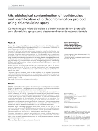
The results of experiment 2 are shown in Figure 1 and
demonstrate an efficient decontamination with 0.12%
chlorhexidine gluconate. Statistical analysis using the
ANOVA-main effects test showed no “carry-over” effect
(or sequence effect) that is common in crossed random tests,
showing that the time used to “wash-out” was enough to
prevent distortion of the results. The treatments with water
Microorganism Group
Range 1
(%/n)
Range 2
(%/n)
Range 3
(%/n)
Total
(%/n)
Streptococci 58/15 31/8 12/3 100/26
Streptococcus mutans 66/10 33/5 0/0 100/15
Staphylococci 50/9 33/6 17/3 100/18
Enterobacteria 29/4 57/8 14/2 100/18
Yeast 100/3 0/0 0/0 100/3
Range 1 corresponds to growth in the range between 100 and 3000 CFU/mL; Range 2 corresponds to growth in
the range between 3,100 and 10,000 CFU/mL; Range 3 corresponds to growth over 10,000 CFU/mL.
Table 3. Frequency of development
within the growth ranges of
microorganisms isolated from
toothbrushes used by Dentistry
students at Unioeste/PR
Table 2. Growth frequency (%) of microorganisms on
toothbrushes used by students from Unioeste/PR
Microorganism group Frequency (%) n
Streptococci 81.3 26
Streptococcus mutans 46.9 15
Staphylococci 56.3 18
Enterobacteria 56.3 18
Yeast 9.4 3
Fig. 1. Growth of total microorganisms in CFU/mL (X102
)
in toothbrushes submitted to the use of 0.12% chlorhexidine
gluconate three times a day, once a day and without the use
of antiseptic (water). Values represent the geometric mean
±95% confidence interval of values for microorganisms’ growth.
The letter “A” represents the presence of statistical significance
(P<0.05) between treatment with chlorhexidine 3 times/day
and water treatment, and the letter “B” indicates the absence
of statistical significance (P>0.05) between treatment with
chlorhexidine once a day and water treatment.
4. 216 Rev Odonto Cienc 2012;27(3):213-217
Microbiological contamination of toothbrushes
and chlorhexidine 1 or 3 times a day were compared using
the ANOVA test. Only the treatment with chlorhexidine
three times a day showed a significantly lower microbial
growth (P<0.05) than the treatment with water. The number
of daily brushings significantly influenced the decrease in the
number of microorganisms on the brushes, and although the
chart shows a sharp drop in the number of microorganisms
after the treatment with chlorhexidine once a day compared
to the treatment with water, this decrease did not reach
statistical significance. No brush from the negative control
group showed microbial growth.
Discussion
It has been shown in the literature that toothbrushes
are excellent locations for the growth of microorganisms
(10,11).
Microbial growth was detected on almost all of the
brushes tested in this study (> 90%), with development
of streptococci observed on the vast majority of the
brushes, which shows that toothbrushes are an excellent
means of transport for bacteria. Nearly half of the brushes
showed growth of mutans streptococci, members of the
oral microbiota that are currently considered to be major
cariogenic agents (1,12). This finding highlights the ability
of mutans streptococci to form biofilms and binding to the
material of the toothbrushes (1).
Different population groups, such as children, adults
or the elderly, may differ in their microbial load (1,13),
particularly in the effectiveness of their brushing. This study
addressed a population with knowledge about the correct
method of brushing, and the results indicate levels of
contamination similar to those obtained by other studies (6,5).
Staphylococci were found in large numbers, on over 50%
of the brushes. Although it belongs to the oral microbiota,
Staphylococcus aureus deserves greater attention because
it is capable of producing many oral infectious diseases.
The contamination by enterobacteria also draws
attention, as it was found on more than 50% of the brushes,
as a result of incorrect storage of brushes, most likely out
of a closet and over the bathroom sink, where it is a target
of aerosols from the toilet (14).
The growth of yeast varies greatly depending on the
methodology and population used in the survey, with the
population surveyed in this study being detected in low
numbers compared to other studies (5,11). The quantification
of these organisms showed heterogeneity in growth for all
types of microorganisms, ranging from a few CFU per
milliliters to a virtually limitless growth depending on the
methodology used. Such differences in growth may be a
result of the different behaviors of the individuals in this
research, including variations in the use of, for example,
mouthwash containing antimicrobial solutions (15), the type
of toothpaste (with or without antibiotics) (16) and the time
of use of these items (17).
This study describes a protocol that is capable of
standardizing the use of chlorhexidine, which is an antiseptic
that is already widely used for the decontamination of the oral
microbiota, for the daily decontamination of toothbrushes.
Several expedients have proven to be effective in
controlling the microbial contamination of toothbrushes
(15,18), and other works (8,19) have shown that chlorhexidine
is effective, but a pattern regarding its periodicity has not
yet been shown.
These results showed that the application of a single
spraying of chlorhexidine per day greatly reduced, although
with no statistical significance, the presence of bacteria,
with an 80.21% decrease comparing the group treated
once per day with the negative control, which suggests that
chlorhexidine at low concentrations (0.12%) can act as an
expedient to avoid a possible source of reinfection.
A significant decrease was found in the spraying of
0.12% chlorhexidine three times a day after every brushing,
with a 90.17% decrease compared to the negative control
and a 63.36% decrease compared to the strategy of daily
spraying with chlorhexidine.
This procedure could become an after-brushing habit
on a day-to-day basis for the general population because
the spraying, a simple and easily understandable act, was
performed with a technique that can be assimilated by any
person without the need for specific skills. This approach
makes this procedure different from other antiseptic brush
techniques that have been tested, which were effective
strategies to prevent microbial growth but were hindered
by their need to be performed on a daily basis (6,9).
One advantage of chlorhexidine gluconate is its cost/
benefit, as it is an inexpensive antiseptic that can be
purchased in specialized pharmacies or in the processed
form (Periogard®
– Colgate Palmolive), with both forms
having similar efficacy (20).
Population groups at risk, such as those that are
immunocompromised, may have serious infections that
are caused by oral microorganisms (21), and these groups
may benefit from the routine use of chlorhexidine on their
brushes, thus preventing contamination caused by daily re-
exposure to microorganisms housed in toothbrushes.
Conclusion
The results of this study showed that the use of 0.12%
chlorhexidine after three daily brushings is effective as an
antiseptic technique for toothbrushes, establishing a still-
lacking protocol for the after-brushing antisepsis procedure
with chlorhexidine. It became clear that contamination of
toothbrushes occurs often, even in individuals who should
know the ideal conditions for the storage of toothbrushes.
5. Rev Odonto Cienc 2012;27(3):213-217 217
Rodrigues et al.
1. Wetzel WE, Schaumburg C, Ansari F, Kroeger T, Sziegoleit A. Microbial contamination
of toothbrushes with different principles of filament anchoring. J Am Dent Assoc
2005;136:758-65.
2. Nelson Filho P, Macari S, Faria G, Assed S, Ito IY. Microbial contamination of toothbrushes
and their decontamination. Pediatric Dent 2000;22:381-4.
3. Goldschmidt MC, Warren DP, Keene HJ, Tate WH, Gowda C. Effects of an antimicrobial
additive to toothbrushes on residual periodontal pathogens. J Clin Dent 2004;15:66-70.
4. Bhat SS, Hedge KS, George RM. Microbial contamination of toothbrushes and their
decontamination. J Ind Soc Pedi Prev Dent 2003;21:108-12.
5. Mehta A, Sequeira PS, Bhat G. Bacterial contamination and decontamination of
toothbrushes after use. N Y State Dent J 2007;73:20-2.
6. Pai V. Effect of a single use toothbrush on plaque microflora. Indian J Dent Res 2009;
20:404-6.
7. Komyiama EY, Back-Brito GN, Balducci I, Koga-Ito CY. Evaluation of alternative methods
for the disinfection methods of toothbrushes. Braz Oral Res 2010;24:28-33.
8. Sato S, Pedrazzi V, Lara EHG, Panzeri H, Albuquerque Jr RF, Ito IY. Antimicrobial spray
for toothbrush disinfection: an in vivo evaluation. Quintessence Int 2005;36:812-16.
9. Bezirtzoglou E, Cretoiu SM, Moldoveanu M, Alexopoulos A, Lazar V, Nakou M. A
quantitative approach to the effectiveness of ozone against microbiota organisms colonizing
toothbrushes. J Dent 2008;36:600-5.
10. Dayoub MB, Rusilko D, Gross A. Microbial contamination of toothbrushes. J Dent Res
1977;56:706.
11. Taji S, Rogers A. The microbial contamination of toothbrushes. A pilot study. Aust Dent J
1998;43:128-30.
12. Napimoga MH, Höfling IF, Klein MI, Kamiya RU, Gonçalves RB. Transmission, diversity
and virulence factors of Streptococcus mutans genotypes. J Oral Sci 2005;47:59-64.
13. Nelson Filho P, Faria G, da Silva RAB, Rossi MA, Ito IY. Evaluation of the contamination
and disinfection methods of toothbrushes used by 24 to 48 month old children. J Dent
Child 2006;73:152-8.
14. Long SR, Dos Santos AS, Nascimento CMO. Avaliação da contaminação de escovas
dentais por enterobactérias. Rev Odontol Univ Santo Amaro 2000;5:21-5.
15. Balappanavara AY, Nagesh L, Ankola AV, Tangade PS, Kakodkar P, Varun S. Antimicrobial
efficacy of various disinfecting solutions in reducing the contamination of the toothbrush
– a comparative study. Oral Health Prev Dent 2009;7:137-45.
16. Lima MVV, Watanabe E, Faria G, Nascimento AP, Verri MP, Ito IY. Biofilme: avaliação
do nível de contaminação de escovas dentais monobloc®
em função do dentifrício. Rev
Odonto Ciênc 2007;22:269-74.
17. Sogi SH, Reddy SD, Kiran SN. Contamination of toothbrush at different time intervals and
effectiveness of various disinfecting solutions in reducing the contamination. J. Indian Soc
Pedod Prev Dent 2002;20:81-5.
18. Ankola AV, Hebbal M, Eshwar S. How clean is the toothbrush that cleans your tooth? Int
J Dent Hygiene 2009;7: 237-40.
19. Ayşegül Ö, Elgin IE, Gulcin A, Nedim S. The effıcacy of chlorhexidine spray vs. mouthwash
in the microbial contamination of child toothbrushes. J Dent Child 2007;74:177-81.
20. Segundo AS, Bosco AF, Semenoff TADV, Rocatto GEGD, Cirilo DM, Buzelle SL et al.
Efetividade do gluconato de clorexidina a 0,12% e do digluconato de clorexidina a
2% adquiridos em diferentes dentais e farmácias na cidade de Cuiabá, sobre Candida
albicans. Rev Period 2007;17:41-5.
21. Kennedy HF, Morrison D, Tomlinson D, Gibson BES, Bagg J, Gemmell CG. Gingivitis and
toothbrushes: potential roles in viridans streptococcal bacteraemia. J Infect 2003;46:67-70.
References