1. MICROSCOPIC FIBER IDENTIFICATION BY REFRACTIVE INDEX
Mark A. Goodman
Determining the identity of a fiber can sometimes be difficult. A method that is useful in
determining the fiber category by refractive index is presented. Application to distinction between
hemp and linen is discussed.
Refractive Index
There is a common misconception that the speed of light is constant. That's right; the speed of light
is only a constant in a vacuum or any other homogeneous material. Light from a microscope enters a
sample the speed of light is slowed down. The common definition of index of refraction (n) for a
fiber is:
speed of light in air
n=
speed of light in fiber
For fibers the refractive index is related to the internal (molecular) structure and is independent of
factors which determine its outer (morphological) features.
Anisotropy
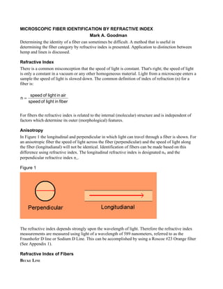
In Figure 1 the longitudinal and perpendicular in which light can travel through a fiber is shown. For
an anisotropic fiber the speed of light across the fiber (perpendicular) and the speed of light along
the fiber (longitudianal) will not be identical. Identification of fibers can be made based on this
difference using refractive index. The longitudinal refractive index is designated n% and the
perpendicular refractive index n⊥.
Figure 1
The refractive index depends strongly upon the wavelength of light. Therefore the refractive index
measurements are measured using light of a wavelength of 589 nanometers, referred to as the
Fraunhofer D line or Sodium D Line. This can be accomplished by using a Roscoe #23 Orange filter
(See Appendix 1).
Refractive Index of Fibers
BECKE LINE
2. A band or halo of light (due to diffraction/refraction) can be seen at the edge of a fiber when the
refractive indices of the fiber and its mounting medium are different. In practice, the Becke Line is
produced by focusing above and below the plane of best focus but is not discernable when the fiber
is in focus. This method is named after the Austrian geologist and petrologist, Friedrich Johann Karl
Becke, 1855-1931, who devised the method.
The Becke Line always moves toward the material of higher refractive index on focusing above the
plane of best focus.
The Becke Line moved inward and outward are presented. Usually it is best to observe the Becke
line at a total magnification of 200X to 500X.
Becke Line Moved Inward (Shown in White)
Becke Line Moved Outward (Shown in White)
DETERMINATION OF PARALLEL AND PERPENDICULAR REFRACTIVE INDICES
MICROSCOPE SET UP
3. Only a linear polarizer and Roscoe #23 filter (Rosco Laboratories Inc., 52 Harbor View, Stamford,
CT 06902) are need to modify a standard light microscope for Becke Line analysis.. The polarizer is
relatively inexpensive and can be purchased at a camera store. Make sure that the polarizer is a
linear polarizer. The microscope set is shown in Figure 5.
Figure 5: Microscope Set Up Diagram
The polarizer must be oriented such that the polarizer direction runs in the east-west direction
relative to the front of the microscope (Figure 6). See Appendix 1 for determination of the polarizer
direction. If you are using a polarizer light microscope, the polarizer should be already oriented
correctly. A discussion of polarization of light and polarizer orientation is presented in Appendix 2.
Figure 6: Polarizer Orientation
4. FIBER ORIENTATION ON SLIDE
For determination of the parallel refractive index (n%), the fiber must be oriented in the parallel
(east-west) direction when viewed in the microscope. In other words the polarization direction of the
light and fiber orientation must be the same as shown below and as discussed in Appendix 2. The
polarization direction and fiber need to be perpendicular for obtaining the perpendicular refractive
index of the fiber (n⊥).
Orientation of Fiber on Slide – as viewed in the microscope
REFRACTIVE INDEX IMMERSION LIQUIDS AND THE BECKE LINE
The iris diaphragm needs to be closed until is it just slightly larger than the microscopes field of
view. The distance between the objective and the fiber sample is reduced (see figure below) using
5. the fine focus knob of the microscope. If the Becke Line moves inward, the fiber has a lower
refractive index than the immersion liquid and is assigned a negative sign (-). If the Becke Line
moves outward from the fiber, the immersion liquid has the higher refractive index than the fiber
and is assigned a positive sign (+).
FIBER IDENTIFICATION
It is recommend that for fiber identification that an immersion liquid of refractive index 1.550 be
used initially. Refractive index immersion liquids can be purchased from Cargille Laboratories, 55
Commerce Rd., Cedar Grove, NJ 07009 USA, (973)-239-6633, cargillelabs@aol.com.
A few fibers are placed on a clean microscope slide and a drop or two of the immersion liquid is
added followed by placing a cover slip on top. Examine the slide on the microscope and observe the
fiber in the parallel orientation, focusing on a sharp edge of the fiber. The iris diaphragm needs to be
closed until is it just slightly larger than the microscopes field of view.
Determine if the immersion liquid has a higher or lower refractive index then the fiber by observing
the Becke line as discussed above. The process can be repeated using other immersion liquids until
the n% of the fiber has been located within an acceptably small range on a trial and error basis. The
fiber is then oriented in the perpendicular direction and the process is repeated until n⊥ is determined.
The Fiber Identification table below has the n% and n⊥ of several common fibers. The fibers are
classified from low to high birefringence. Birefringence is defined below:
birefringence = n% - n⊥
6.
7. STANDORT PLOT
The Standort Plot shown below is useful in determining different basic types of fibers. For example
to know if a fiber is polyester, the Standort Plot shows that the parallel refractive index (n%) is
greater than 1.60. Since there are no other fiber classes that have a refractive index that high. An
immersion liquid of refractive index 1.550 could be used and if the n% is determined to be greater
than the immersion liquid and then the fiber is identified as polyester. This can be done without
performing the n⊥. This is a basic example similar schemes could be used to perform the fiber
identity for other fiber classes.
8.
9. Linen and Hemp Fiber Identification
The Becke Line can be used to determine if a fiber is linen or hemp. Refractive indexes for some
fibers presented below.
In most cases ramie can be distinguished from linen or hemp based on the fact that ramie has a
readily identifiable twist which is not present in hemp or linen fibers. Based on the n⊥ values
presented in table, it would be impossible to tell the difference between ramie, jute, linen, or hemp.
Fiber n% n⊥
Wool 1.553-1.556 1.542-1.547
Silk 1.591-1.595 1.538-1.543
Cotton 1.573-1.581 1.529-1.534
Ramie 1.595-1.599 1.527-1.540
Linen 1.594-1.596 1.528 -1.532
Hemp 1.585-1.591 1.526-1.530
Jute 1.577 1.536
Viscose rayon 1.539-1.550 1.514-1.523
Cellulose acetate 1.476-1.478 1.470-1473
Polyethylene 1.556 1.512
Refractive indices of fibers using immersion oils can be determined to within 0.002. Since this value
is than the reported difference between hemp and linen use of refractive index method was
investigated.
Initial investigation revealed that the published values for the refractive index of hemp and linen
given above were inaccurate. From this investigation the refractive index of linen (n%) was ~1,579
while that of hemp was ~1.582.
The refractive index data for different sources of hemp and linen are presented below. Based on this
data the identification hemp or linen based on refractive index is possible using the Becke Line
method for determination of refractive index.
10. Parallel Refractive Index Data for Different Sources of Hemp and Linen
Refractive Index Oil
Sample Comments
1.576 1.578 1.580 1.584
Raw & Unprocessed + + - Using Polarized Light Microscope
Raw & Unprocessed + + - Using Polarized Light Microscope
Standard Microscope using Nikon
Raw Linen - Unprocessed + + - Polarizer
Linen Suit Collar 1 + + - Using Polarized Light Microscope
cloth blended with cashmere + + - Using Polarized Light Microscope
dry spun yarn + + - Using Polarized Light Microscope
Linen
blue yarn from Belgium + + - Using Polarized Light Microscope
Using Polarized Light Microscope. Slight
wet spun yarn + + - Becke line
Jones NY collar - made in China + + - Using Polarized Light Microscope
Bleached Linen + + - Using Polarized Light Microscope
White Linen Cord + + -
Hemp Cord – Mislabeled + + -
Hemp Cord Source 1 + + + - Using Polarized Light Microscope
Green Hemp Cord + + + - Using Polarized Light Microscope
Hemp Cord Source 2 + + + - Using Polarized Light Microscope
Hemp
Hemp/Wool Sweater + + + - Using Polarized Light Microscope
72% Wool/28% Hemp cloth + + + - Using Polarized Light Microscope
Hemp Rope 1 (Mystic Seaport) + + - Using Polarized Light Microscope
Hemp Rope 2 (Mystic Seaport) + + - Using Polarized Light Microscope
11. APPENDIX 1: Determination of Polarizer Orientation.
To determine the orientation of a polarizer hold the polarizer in your hand and observe a reflect spot
on a glass surface or a shiny black (non-metallic) flat surface. Reflected light from these surfaces
can produce nearly polarized light. While observing reflection turn the polarizer slowly until the you
observe a spot in which the light is reduced to a minimum or is extinct. This will mean that the
orientation of the polarizer will be from the top to bottom. You can usually place a small mark at the
top and bottom to indicate orientation direction.
12. APPENDIX 2: Polarization of Light
The light source of a microscope produces unpolarized light which is made up of light waves
vibrating in all directions. When unpolarized light passes through a polarizer, only light in the
polarization direction passes trough. This is diagrammatically shown below. The incident beam is
made of light waves in every 360 degree orientation. The polarized light shown exiting the polarizer
in only in one direction called the polarization direction.
The microscope from the front view is shown below. When the polarizer is oriented in the east-west
direction (in the plane of the paper) the polarized light emerging from the polarizer, represented in
the blue box is also in the plane of the paper. This is important, because the polarization direction
and fiber orientation must be the same for determination of the parallel refractive index.
Figure 8: Front View of Microscope with Polarized Light Orientation
13. APPENDIX 3 – Use of Filter for Refractive Index Measurements.
The light source in a microscope is generated from a bulb which produces white light. This light is
made up of a mixture of wavelengths in the visible spectrum. In the refractive index depends
strongly upon the wavelength of light. In other words small changes in wavelength will cause large
changes in the refractive index (see figure below). Refractive index measurements are performed at
a specified wavelength. The most common measurement wavelength is the Fraunhofer D line at 589
nanometers. The Fraunhofer F, D and C lines are shown in the figure.
The importance of acquiring refractive index at a known wavelength is graphically demonstrated in
the figure below. The graph shows refractive index of two fibers (blue and red lines) which have
different dependences on wavelength. At the Fraunhofer D wavelength the fibers have the same
refractive index of 1.50. A small change in wavelength results will change the refractive index of the
fiber. A wavelength change from 589 nm to 600 nm results in a refractive index of 1.498 for the blue
fiber and the red fiber will have a refractive index of 1.496. This demonstrates that accurate
refractive index measurements require a filter to insure that the wavelength does not varry. Optical
filters are usually very expensive. An inexpensive solution is to use Roscoe #23 Orange filter (Rosco
Laboratories Inc., 52 Harbor View, Stamford, CT 06902) which will provide light equivalent to the
Fraunhoffer D line. The importance of using a filter when obtaining Becke lines for refractive index
measures was reported by R.C. Emmons and R.M. Gates, American Mineralogist. 1948; Vol:33,
612.
Dependence of Refractive Index on Wavelength of Light for Two Fibers.
F D C
1.58
1.56
Refractive Index
1.54
1.52
1.50
1.48
1.46
1.44
450 500 550 600 650 700
Wavelength (nm)