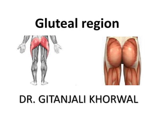
2. Gluteal region
• The transitional area between the trunk and
the lower extremity.
• The gluteal region includes the rounded,
posterior buttocks and the laterally placed hip
region.
5. Gluteal Aponeurosis
• This is attached to the
lateral border of the
iliac crest superiorly,
and
• splits anteriorly to
enclose tensor fasciae
latae and posteriorly
to enclose gluteus
maximus.
6.
7. Muscles of Gluteal region
Superficial Layer
• Gluteus maximus
• Tensor fasciae latae
8. Muscles of Gluteal region
Intermediate layer
• Gluteus medius
• Piriformis
• Superior gemellus.
• Tendon of obturator
internus.
• Inferior gemellus
• Quadratus femoris
• Upper part of
Adductor magnus
• And Hamstrings
9. Muscles of Gluteal region
Deep layer
• Gluteus minimus
• Reflected head of
rectus femoris
• Tendinous insertion
of obturator
externus
10. Gluteus Maximus
Origins: posterior end of the iliac crest,
posterior surface of the sacrum, coccyx and
sacrotuberous ligament.
Insertions: ilio-tibial tract( 3/4)and gluteal
tuberosity.(1/4 )
Innervation: inferior gluteal nerve - [ Ventral
rami of L5, S1,2] - emerges below the
piriformis muscle to penetrate the deep
surface of the gluteus maximus with
accompanying vessels.
11. Actions
• Extensor at hip joint during
running and climbing upstairs.
• Chief antigravity muscle in the
standing up from a seated position.
• Strong lateral rotation of the thigh.
Its upper fibres are active in
powerful abduction of the thigh.
• It is a tensor of the fascia lata, and
through the iliotibial tract it
stabilizes the femur on the tibia
when the extensor muscles of the
knee are relaxed.
12. Tensor Fascia Lata
Small muscle close to the anterior
border of the gluteus medius, at the dorsal
surface of the ASIS.
Origin: outer lip of iliac crest from ASIS to
tubercle of iliac crest.
Insertion: ilio-tibial tract.
Innervation - superior gluteal nerve.
Action - helps in flexion and abduction of the
thigh. Maintains extension of knee joint.
13. Structures under cover of gluteus maximus
• Bones
• Ligaments
• Bursae
Trochanteric
Gluteofemoral
Ischial
• Muscles
• Blood vessels and
• Nerves
• Arterial Anastomosis
Trochanteric
cruciate
16. GLUTEUS MEDIUS
Covered partially by Gluteus maximus
Origins: dorsal surface of the ilium
between the anterior and posterior
gluteal lines and from the gluteal
aponeurosis.
Insertion: lateral surface of the greater trochanter on an
oblique ridge.
17. GLUTEUS MINIMUS
Covered completely by Gluteus
medius.
Origins: gluteal surface of the ilium
between the anterior and inferior
gluteal lines upto margin of greater
sciatic notch.
Insertion: lateral part of anterior surface of the greater
trochanter.
18. • Innervation of Gluteus medius and minimus:
superior gluteal nerve [L4, 5, S1] – that emerges
above the piriformis muscle, with accompanying
vessels, to penetrate the deep surface of the
muscle.
• Actions
Abduction of the thigh and medial rotation.
Preventing the unsupported side of pelvis from
sagging downward during locomotion.
Lurching Gait
20. • Trendelenburgs sign is positive in
paralysis of gluteus medius & minimus,
congenital dislocation of hip joint,
fracture of the neck of femur
21. Piriformis
Origin: antero-lateral
surface and border of the
sacrum.
Insertion: the fibers are emerge laterally through the
greater sciatic foramen as a narrow tendon attached to
the posterior inturned upper border of the greater
trochanter.
Innervation - “nerve to the piriformis” [S1, 2.]
Action - lateral rotator and abductor of the thigh.
23. Obturator internus
• Origin : inner surface
of obturator membrane and
Adjoining ischio-pubic ramus.
• Insertion: Tendon makes a right
angle bend at lesser sciatic foramen
to insert to the medial surface of
greater trochanter above and in
front of the trochanteric fossa
24. Obturator internus
• It is accompanied by Superior and
Inferior Gemelli and insert at
superior and inferior margin of
the insertion of obturator
internus.
• Superior Gemellus from ischial
spine.
• Inferior Gemellus from lower
margin of lesser sciatic notch.
25. Obturator internus
• Nerve supply :
Nerve to obturator internus also
supplies Sup. Gemellus (L5,S1,S2)
Inf. Gemellus is supplied by nerve
to Quadratus femoris (L4,L5,S1)
• Action:
Lateral rotation at Hip joint
26. Quadratus Femoris
• Origin:
Linear origin from external surface
of ischial tuberosity.
• Insertion:
Quadrate tubercle near middle of
intertrochanteric crest.
• Innervation: nerve to Quadratus
femoris (L4,L5,S1)
• Action: Lateral rotation of hip
32. Superior gluteal nerve
Ventral rami of
L4, L5, S1
Inferior gluteal nerve
Ventral rami of
L5, S1, S2
Pudendal nerve
Ventral rami of
S2, S3, S4
33. Posterior femoral cutaneous nerve
D (S1, S2)
V (S2, S3)
It descends on the back of
the thigh, and in the popliteal
fossa it pierces the deep fascia
and supplies the skin on the back
of the thigh and the upper part
of the leg
Branches:
a) Gluteal
b) Perineal
c) Perforating
34. • Nerve to obturator
internus
Ventral rami of
V (L5, S1, S2)
• Nerve to quadrator
femoris
Ventral rami of
V (L4, L5, S1)
37. • Superior gluteal artery divides into
Superficial branch
Deep branch- upper branch and lower branch
SPINOUS ANASTOMOSIS
1. Upper branch of Superior
gluteal artery
2. Superficial and deep
circumflex iliac arteries
3. Ascending branch of lateral
circumflex femoral artery
4. Iliac branch of ilio-lumbar
artery.
38. • Superior gluteal artery
Branch of posterior division of Internal Iliac artery.
Branches:-
a) Muscular branches
b) Anastomotic branches
c) Arteria nervi ischiadici
• Internal pudendal artery
Branch of anterior
division of Internal Iliac artery
39.
40. CRUCIATE ANASTOMOSIS
•Descending branch of inferior
gluteal artery
•Ascending branch of 1st
perforating artery
•Medially- transverse branch of
medial circumflex femoral
artery
•Laterally- transverse branch of
lateral circumflex femoral artery
43. Trochanteric anastomosis
Descending branch of superior gluteal artery
ascending branches of medial & lateral
circumflex femoral artery
Branch from inferior gluteal artery
situated near the trochanteric fossa of the
femur & supplies the head of femur and
retinacular fibers of neck
45. Hamstring Muscles
Common name applied to the muscles in the
Posterior compartment.
They have a common origin from the ischial
tuberosity and crosses knee joint to insert on tibia
or fibula.
They are innervated by the tibial component of
sciatic nerve.
They also have a common primary function of
flexing the leg, but they also help to extend and
adduct the thigh.
Their blood supply comes principally from the
perforating branches of the deep femoral artery.
46. adductor magnus
long head of biceps femoris
semitendinosus
semimembranosus short head of biceps femoris
popliteal vessels in the
popliteal fossa
sartorius
gracilis
47. Origin - Common from Ischial tuberosity.
Insertion - One of the leg bones.
Nerve supply-Tibial part of sciatic nerve.
Common action Extensors of hip joint.
Flexors of knee joint.
48. True hamstrings-
• Semimembranosus
• Semitendinosus
Modified hamstrings
•Long head of biceps femoris-
•Sacrotuberous ligament morphologically
degenerated part.
•Ischial head of adductor magnus-
Tibial collateral ligament represents the
morphological degenerated part of adductor magnus
49. Biceps Femoris - most lateral muscled with
a “long head” from the ischial tuberosity,
and a “short head” from the middle of the
linea aspera and the lateral supracondylar
ridge.
*The two heads unite to form a common
tendon, which deviates lateral to its
insertion into the apex of the head of the
fibula where it is joined by an extension of
the iliotibial tract.
•The short head receives a branch from
the common peroneal nerve; it also helps
in the lateral rotation of the leg.
50. Semitendinosus - usually fusiform tapering
distally into a long cylindrical tendon at the
popliteal region to be inserted to the upper
medial surface of the tibia, adjacent to the
attachments of the sartorius and gracilis.
Semimembranosus - usually has a fleshy belly
that form a thick flattened tendon that inserts at
the back of the medial condyle of the tibia, the
tendon contributes to the formation of the
“oblique popliteal ligament” of the knee joint,
which reinforces the posterior capsule of the
joint.