Contenu connexe
Similaire à 5 linfócitos b e receptores do tipo toll (20)
Plus de ufamimunologia (19)
5 linfócitos b e receptores do tipo toll
- 1. REVIEWS
Integration of B cell responses through
Toll-like receptors and antigen receptors
David J. Rawlings1,2,3, Marc A. Schwartz2,3, Shaun W. Jackson1,3 and
Almut Meyer-Bahlburg4
Abstract | Unlike other immune cells, B cells express both an antigen-specific B cell receptor
(BCR) and Toll-like receptors (TLRs). Dual BCR and TLR engagement can fine-tune functional
B cell responses, directly linking cell-intrinsic innate and adaptive immune programmes.
Although most data regarding B cell-specific functions of the TLR signalling pathway have
been obtained in mice, the discovery of patients with a deficiency in this pathway has
recently provided an insight into human B cell responses. Here, we highlight the importance
of the integration of signalling pathways downstream of BCRs and TLRs in modulating
B cell function, focusing when possible on B cell-intrinsic roles.
Over the last decade, the unexpected success of B cell Notably, based on their functional responses as well
B‑1 cells
IgMhiIgDlowMAC1+B220lowCD23– depletion therapies in treating human autoimmunity, as their BCR repertoire, naive mature B cell populations
cells that are dominant in the combined with a growing recognition of the importance have been defined as either innate-like cells or adaptive
peritoneal and pleural cavities. of neutralizing antibody responses in host defence, has cells (BOX 1). Innate-like B‑1 cells and marginal zone B cells
The size of the B‑1 cell led to an increased focus on understanding the role(s) generate rapid antibody responses independently of T cell
population is kept constant
owing to the self-renewing
of B cells in human immune function. B cells do not help. By contrast, adaptive follicular B cells primarily par-
capacity of these cells. B‑1 merely produce immunoglobulins; they can also secrete ticipate in T cell-dependent (referred to as T‑dependent
cells recognize self components, cytokines and serve as antigen-presenting cells, and throughout this article) responses that lead to the genera-
as well as common bacterial therefore B cells have a multifaceted involvement in tion of high-affinity antibodies and long-term memory.
antigens, and they secrete
distinct immune responses. Importantly, the expression of distinct profiles of TLRs
antibodies that tend to have low
affinity and broad specificity. A striking characteristic of B cells is the expression of a and specific BCR profiles probably helps to specify the
clonally rearranged, antigen-specific B cell receptor (BCR) differentiation and function of these key innate-like
in conjunction with the expression of one or more mem- versus adaptive B cell populations. During T‑independent
1
Department of Pediatrics, bers of a family of germline-encoded receptors termed immune responses, dual BCR and TLR signalling rapidly
University of Washington Toll-like receptors (TLRs), which are capable of recogniz- induces marginal zone B cell and B‑1 cell migration and
School of Medicine, Seattle,
ing discrete microbial ligands. This dual expression pattern antibody production. In addition, following the trigger-
Washington 98195, USA.
2
Department of Immunology, permits B cells to uniquely integrate both antigen-specific ing of T‑dependent immune responses, TLR responsive-
University of Washington signals and ‘danger’ signals via these key receptor systems. ness is directly modulated in activated follicular B cells,
School of Medicine, Seattle, Although B cell development and survival both appear thereby affecting germinal centre responses. TLR engage-
Washington 98195, USA. phenotypically unperturbed in the absence of TLR sig- ment, in conjunction with BCR ligation, also provides
3
Center for Immunity and
Immunotherapies, Seattle
nals1, patients with a deficiency in a TLR signalling mol- a bridge between the innate and adaptive immune sys-
Children’s Research Institute, ecule — either myeloid differentiation primary-response tems that may have an impact on antigen presentation,
1900 Ninth Avenue, Seattle, protein 88 (MYD88) or IL‑1R‑associated kinase 4 (IRAK4) primary antibody responses, class-switch recombination
Washington 98101, USA. — possess an altered BCR repertoire with an increased (CSR) and subsequent memory responses.
4
Department of Pediatric
proportion of autoreactive B cells, presumably owing to All TLRs, with the exception of TLR3, require the
Pneumology and Neonatology,
Hannover Medical School, alterations in B cell selection processes2. Different B cell signalling adaptor MYD88 to mediate activation signals,
Hannover, Germany. subsets exhibit variations in TLR expression patterns, and although TLR4 is unique in that it can signal through both
Correspondence to D.J.R. signalling via TLRs can modify B cell responses such as the MYD88‑dependent and the MYD88‑independent
e‑mail: antibody production, antigen presentation and cytokine pathway 5. Therefore, analyses of animal models and
drawling@u.washington.edu
doi:10.1038/nri3190
secretion. Therefore, individual TLR expression profiles humans with deficient function of this adaptor have
Published online permit various effector B cell populations to manifest begun to provide important new insights into how signals
16 March 2012 specific response profiles following TLR engagement3,4. via TLRs affect B cell function and immune responses.
282 | APRIL 2012 | VOLUME 12 www.nature.com/reviews/immunol
© 2012 Macmillan Publishers Limited. All rights reserved
- 2. REVIEWS
Box 1 | Innate-like and adaptive B cell subsets
This Review focuses on the role of dual BCR and
TLR signalling (as well as other MYD88‑dependent
Based on phenotypical, functional and topographical characteristics, B cells can be signals) in normal and dysregulated B cell immune
divided into innate-like and adaptive immune cells109. Follicular B cells are the main function, with a particular emphasis on B cell-intrinsic
players during T‑dependent immune responses and belong to the adaptive arm of the events. We first summarize the specific roles for
immune system. They generate a clonally rearranged antigen-specific B cell receptor
MYD88 in T‑independent versus T‑dependent B cell
(BCR) and form memory responses that are dependent on T cell help. By contrast, B‑1
and marginal zone B cells are usually considered to be innate-like immune cells and
immune responses. We highlight work that demon-
generate rapid but lower affinity antibody responses that are independent of T cell help. strates a non-redundant role for B cell-intrinsic MYD88
The term ‘B‑1’ refers to the idea that this population develops earlier during ontogeny signals in antiviral immune responses, describe how
than conventional B‑2 cells110 (see below). B‑1 cells are enriched in the peritoneal and MYD88 controls a B cell-intrinsic, TLR-independent
pleural cavities but can also be found in the spleen. CD5 expression further subdivides pathway for immunoglobulin diversification, and
mouse B‑1 cells into CD5+ B‑1a and CD5– B‑1b cells. Recently, a B‑1 cell progenitor was review new information (gained from the analysis of
identified in the bone marrow of adult mice111. The term ‘B‑2’ has traditionally been patients with MYD88 or IRAK4 deficiencies) regard-
used to describe the main population of mature B cells that develop from common ing the role for TLR signals in human immune func-
bone marrow precursors and are located in the bone marrow, spleen and lymph nodes. tion. Next, we describe how dual BCR and TLR signals
B‑2 cells therefore include both follicular and marginal zone subsets, which currently
may potentiate the risk for autoimmunity and discuss
are referred to as separate populations because of their distinct phenotypical and
functional characteristics.
recent findings regarding how regulatory B cell func-
Recent work has defined a B cell subset in human peripheral blood with functional tion also requires TLR–MYD88 signals. Finally, we
responses similar to those of mouse B‑1 cells112. Consistent with their innate-like discuss emerging data indicating that dysregulated
immune cell phenotype, B‑1 and marginal zone B cells mainly express germline- TLR and BCR signals collaborate during the devel-
encoded antigen receptors that have limited diversity and are enriched for opment of B cell malignancies and, possibly, in other
specificities that recognize microbial and self antigens. In addition to BCR ligation, pathogenic B cell states.
the activation of pattern-recognition receptors, including Toll-like receptors (TLRs),
on these cells is important for their immune responses. Moreover, B‑1 and marginal T-independent B cell immune responses
zone B cells are the primary producers of natural IgM antibodies113. Through these As innate-like cells, B‑1 and marginal zone B cells
characteristics, both subsets are crucial in the early phase of T‑independent immune
are the main players during T‑independent immune
responses10, as they link innate and adaptive immune mechanisms. Moreover, owing to
their polyspecific BCR repertoire, B‑1 and marginal zone B cells have been implicated
responses. Innate-like B cells rapidly develop into
in driving autoimmune processes109. IgM-producing plasmablasts during the early primary
immune response10, and the importance of these subsets
in T‑independent responses to encapsulated bacteria has
Furthermore, recent work indicates that MYD88 also been demonstrated in models of infection, most notably
orchestrates B cell-activating factor (BAFF)-dependent with Streptococcus pneumoniae11. Both mice and humans
signals through TACI (transmembrane activator and with deficiencies in B‑1 and marginal zone B cells —
CAML interactor; also known as TNFRSF13B). including patients with hypogammaglobulinemia,
Marginal zone B cells BAFF and the related cytokine APRIL (a proliferation- agammaglobulinemia12 or Wiskott–Aldrich syndrome13 —
Mature B cells that are
inducing ligand) have a major role in peripheral B cell also exhibit increased susceptibility to infections with
enriched mainly in the spleen
marginal zone, which is located homeostasis and survival6. These cytokines bind to a fam- encapsulated bacteria. In addition, young children (who
at the border of the white pulp. ily of receptors expressed primarily on B cells, including lack marginal zone B cells because the formation of this
the BAFF receptor (BAFFR), B cell maturation antigen compartment is physiologically delayed until about
Follicular B cells (BCMA) and TACI. BAFF levels are modulated by a 2 years of age), as well as splenectomized individuals,
A recirculating mature B cell
subset that populates the
complex interplay of factors, which include the num- are at very high risk for such infections14.
follicles of the spleen and ber of B cells competing for ligand; the relative level of
lymph nodes. BAFF-binding receptors on specific B cell subsets; and In vitro analyses of TLR-modulated T‑independent
pro-inflammatory (including TLR-driven) signals that responses. Stimulation of B cells via TLR ligands
Germinal centre
modulate BAFF production by myeloid and/or stromal has been used extensively as an in vitro model of
A lymphoid structure that
arises in follicles after cell populations. BAFF- or BAFFR-deficient mice have T‑independent immune responses. In vitro, both B‑1
immunization with, or exposure severely decreased numbers of B cells, whereas over- and marginal zone B cells rapidly proliferate and secrete
to, a T-dependent antigen. expression of BAFF increases B cell numbers and pro- antibodies in response to the engagement of TLRs,
This structure is specialized for motes systemic lupus erythematosus (SLE)-like disease. including TLR1–TLR2, TLR4, TLR6, TLR7 and TLR9
facilitating the development of
high-affinity, long-lived plasma
Both BCR-mediated and TLR-mediated stimulation of (REFS 3,15–18). Interestingly, B‑1 cells (predominantly
cells and memory B cells. mouse marginal zone B cells7,8 or B‑1 cells9 upregulates B‑1b cells; see BOX 1) stimulated in this manner secrete
the expression of BAFFR and TACI, and thus increases the large amounts of IgA, whereas marginal zone B cells pri-
Class-switch recombination sensitivity of these cells to BAFF and APRIL. As a result, marily produce IgM4,15; these features correlate with their
(CSR). The process by which a
B cell activation during immune responses directly modu- in vivo functional roles against mucosal and blood-borne
heavy-chain variable-region
gene segment that is attached lates the outcome of BAFF signalling by regulating the infections, respectively. Notably, both B‑1 and marginal
to one heavy-chain constant- expression of BAFF family receptors. Finally, an additional zone B cells exhibit stronger functional responses to
region gene segment in the level of control is provided through the direct participa- TLR ligands than follicular B cells, as measured by the
expressed heavy-chain gene is tion of MYD88 in BAFF-triggered TACI signalling. This upregulation of activation markers15,18 and the produc-
recombined with a downstream
constant-region gene segment,
dual role for MYD88 in TLR and TACI signalling permits tion of interleukin‑10 (IL‑10)19,20. Marginal zone B cells
leading to the expression of a the precise regulation of peripheral B cell responses to have also been shown to exhibit a greater potential to
new antibody class. infectious challenge and other microenvironmental cues. act as antigen-presenting cells than follicular B cells in
NATURE REVIEWS | IMMUNOLOGY VOLUME 12 | APRIL 2012 | 283
© 2012 Macmillan Publishers Limited. All rights reserved
- 3. REVIEWS
a Redistribution of B-1 and b Direct functional responses c Extrafollicular CSR
marginal zone B cells TLR
BAFF or
APRIL
Marginal zone TACI
B cell IgM, IgA and IgG3
Proliferation TLR BCR production
Blood
vessel
MYD88
IRAK1 IRAK4
B-1 or TRAF6
marginal
zone B cell
Cytokine Survival:
TLR Follicle IKKγ
secretion BAFF receptor
Marginal expression IKKα IKKβ
B-1 cell
zone
Spleen
Peritoneal Antigen p50 p65
cavity presentation NF-κB CSR
p50 p65 AID
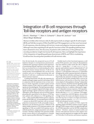
Figure 1 | The role of BCR and MYD88 signalling in B cells during T‑independent responses. Innate-like B cells —
that is, B‑1 and marginal zone B cells — are most crucial for T‑independent immune responses. a | Following the ligation
of Toll-like receptors (TLRs; specifically TLR2, TLR3 (which is MYD88 independent), TLR4 and TLR7), B-1 and marginal
Nature Reviews | Immunology
zone B cells downregulate integrin receptor expression, resulting in their redistribution to the blood, lymph nodes and
spleen follicles. b | Subsequently, B‑1 and marginal zone B cells start to proliferate and secrete immunoglobulins and
cytokines. Upregulation of the B cell-activating factor (BAFF) receptor leads to the prolonged survival of these cells.
Increased expression of co-stimulatory molecules also enables activated innate-like B cells to function as antigen-presenting
cells, thereby linking T‑independent and T‑dependent immune responses. Most of these functional responses occur in
response to direct TLR engagement in vitro; however, in vivo, these events probably require synergistic B cell receptor
(BCR) and TLR engagement. c | Recent data demonstrate the direct involvement of myeloid differentiation primary-
response protein 88 (MYD88) in generating nuclear factor-κB (NF-κB) signals that are dependent on TACI (transmembrane
activator and CAML interactor; also known as TNFRSF13B). These signals can result in the expression of activation-induced
cytidine deaminase (AID; an enzyme that is required for somatic hypermutation and class-switch recombination (CSR) in
germinal centres) and extrafollicular CSR, independently of T cell help. This pathway might also be directly activated by
TLR signals, as in vitro stimulation of B cells with TLR ligands induces AID expression. Moreover, because TLR stimulation
leads to the upregulation of receptors for BAFF, signalling via this pathway and the induction of extrafollicular
CSR might be enhanced by dual TACI and TLR engagement. APRIL, a proliferation-inducing ligand; IKK, IκB kinase;
IRAK, IL‑1R‑associated kinase; TRAF6, TNFR-associated factor 6.
response to TLR stimulation18, allowing this unique sub- Thus, consistent with their characterization as innate-
Systemic lupus
erythematosus
set to efficiently bridge T‑independent and T‑dependent like immune cells, B‑1 and marginal zone B cells gener-
(SLE). An autoimmune disease responses by shuttling antigens from the marginal zone ally exhibit stronger in vitro responses to TLR signalling
in which autoantibodies that and presenting them in the T cell zones (FIG. 1b). than follicular mature B cells, and the regulation of MYC
are specific for DNA, RNA or Our group has investigated the mechanism(s) expression levels contributes, in part, to this differential
proteins associated with
accounting for the differential responsiveness of adap- response profile.
nucleic acids form immune
complexes that damage small tive (follicular) and innate-like (marginal zone) B cells
blood vessels, especially to the TLR4 ligand lipopolysaccharide (LPS)3, which is In vivo analyses of TLR-modulated T‑independent
in the kidney. Patients with a classical T‑independent type 1 (polyclonal) stimulus. responses. Signalling via both MYD88 and Bruton’s tyro
SLE generally have abnormal We confirmed an increase in cell cycle entry (prolifera- sine kinase (BTK; a kinase essential for BCR signalling) is
B and T cell function.
tion) in marginal zone compared with follicular B cells required for T‑independent pathogen-specific IgM pro-
Wiskott–Aldrich syndrome in response to LPS, but could not identify significant dif- duction in a mouse model of Borrelia hermsii infection21.
A life-threatening X‑linked ferences in proximal TLR-driven biochemical signalling Mice deficient in both BTK and MYD88 failed to generate
immunodeficiency caused by events, such as the activation of the nuclear factor-κB pathogen-specific IgM21, whereas mice deficient in BTK
mutations in the gene encoding
(NF-κB) and mammalian target of rapamycin (mTOR) alone generated specific antibodies and resolved bacterae-
Wiskott–Aldrich syndrome
protein. It is characterized by pathways. Notably, follicular B cells had reduced basal mia, but with delayed kinetics compared with wild-type
thrombocytopenia with small and inducible levels of the cell cycle and growth regula- mice. Mice deficient in TLR1, TLR2 or MYD88 generated
platelets, eczema, recurrent tor MYC. Consistent with a key role for MYC in modu- pathogen-specific antibodies with delayed kinetics and
infections caused by lating LPS responsiveness, enforced expression of MYC suffered more severe episodes of bacteraemia compared
immunodeficiency and an
increased incidence of
in follicular B cells eliminated the delay in cell cycle entry with control mice, suggesting that MYD88 synergizes
autoimmune manifestations and promoted increased immune responses that mim- with BCR signals and that, in Btk–/– B cells, TLR-mediated
and malignancies. icked the functional responses of marginal zone B cells. stimulation is required to rescue the defective BCR signal.
284 | APRIL 2012 | VOLUME 12 www.nature.com/reviews/immunol
© 2012 Macmillan Publishers Limited. All rights reserved
- 4. REVIEWS
Two groups have analysed in vivo B cell responses to expression by marginal zone B cells, and thus reduces the
Salmonella enterica subsp. enterica serovar Typhimurium chemotactic responsiveness of these cells to S1P, possibly
in the setting of a MYD88 deficiency restricted to the explaining the effect of LPS on B cell localization. Of
B cell compartment 22,23. S. Typhimurium induces both note, TLR2, TLR3 and TLR7 ligands also promote mar-
T‑independent and T‑dependent immune responses. ginal zone B cell migration28. However, whereas in vivo
Following S. Typhimurium infection, S. Typhimurium- treatment with TLR2, TLR4 and TLR7 ligands resulted
specific IgM and IgG levels were initially lower in mice in the downregulation of S1pr1 mRNA levels, no change
with Myd88–/– B cells than in control mice, but no differ- was seen after stimulation via TLR3, implying that at
ence was observed at later time points (>4 weeks) after least two distinct TLR-dependent signalling pathways
infection. Moreover, MYD88 signalling in B cells mark- can promote the migration of marginal zone B cells.
edly inhibited both neutrophil and natural killer (NK) Similarly, B‑1 cells express very high levels of integ-
cell responses to S. Typhimurium23. Based on these lim- rins, and stimulation via TLRs induces a massive egress
ited in vivo data, MYD88 signalling in B cells in conjunc- of B‑1 cells from the peritoneal cavity that is associated
tion with BCR engagement appears to have an important with the coordinated downregulation of integrin and
role during early T‑independent immune responses to CD9 expression29. Thus, B cell-intrinsic TLR signalling
bacteria, leading to the rapid production of protective in vivo can directly alter B‑1 cell responsiveness, thereby
IgM and IgG, as well as the modulation of other innate directing the migration of these cells to sites where rapid
effector cell populations (presumably via cytokine and/or local antibody responses help to limit pathogen growth.
chemokine production). This probably also allows innate-like B cells to modulate
the local effector functions of other immune cell types.
TLR signals in innate-like B cell positioning. TLR sig-
nals have an important role in the regulation of the local- MYD88 signals in human innate-like B cell responses.
ization and migration of B‑1 and marginal zone B cells Although it is difficult to judge how findings generated
(FIG. 1a). Although they are enriched in the splenic mar- using mouse models can be translated into the human
ginal zone, marginal zone B cells continuously circulate system, some insight into the role of B cell-intrinsic TLR
between the marginal zone and the splenic follicles, a signals can be obtained from the analysis of patients
property that allows this population to capture and shut- with inborn errors in one of the downstream TLR
tle blood-borne antigens to follicular dendritic cells. signalling effectors MYD88 and IRAK4 (REFS 30–32)
In vivo treatment with LPS promotes near-complete (BOX 2). In these patients, S. pneumoniae is the most
relocalization of marginal zone B cells into the splenic common invasive pathogen, followed by Staphylococcus
white pulp24–26. Sphingosine-1‑phosphate receptor 1 aureus and Pseudomonas aeruginosa — all of which
(S1PR1; also known as S1P1) is an important regulator trigger T‑independent responses31. By contrast, anti-
of marginal zone B cell retention, and blocking S1PR1 body responses to protein antigens are normal in these
with the inhibitor FTY720 in vivo leads to the migration patients, and no severe viral, parasitic or fungal diseases
of marginal zone B cells into follicles27. Interestingly, LPS have been observed. The predominance of infections
administration results in the downregulation of S1PR1 with encapsulated bacteria suggests that TLR signalling
Box 2 | A requirement for TLR responses in humans is revealed by primary immunodeficiency disorders
Over the last decade, several monogenic primary immunodeficiencies affecting the Toll-like receptor (TLR) signalling
pathway have been identified. Inborn errors of the TLR and interleukin‑1 receptor (IL‑1R) pathway include the
deficiencies in IL‑1R‑associated kinase 4 (IRAK4) and myeloid differentiation primary-response protein 88 (MYD88) that
were identified in patients in 2003 (REF. 32) and 2008 (REF. 30), respectively. MYD88 and the serine/threonine kinase
IRAK4 are key adaptor molecules that function downstream of TLRs and IL‑1R. Both inborn errors are inherited in an
autosomal recessive manner and result in indistinguishable clinical diseases31. Patients with these inborn errors are
typically predisposed during infancy and early childhood to recurrent life-threatening bacterial infections, which most
commonly involve invasive pneumococcal disease with only limited signs of systemic inflammation. Interestingly, no
deaths or invasive infectious episodes have been reported in patients beyond 14 years of age. Moreover, no severe viral,
parasitic or fungal diseases have been reported in these patients, and the range of bacterial pathogens that they are
susceptible to seems to be restricted to Streptococcus pneumoniae, Pseudomonas aeruginosa and Staphylococcus aureus.
Immune responses to protein antigens were also mainly normal. Treatment of these patients with prophylactic
antibiotics, anti-pneumococcal vaccination and/or intravenous immunoglobulins seems to have a beneficial impact on
the disease. The absence of severe infections in older patients may result from maturation of other signalling cascades
and/or adaptive immune function that gradually compensates for the lack of TLR signalling.
Another group of inborn errors have been shown to affect the induction of interferon-β (IFNβ) and IFNλ in response to
TLR3 signalling (which is independent of MYD88) or in response to TLR7, TLR8 and/or TLR9 signalling. These disorders are
caused by an autosomal recessive deficiency of UNC93B (an endoplasmic reticulum‑resident transmembrane protein
deficiency that affects TLR3, TLR7, TLR8 and TLR9)114 and autosomal dominant TLR3 deficiency115. Both deficiencies are
associated with the occurrence of herpes simplex virus-mediated encephalitis.
Overall, these data suggest that deficits in TLR signalling increase the susceptibility of the host to specific pathogens
without obviously affecting immune responses to T‑dependent immunization or immune cell development. However,
further studies are required to fully understand the precise contributions of TLRs and IL‑1R to human host defence.
NATURE REVIEWS | IMMUNOLOGY VOLUME 12 | APRIL 2012 | 285
© 2012 Macmillan Publishers Limited. All rights reserved
- 5. REVIEWS
Common variable immune in human B cells is likely to be crucial for triggering clinical observations in individuals with MYD88 or
deficiency syndrome T‑independent immune responses that require B‑1 and/ IRAK4 deficiencies, as well as those in patients with
(CVID syndrome). The most or marginal zone B cells. A detailed analysis of the in vivo common variable immune deficiency syndrome owing to
common symptomatic responses to polysaccharide immunization in such indi- mutations in TACI, who present with hypogammaglob-
primary antibody deficiency,
characterized by decreased
viduals would be a helpful means to begin to directly test ulinaemia and impaired IgG responses to T‑independent
levels of serum immuno lobulins
g this interpretation. antigens34,35.
and a low or normal number In summary, MYD88 signalling in B cells is crucial
of B cells. Most patients A unique role for MYD88 in TACI signalling. Recent for T‑independent B cell responses, as these signals
suffer from recurrent infections,
work has identified an unexpected role for MYD88 in activate and position B cells, thereby promoting rapid
predominantly of the respiratory
and gastrointestinal tracts.
signal transduction via the BAFF- and APRIL-binding and appropriately localized production of pathogen-
The incidence of malignancies, cell-surface receptor TACI33 (FIG. 1c). After binding to specific antibodies. In addition, B cell-intrinsic MYD88
such as gastric carcinoma or BAFF, TACI directly interacts with MYD88 (REF. 33), signals triggered by TACI appear to promote extra
lymphoma, is increased in resulting in the activation of NF‑κB via a signalling follicular CSR, facilitating improved primary protective
patients with CVID syndrome.
cascade that is dependent on IRAK1, IRAK4, TNFR- T‑independent antibody responses.
associated factor 6 (TRAF6), TGFβ-activated kinase 1
(TAK1) and the IκB kinase (IKK) complex. Strikingly, MYD88 signals in T‑dependent B cell responses
TACI engagement, in conjunction with stimulation by B cells are recruited into T‑dependent immune responses
cytokines or TLR ligands, markedly facilitates CSR33, primarily from the follicular subset, after which they
suggesting that TACI signalling via MYD88 may help to enter a germinal centre and differentiate into either
promote extrafollicular B cell responses that lead to rapid antibody-secreting plasma cells or memory B cells.
and sustained IgG and IgA responses to T‑independent Although innate-like B cells generally display stronger
antigens (including polysaccharides from encapsulated responses to TLR engagement alone, antigen-mediated
bacteria). TACI–MYD88 interactions may also enhance follicular B cell responses can be modulated by simul-
antibody responses by delivering survival signals to acti- taneous BCR and TLR engagement (FIG. 2). In vitro,
vated extrafollicular B cells, including class-switched follicular B cells proliferate, produce cytokines and
plasmablasts. This mechanism may help to explain the secrete antibodies following stimulation by various
• Virus
• VLP containing a
TLR ligand
• Soluble TLR ligand
Plasma ↑ Antibody
BCR Endosome cell production
↑↑ MYD88
↑ Migration and Germinal
differentiation centre Maintenance
MYD88 B cell of long-term
humoral
MHC class II CSR to immunity
Follicular IgG2a/c
B cell Peptide
TCR
Memory
Cytokine B cell
CD4+ T cell Proliferation
TH cell differentiation
Figure 2 | The role of BCR and MYD88 signalling in B cells during T‑dependent immune responses. Follicular B cells
are activated following B cell receptor (BCR) engagement and interaction with cognate CD4+ T cells. Signalling through
Nature Reviews | Immunology
myeloid differentiation primary-response protein 88 (MYD88) in B cells during the primary response may enhance antigen
presentation by B cells to T cells, as well as the secretion of cytokines such as interleukin‑6 (IL‑6) and interferon‑γ (IFNγ),
which drive T cell differentiation to T helper 17 (TH17) and TH1 cells, respectively. Recent data suggest that Toll-like receptor
(TLR) ligands that are present in viruses or virus-like particles (VLPs) can directly stimulate B cells and that TLR ligands in this
form may be necessary to fully elicit the B cell-intrinsic TLR response programme. After entering a germinal centre, B cells
upregulate MYD88 expression and become more responsive to TLR ligands. Signalling through MYD88 at this stage can
drive increased proliferation of germinal centre B cells and can promote class-switch recombination (CSR), especially to the
IgG2a subclass (or to IgG2c in C57BL/6 mice). Soluble TLR ligands may increase B cell migration and entrance into ongoing
germinal centre reactions. Finally, MYD88‑dependent signals can drive the differentiation of B cells into antibody-secreting
plasma cells, helping to generate primary and sustain memory humoral immune responses. TCR, T cell receptor.
286 | APRIL 2012 | VOLUME 12 www.nature.com/reviews/immunol
© 2012 Macmillan Publishers Limited. All rights reserved
- 6. REVIEWS
TLRligands3,4,15,36. Purification of mouse germinal centre mice in response to TNP–KLH in Ribi adjuvant. In a
B cells revealed an enhanced responsiveness of these cells third study, wild-type or MYD88‑deficient B cells were
to TLR ligands and increased expression of Myd88 mRNA transferred to μMT mice, which were then immunized
compared with follicular B cells, suggesting that follicular with the antigen 4‑hydroxy-3‑nitrophenylacetyl–chicken
B cells may become more sensitive to TLR engagement γ‑globulin (NP–CGG) in alum16. During both the
during the course of a T‑dependent immune response16. primary immune response and the re-challenge with NP–
In addition, TLR4 signalling in B cells has been proposed CGG 4 months after initial immunization, NP‑specific
to increase their migration to lymph nodes and accumu- IgM and IgG levels in the recipients of MYD88‑
lation in germinal centre dark zones, perhaps helping to deficient B cells were the same as those in the recipients
sustain ongoing germinal centre reactions37. of wild-type B cells. However, the addition of LPS during
A role for TLRs in promoting plasma cell differentia- the primary immunization greatly increased the antibody
tion has been proposed. This is based on the finding that response to NP–CGG, and the increase in NP‑specific
follicular B cells secrete immunoglobulins and express the IgM and IgG2a levels required MYD88 expression in
differentiation factors B lymphocyte-induced maturation B cells, whereas the increase in NP‑specific total IgG
protein 1 (BLIMP1; also known as PRDM1) and X‑box- levels did not. The ability of TLR signals to activate
binding protein 1 spliced isoform (XBP1s) in response to memory B cells was also tested in μMT mice that had
TLR ligands, although in the absence of co-stimulation received wild-type or MYD88‑deficient B cells by
these responses were primarily elicited in B‑1 and mar- injecting LPS several months after a series of NP–CGG
ginal zone B cells15. In humans, naive B cells are minimally immunizations. LPS induced a transient increase in both
responsive to TLR ligands, but memory B cells proliferate total and NP‑specific IgM and IgG levels in recipients of
and differentiate into antibody-secreting plasma cells in wild-type B cells but not in recipients of MYD88-deficient
response to CpG oligodeoxynucleotides (ODNs), which B cells. Together, the last two studies support a model
are ligands for TLR9 (REF. 38). These data led to the sug- in which B cell MYD88 signalling is not required to
gestion that the stimulation of human memory B cells generate T‑dependent antigen-specific antibody
through TLRs may contribute to the long-term main- responses, but such signals can augment early anti-
tenance of serological memory 38. A later study showed body production, influence CSR and promote the
that maximal activation of naive human B cells requires differentiation of memory B cells into plasma cells.
a combination of BCR engagement, T cell help via Several factors may help to explain the discrepancy
CD40 signalling and TLR stimulation39. in the results from these studies. The engagement of
B cell TLRs might be required in settings where BCR
B cell-intrinsic MYD88 signals in response to engagement or perhaps T cell co-stimulation is limited.
T‑dependent antigens. Together, the above data suggest Thus, it is possible that the lower levels of BCR cross
that, in both mice and humans, B cell-intrinsic TLR sig- linking mediated by HSA compared with TNP–KLH
μMT mice nals can promote events associated with T‑dependent (or NP–CGG) may affect the requirement for MYD88
These mice carry a stop codon immune responses. This has led to significant interest signals in B cells41. In addition, a comparison of responses
in the first membrane exon of
in determining the in vivo role of MYD88 in B cells in to HSA with or without the hapten dinitrophenol (DNP)
the immunoglobulin μ‑chain
constant region. They lack the response to protein antigens. This question was indicated that the antibody response to HSA with
IgM+ B cells, and B cell first addressed by transferring either wild-type or LPS requires MYD88 signalling, whereas DNP–HSA
development is arrested before MYD88‑deficient B cells into μMT mice, which lack in incomplete Freund’s adjuvant elicited equivalent
the differentiation stage at functional B cells, and analysing the response to intra- responses in wild-type and Myd88–/–Trif–/– mice. These
which IgD can be expressed.
peritoneal immunization with human serum albumin findings led to the suggestion that haptenated antigens
Ribi adjuvant (HSA) and LPS co-adsorbed onto the adjuvant alum40. are inherently immunogenic and that this limits the
An emulsion containing a In this setting, the production of HSA-specific IgM and requirement for MYD88 signals42.
metabolizable oil, a detergent IgG1 antibodies was severely impaired in mice with In addition to the variation in the antigens used in
and bacterial products,
MYD88‑deficient B cells. Similar results were obtained these studies, recent reports suggest that the physi-
including the TLR4 ligand
monophosphoryl lipid A. after immunization with flagellin, and the authors cal form of TLR ligands has an important role during
provided evidence that MYD88 signalling promotes adaptive immune responses. Using either a myeloid
Hapten enhanced antigen presentation by B cells, as well as the cell- or B cell-specific deletion of Myd88, DeFranco
A molecule that can bind to differentiation of germinal centre B cells into plasma and colleagues investigated how various forms of a
antibodies but cannot by itself
elicit an immune response.
cells. This study concluded that the activation of TLR TLR ligand influence T‑dependent immune responses43.
Antibodies that are specific for signalling in B cells is necessary for antibody responses Ovalbumin-specific antibody responses to ovalbumin
a hapten can be generated to T‑dependent antigens. with soluble CpG ODNs, to ovalbumin with an aggre-
when the hapten is chemically A subsequent study further tested this idea by immu- gated form of CpG ODNs and to ovalbumin covalently
linked to a protein carrier that
nizing wild-type mice and mice deficient in both MYD88 linked to CpG ODNs were dependent on MYD88
can elicit a T cell response.
and TIR-domain-containing adaptor protein induc- expression in dendritic cells (DCs) but not B cells. By
Virus-like particles ing IFNβ (TRIF) with trinitrophenol–haemocyanin contrast, antigen-specific IgG responses to immuniza-
(VLPs). Virion-like structures (TNP–Hy) or trinitrophenol–keyhole limpet haemocya- tion with CpG ODNs incorporated in proteinaceous
that are formed from the nin (TNP–KLH) as antigens in combination with various virus-like particles (VLPs) that were derived from bac-
self-assembly of viral envelope
or capsid proteins in vitro. VLPs
adjuvants1. TNP-specific antibody responses were largely teriophage Qβ depended largely on MYD88 expres-
are not infectious because they intact in the double-deficient mice, although IgG2b and sion in B cells but not DCs. Mice that lacked MYD88
do not contain a viral genome. IgG2c titres were slightly lower than those of wild-type in B cells were deficient in bacteriophage Qβ-specific
NATURE REVIEWS | IMMUNOLOGY VOLUME 12 | APRIL 2012 | 287
© 2012 Macmillan Publishers Limited. All rights reserved
- 7. REVIEWS
IgG2b and IgG2c antibodies, and this correlated with B cell-intrinsic MYD88 signalling appears be involved
a dramatic defect in the differentiation of germinal in: driving class switching to IgG2a (or its equivalent
centre B cells. Consistent with a crucial role for B cell- (IgG2c) in C57BL/6 mice) during primary T‑dependent
intrinsic MYD88 signals in the humoral response to responses; promoting the differentiation of germinal cen-
viruses, the influenza virus-specific IgG response gen- tre and memory B cells into antibody-secreting plasma
erated following immunization with inactivated H1N1 cells; and supporting effector T cell differentiation
influenza virus was substantially reduced in mice with through cytokine secretion (FIG. 2).
MYD88‑deficient B cells43.
These data are consistent with the previous demon- MYD88 and B cells in autoimmunity
stration that VLPs containing the human papilloma Recent work demonstrates that MYD88 signals and,
virus 16 (HPV16) major capsid protein L1 can directly most notably, dual TLR and BCR engagement influence
induce CSR in B cells in a manner dependent on intrin- mechanisms of B cell tolerance (FIG. 3). Furthermore, new
sic MYD88 expression44. In another study, μMT mice data suggest that genetic changes that alter BCR and/
reconstituted with TLR9‑deficient B cells and challenged or TLR signalling thresholds probably promote these
with bacteriophage Qβ VLPs containing CpG ODNs signalling events and that autoreactive B cells triggered
had substantially lower VLP-specific IgG2a titres than in this way may directly break T cell tolerance, thereby
recipients of wild-type B cells, further supporting a role facilitating germinal centre reactions that lead to the
for B cell-intrinsic MYD88 signalling in driving CSR45. production of pathogenic autoantibodies.
Of note, major T‑dependent immune deficits have not
been identified in MYD88- or IRAK4‑deficient patients; Dual BCR and TLR activation in promoting B cell-
however, published data are largely limited to total IgG mediated autoimmunity. Despite a diverse range of
responses to robust T‑dependent antigens (for example, potential autoantigens, many autoimmune diseases are
tetanus toxoid- and diphtheria toxin-derived antigens)31. characterized by a restricted autoantibody repertoire.
In addition to recognizing pathogens, TLRs can recog-
MYD88‑dependent T‑dependent B cell responses in nize self ligands, in particular nuclear antigens that are
infection. Studies involving the infection of mice with released from apoptotic cells. Furthermore, dual TLR
Antinuclear antibodies
live pathogens have supported a role for MYD88 in and BCR engagement in B cells has been implicated
(ANAs). Heterogeneous
autoantibodies that are modulating humoral immunity, although this role has in the initial activation of autoreactive B cells, helping
specific for one or more not been shown to be B cell intrinsic in all cases. Both to explain the propensity of these cells to develop
antigens present in the TLR7 and MYD88 were shown to be important for class antinuclear antibodies (ANAs)49–51.
nucleus, including chromatin, switching to IgG2a/c in response to influenza virus infec- The importance of dual BCR and TLR signalling
nucleosomes and ribonuclear
proteins. ANAs are found in
tion46. Furthermore, although B cell-specific expression in the activation of autoreactive B cells was initially
association with many different of MYD88 was dispensable for the early T‑dependent demonstrated in vitro using B cells expressing the
autoimmune diseases. antibody response to mouse polyoma virus infection, it AM14 BCR, which is a low-affinity BCR specific for
was required for the maintenance of long-term antibody autologous IgG2a52. Stimulation of AM14 B cells with
Immune complexes
production47. A recent study examining the failure of res- DNA–IgG2a or DNA-associated protein–IgG2a immune
Complexes of antigen bound to
antibody and, sometimes, piratory syncytial virus (RSV) vaccines in infants con- complexes resulted in B cell proliferation, whereas
components of the cluded that insufficient TLR signalling in B cells resulted IgG2a immune complexes containing foreign protein,
complement system. The levels in the production of low-affinity, non-protective antibod- but no TLR ligand, did not. MYD88 signalling down-
of immune complexes are ies48. Moreover, infection with live RSV failed to elicit stream of TLR9 was crucial for the activation of AM14
increased in many autoimmune
disorders, in which they
virus-specific IgG responses in μMT mice reconstituted B cells, suggesting the involvement of dual BCR and
become deposited in tissues with MYD88‑deficient B cells48. TLR signals52. Similarly, immune complexes contain-
and cause tissue damage. These findings are not limited to viral infections; one ing RNA or RNA-associated protein activated AM14
report described a B cell-intrinsic role for MYD88 sig- B cells via TLR7 (REF. 53). Furthermore, a crucial role
MRL–lpr mice
nalling in generating an appropriate primary T helper 1 for MYD88 signalling in the generation of RNA- and
A mouse strain that
spontaneously develops (TH1) cell response to S. enterica 36. This study used DNA-specific autoantibodies in vivo was demonstrated
glomerulonephritis and other mixed bone marrow chimaeras in which the B cell in MYD88‑deficient MRL–lpr mice and MRL–gld mice. In
symptoms of systemic lupus compartment was deficient in MYD88. At 1 week post- contrast to littermate controls, these MYD88‑deficient
erythematosus. The lpr infection, T cells isolated from these chimeric mice mice do not have detectable levels of ANAs, and they
mutation causes a defect in
CD95 (also known as FAS),
exhibited deficient interferon-γ (IFNγ) and IL‑10 secre- have markedly reduced levels of RNA- and DNA-specific
preventing apoptosis of tion following challenge with an S. enterica antigen. autoantibodies and less severe glomerulonephritis53,54.
activated lymphocytes. The Furthermore, using mixed bone marrow chimaeras in Two distinct mechanisms could explain the impor-
MRL strain contributes which B cells lacked expression of specific cytokines, the tance of MYD88‑dependent signalling for autoanti-
disease-associated mutations
authors identified a supporting role for B cell-mediated body production in vivo in these autoimmune models.
that have yet to be identified.
secretion of IL‑6 and IFNγ in the development of TH17 First, autoantibody production could be induced by
MRL–gld mice and TH1 responses, respectively 36. direct B cell-intrinsic signals mediated by dual BCR
A mouse strain that has a Collectively, these data demonstrate that MYD88 and TLR activation or, second, this could be an indirect
naturally occurring mutation in signalling in B cells can make important contributions effect of TLR- and immune complex-mediated activa-
CD95 ligand that causes a
generalized lymphoprolifera‑
to T‑dependent antibody responses, and the require- tion of plasmacytoid DCs, as the activation of these
tive disease, similar to that of ment for B cell MYD88 varies depending on the nature cells leads to increased type I IFN production, which
MRL–lpr mice. of the protein antigen and the TLR ligand. In particular, has an important role in the pathogenesis of SLE55.
288 | APRIL 2012 | VOLUME 12 www.nature.com/reviews/immunol
© 2012 Macmillan Publishers Limited. All rights reserved
- 8. REVIEWS
BCR
Autoreactive New emigrant Mature naive internalization Endosome
Apoptotic
BCR B cells B cells cell
Non-autoreactive Self
BCR TLR7 or TLR9
antigen
(other TLRs?)
BCR signals
MYD88
Autoreactive
Central tolerance Peripheral tolerance B cell activation
• Clonal deletion* • Clonal deletion*
Recruitment
BAFF to germinal centre
• Receptor editing* • Anergy
BAFF-enhanced, T-dependent CSR,
T-independent CSR affinity maturation
and autoantibody and autoantibody
production production
Figure 3 | The role of B cell-intrinsic MYD88 signalling in B cell tolerance and autoimmunity. B cell
development frequently results in the generation of autoreactive B cells. Such cells are removed at distinct
checkpoints in the bone marrow (central tolerance) and periphery (peripheral tolerance) Nature Reviews | Immunology
via a combination of
mechanisms, including B cell clonal deletion, receptor editing and functional anergy116. Although the mechanisms
remain to be determined, signalling through myeloid differentiation primary-response protein 88 (MYD88) may
have an impact on tolerance mechanisms, as greater autoreactivity is noted in both new emigrant and mature naive
B cell populations in patients with inborn errors in MYD88, IL‑1R‑associated kinase 4 (IRAK4) or UNC93B2. Despite
intact tolerance mechanisms, autoreactive B cells also enter the mature compartment in healthy individuals.
Mouse models have demonstrated the crucial importance of dual B cell receptor (BCR) and Toll-like receptor (TLR)
signalling in the activation of autoreactive B cells. Antigen receptors on DNA- or RNA-reactive B cells can be
engaged either directly by self antigens on the surface of apoptotic cells and apoptotic debris or indirectly by self
antigens on antigen-presenting cells in the context of MHC class II molecules (not shown). Following engagement,
BCR internalization shuttles DNA- or RNA-associated antigens to TLR7- and TLR9‑containing intracellular
compartments, resulting in MYD88‑dependent B cell activation. The potential requirement for additional TLRs in
activating autoreactive B cells with different specificities has not yet been addressed. Activated autoreactive B cells
either undergo T‑independent class-switch recombination (CSR) and produce autoantibodies (processes that are
enhanced by B cell-activating factor (BAFF)), or recruit autoreactive T cells to germinal centres, where the
autoreactive B cells undergo T‑dependent CSR and affinity maturation and produce pathogenic autoantibodies.
*A role for MYD88 in these processes has been implicated in human studies.
To date, the requirement for B cell-intrinsic MYD88 autoantibodies or signs of autoimmune disease, demon-
signals has been investigated in only a limited number strating a crucial role for B cell-intrinsic MYD88 signal-
of autoimmune models. Using a mixed bone marrow ling in this model. Disease development also required
chimaera strategy, Groom et al. demonstrated that wild-type T cells, which suggests that enhanced BCR and
B cell-specific MYD88 deficiency decreases auto TLR signals are sufficient to drive a loss of T cell tolerance
antibody production and the glomerular deposition through B cell-intrinsic MYD88 signalling. However,
of immunoglobulins and complement components in other models, type I IFNs derived from immune
in transgenic mice overexpressing BAFF, stressing complex-activated plasma ytoid DCs may still have a
c
the importance of B cell-intrinsic MYD88 signalling dominant role in disease development.
in BAFF-triggered autoimmunity 56. The deletion of The relative contribution of specific TLR signals
Myd88 also prevented class switching to pathogenic to MYD88‑dependent autoimmune disease has been
IgG2a and IgG2b in FcγRIIB (IgG Fc receptor IIB)- addressed by several groups50,51. Notably, TLR7 and
deficient B cells that expressed a DNA-specific BCR TLR9 exhibit divergent roles in mouse autoimmune
heavy chain, suggesting that the role of MYD88 is B cell disease. Briefly, TLR7‑deficient MRL–lpr mice54 have
intrinsic in this model57. decreased RNA-specific autoantibody titres but pre-
Furthermore, our group recently characterized a served autoimmune responses to double-stranded
model of autoimmunity in which B cells, but not other DNA (dsDNA) compared with control MRL–lpr
haematopoietic lineages, lack Wiskott–Aldrich syndrome mice. By contrast, TLR9‑deficient MRL–lpr mice
protein (WASP)49. In the absence of WASP, peripheral have markedly decreased levels of autoantibodies spe-
B cells are mildly hyperresponsive to BCR and TLR cific for dsDNA and chromatin, but elevated titres of
ligands, and mice with WASP-deficient B cells develop autoantibodies specific for RNA and RNA-associated
SLE-like autoimmunity characterized by spontaneous protein58,59. Tlr7–/– MRL–lpr mice are protected from
germinal centre formation, the generation of pathogenic immune complex-mediated glomerulonephritis,
IgG2b and IgG2c autoantibodies, glomerulonephritis whereas glomerulonephritis and mortality are exacer-
and early mortality. However, chimeric mice with B cells bated in the absence of TLR9 (REFS 58,60). Although the
lacking both WASP and MYD88 fail to develop any events responsible for this TLR9‑mediated protective
NATURE REVIEWS | IMMUNOLOGY VOLUME 12 | APRIL 2012 | 289
© 2012 Macmillan Publishers Limited. All rights reserved
- 9. REVIEWS
effect remain unclear, it is interesting that the accelerated these are mutations in IFN-regulatory factor 5 (IRF5)62
autoimmunity in Tlr9–/– mice depends on intact TLR7 — which encodes a transcription factor involved in the
signalling 54,60. Importantly, whether TLR7 and TLR9 production of IFNα following TLR ligation — and in
signalling influences autoimmunity in a B cell‑intrinsic IRAK1 (REF. 63). Two proteins involved in ubiquitin-
manner has not been addressed. mediated downregulation of NF‑κB signalling — namely,
In addition, MYD88 signalling may affect auto TNF-induced protein 3 (TNFAIP3; also known as A20)
immunity independently of TLR7 and TLR9, via TACI and its binding partner TNFAIP3‑interacting protein 1
signalling 33 or, alternatively, via its role in IL‑1 or IL‑18 (TNIP1; also known as ABIN1) — have also been asso-
signalling. TACI-deficient mice develop a spontane- ciated with human SLE, and this might implicate TLR-
ous lymphoproliferative and autoimmune disease, and mediated NF‑κB activation in disease pathogenesis64,65.
patients with common variable immune deficiency B cells from mice with a B cell-specific Tnfaip3 dele-
syndrome owing to TACI mutations exhibit a higher risk tion66 or from mice with mutant TNIP1 that is unable
for autoimmunity 61; this suggests a protective role for to bind polyubiquitin (owing to a D485N substitution)67
TACI in autoimmunity. Overall, both B cell-intrinsic have enhanced TLR-mediated NF‑κB activation, and
and B cell-extrinsic MYD88‑dependent TLR signals both groups of mice develop lupus-like systemic auto
are likely to promote autoimmune disease, with TLR7 immunity. Disease development in Tnip1D485N mice
and TLR9 having opposing roles. However, the B cell- was shown to be MYD88 dependent, which implicates
intrinsic roles of individual TLR signals in autoimmunity TLR-mediated NF‑κB activation in the pathogenesis
remain to be defined. of human SLE. Although not validated via genome-
wide association studies, candidate-gene approaches
Roles for MYD88 signals in human autoimmunity. have suggested associations between TLR7 or TLR9
Direct evidence for B cell-intrinsic MYD88 signalling polymorphisms and autoimmune disease68–70.
in the pathogenesis of human autoimmune disease is Interestingly, despite the role for TLR–MYD88
lacking. However, genome-wide association studies have signalling in the activation of autoreactive B cells, an
implicated genetic polymorphisms that affect TLR sig- unanticipated increase in autoreactive naive B cells
nalling pathways in susceptibility to SLE. Notable among was observed in patients with MYD88 or IRAK4 defi-
ciency 2. A high number of immature B cells from these
patients express BCRs that bind to nuclear antigens, and
Mouse the deletion of these autoreactive B cells before entry
EAE into the mature peripheral compartment is defective.
Colitis* Together, these data suggest that the normal mecha-
Collagen-induced arthritis*
S. Typhimurium infection nisms of central and peripheral B cell tolerance may
TLR ligand
H. pylori infection depend on intact MYD88 signalling. However, despite
the increase in autoreactive B cells, patients with
BCR
TLR IL-10 MYD88 or IRAK4 mutations do not have increased
Human
serum ANA titres, which is perhaps consistent with a
TNF
requirement for MYD88 also in the activation of such
MYD88 Monocyte autoreactive B cells.
Differentiation
and clonal
expansion
MYD88 signals in regulatory B cell function
(in vitro) IL-10-producing Functional and phenotypical definitions of regula-
TH1 cell*
B cell tory B cells. Although B cell-intrinsic TLR signals can
Mouse potentially break tolerance and trigger autoimmune dis-
• CD1dhiCD5+ Defects in these cells in ease, these same signals also have a role in the function
• CD1dhiCD23+IgMhi some patients with SLE
Human of regulatory B cells, which help to suppress immune
• CD24hiCD27+ responses and maintain self-tolerance (FIG. 4). It has long
• CD24hiCD38hi* been recognized that both mouse and human B cells
Figure 4 | The role of MYD88 signalling in B cells with a regulatory function. can produce IL‑10 (REFS 71,72). Recently, this capacity
B cells with a regulatory function can be identified in both humans and mice based on to generate high levels of IL‑10 has defined popula-
Nature Reviews | Immunology
the expression of various cell-surface molecules. Toll-like receptor (TLR) signals are not tions of B cells with regulatory function. Two distinct
required for the development of these cells, but stimulation via myeloid differentiation phenotypical definitions for regulatory B cells have
primary-response protein 88 (MYD88) leads to the differentiation and expansion of been described in mice: CD19+CD21hiCD23hiCD1dhi
interleukin‑10 (IL‑10)-expressing B cell populations in vitro. In mice, the suppressor marginal zone precursor B cells 73 and CD1dhiCD5+
function of B cells is evident in several autoimmune and infectious models. Human B cells74. Based on their function, these populations
B cells with a regulatory function can suppress the production of tumour necrosis factor have been termed regulatory B cells (BReg cells)75,76 and
(TNF) by monocytes and of interferon-γ (IFNγ) by T helper 1 (TH1) cells. Functional defects
B10 B cells74, respectively. As IL‑10 production can be
in human regulatory B cell function have been identified in patients with systemic lupus
erythematosus (SLE). By contrast, increased numbers of IL‑10‑producing B cells have triggered by a variety of stimuli, it remains unlikely that
also been observed in some patients with autoimmune diseases (including rheumatoid these cells comprise a distinct B cell developmental sub-
arthritis, SLE and Sjögren’s syndrome)86 and in autoimmune mouse models of SLE and set. Also, although not discussed here, B cells may also
type 1 diabetes74. BCR, B cell receptor. *Dependency on MYD88 signalling has not been use transforming growth factor‑β (TGFβ) secretion to
demonstrated to date. exert their regulatory activity 77,78.
290 | APRIL 2012 | VOLUME 12 www.nature.com/reviews/immunol
© 2012 Macmillan Publishers Limited. All rights reserved
- 10. REVIEWS
The importance of IL‑10‑producing B cells has been IL‑10‑producing B cells with a regulatory function
demonstrated in a range of mouse models of autoimmune have also been identified in humans by two independ-
diseases, including inflammatory bowel disease75, ent groups86,87. IL‑10 expression was identified in B cells
collagen-induced arthritis73, experimental autommune
i derived from the peripheral blood, spleen and tonsils
encephalomyelitis (EAE)79 and SLE20. Additional data following stimulation either by LPS or by CpG DNA
indicate a regulatory function for B cells in models plus CD40 ligation86,87. Notably, suppressive function
of infection with Leishmania major 80, Schistosoma and IL‑10 production were impaired in peripheral
mansoni 81, Brugia pahangi 82, S. Typhimurium23 and B cells derived from patients with SLE87.
Helicobacter pylori 83. In summary, B cells with regulatory function dif-
ferentiate and exert their suppressor function mainly
Roles for TLR signals in modulating regulatory B cell through IL‑10 production following TLR signalling.
activity. Intrinsic TLR stimulation appears to be cru- So far, most data have been obtained from mouse
cial for modulating the activity of IL‑10‑producing studies, and the importance of MYD88 signals in
B cells, predominantly by promoting their differ- initiating suppressive B cell function has been demon-
entiation and clonal expansion. In vitro, stimula- strated in at least two models of infection. Additional
tion of mouse splenic B cells through TLR9 results studies are required to determine whether MYD88
in high levels of IL‑10 production and, interestingly, signalling is required for the generation or function of
the amount of IL‑10 is substantially higher in B cells IL‑10‑producing B cells in humans.
from lupus-prone mice than in B cells from wild-type
mice84. In splenic marginal zone and marginal zone MYD88 in additional pathogenic B cell responses
precursor B cells, IL‑10 production can be triggered Roles for BCR and MYD88 signals in B lymphoid malig-
by TLR4 or TLR9 engagement 20. TLR4 ligation, as well nancies. Approximately 90% of human lymphomas arise
as polyclonal B cell stimulation, induced cytoplasmic from B cells, and diffuse large B cell lymphoma (DLBCL)
IL‑10 expression and rapid clonal expansion of splenic is the most common type of non-Hodgkin’s lymphoma.
CD1dhiCD5+ B cells74. Although the development of Several distinct DLBCL subtypes have been defined
IL‑10‑producing CD1dhiCD5+ B cells was normal in based on genetic signatures that reflect their putative
Myd88–/– mice, direct LPS-induced IL‑10 production origins; one such subtype is activated B cell-like (ABC)
and secretion were markedly reduced. In a mouse DLBCL, which is treatment resistant and has a poor
model of EAE, B cell-intrinsic TLR signals resulted in prognosis88. ABC DLBCL probably originates from the
the suppression of both TH1 cell-mediated and TH17 early stages of a germinal centre reaction and this tumour
cell-mediated inflammatory responses and facilitated type is characterized by high-level constitutive activa-
recovery from disease85. tion of NF‑κB89 and coordinate high-level expression of
The MYD88‑dependent production of IL‑10 by protein kinase Cβ (PKCβ), which is the BCR-dependent
B cells can also modulate the response to infectious upstream activator of the NF-κB signalling cascade90,91.
challenge. For example, B cells exert a regulatory func- RNA interference screens of ABC DLBCL tumours
tion during S. Typhimurium infection that requires identified the importance of the CARD11–BCL‑10–
B cell-intrinsic MYD88 signalling and IL‑10 secre- MALT1 complex downstream of the BCR–PKCβ path-
tion23. MYD88‑deficient mice infected intravenously way in driving NF‑κB activation92. Consistent with
with virulent S. Typhimurium exhibit increased mor- this, somatic mutations in the genes encoding the scaf-
tality, whereas mice with a B cell-specific MYD88 fold protein CARD11 (caspase recruitment domain-
deficiency exhibit prolonged survival. Notably, B cell- containing protein 11; also known as CARMA1)93 and the
intrinsic MYD88 signals appear to drive two distinct pro- BCR-associated signalling effector molecules CD79a and
grammes in this setting. Mice with Myd88–/– B cells have CD79b94 are present in ~10% and 20% of ABC DLBCL
lower basal levels of total and S. Typhimurium-specific tumours, respectively. Recently, MYD88 mutations have
natural IgM and delayed S. Typhimurium-specific IgM also been identified in 39% of ABC DLBCL tumours,
and IgG responses after infection, indicating that thereby also implicating TLR-mediated activation of
MYD88 signals accelerate humoral immunity. By NF‑κB in lymphomagenesis95. Strikingly, a single substitu-
contrast, bone marrow chimaeras with a B cell- tion (L256P) within the Toll/IL‑1‑receptor (TIR) domain
restricted IL‑10 deficiency exhibit increased survival of MYD88 accounted for most of these mutations. L256P
after S. Typhimurium infection 23, suggesting that MYD88 was noted to be a gain-of-function mutant that
B cell-derived IL‑10 limits immune responses to spontaneously forms a signalling complex with IRAK1
S. Typhimurium. and IRAK4, and thus causes enhanced NF‑κB activation.
In a model of H. pylori-induced gastric immuno- Interestingly, L256P MYD88 also activated signalling
T regulatory type 1 cells
pathology, infection activates B cells in a MYD88- and through the JAK–STAT3 (Janus kinase–signal transducer
A subset of CD4+ regulatory
T cells that secrete high levels TLR2‑dependent manner 83. Activated B cells subse- and activator of transcription 3) pathway, resulting in
of IL‑10 and that downregulate quently promote IL‑10 production by CD4+CD25+ enhanced production of IL‑6 and IL‑10, which probably
TH1 and TH2 cell responses T regulatory type 1 cells in vitro and in vivo, and this promotes autocrine survival signals96,97.
in vitro and in vivo through one suppresses excessive H. pylori-associated gastric pathol- Most remarkably, MYD88 mutations were noted in
or more contact-independent
mechanisms that are mediated
ogy. In contrast to the S. Typhimurium infection model, ABC DLBCL tumours that also contained CARD11 or
by the secretion of soluble however, these events do not require B cell-intrinsic CD79A and CD79B mutations that affect BCR signal-
IL‑10 and TGFβ1. IL‑10 expression. ling 95. Thus, in a manner analogous to both virus-driven
NATURE REVIEWS | IMMUNOLOGY VOLUME 12 | APRIL 2012 | 291
© 2012 Macmillan Publishers Limited. All rights reserved