Fore-brain it's structure functions and related Disorders.
•Télécharger en tant que PPTX, PDF•
4 j'aime•2,348 vues

Also having the description of its parts

Recommandé
Contenu connexe
Tendances
Tendances (20)
Similaire à Fore-brain it's structure functions and related Disorders.
Similaire à Fore-brain it's structure functions and related Disorders. (20)
Dernier
Dernier (20)
Fore-brain it's structure functions and related Disorders.
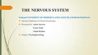
- 1. THE NERVOUS SYSTEM National UNIVERSITY OF MODERN LANGUAGES ISLAMABAD PAKISTAN. Advance Diploma in Clinical Psychology. Presented by: Anisa Sarwar. Umar Zada Abdul Rahim Subject: Psychophysiology
- 2. The Nervous System The nervous system is a complex network of interconnected nerve fibers. Sensory nerve fibers provide input to the brain and spinal cord by carrying signals from sensory receptors. Motor nerve fibers provide output from the brain of spinal cord and other organs. And that resulting in voluntary and involuntary movement.
- 3. Central nervous system The nervous system is made up of central nervous system, which consists of brain and spinal cord. CNS carries voluntary nerve impulses to skeletal muscles and skin. CNS carries involuntary impulses to muscles and glands.
- 4. Peripheral Nervous system And peripheral nervous system, which consists of the rest of the nerves in the body, including those that connect to the brain and spinal cord. The PNS is itself made up of the somatic nervous system and autonomic nervous system.
- 5. Somatic Nervous System The somatic or voluntary nervous system connects nerve fibers to voluntary muscles and provides the brain with feedback in the form of sensory information about voluntary movements. SNS controls voluntary movements.
- 6. Autonomic Nervous System The autonomic or involuntary nervous system connects the central nervous system to all integral organs over which people do not usually have control. ANS controls the organs that operate involuntarily. Regulation of autonomic nervous system occurs via the sympathetic nervous system and the parasympathetic nervous system.
- 7. Sympathetic Nervous System The sympathetic nervous system prepares the body to respond to emergencies. To strong emotions such as anger or fear, and to strenuous activity. As such, it plays an important role in reactions to the stress. Because it concerned with the mobilization and exertion of energy, it is called catabolic system.
- 8. Parasympathetic Nervous System Parasympathetic nervous system controls the activities of organs under normal circumstances and acts antagonistically to the sympathetic nervous system. When an emergency has passed, the parasympathetic nervous system helps to restore the body to the normal state. Because it is concerned with the conservation of the body, it is called anabolic system
- 9. The components of the Nervous system
- 10. Brain The brain is an organ that serves as the center of the nervous system in all vertebrate and most invertebrate animals. The brain is located in the head, usually close to the sensory organs for senses such as vision. The brain is the most complex organ in a vertebrate's body.
- 11. What is the brain's function. It assembles the messages in a way that has meaning for us, and can store that information in our memory. The brain controls our thoughts, memory and speech, movement of the arms and legs, and the function of many organs within our body. The central nervous system (CNS) is composed of the brain and spinal cord.
- 12. Parts of brain. The brain can be divided into three basic units: the forebrain, the midbrain and the hindbrain. These basic units consist of Occipital lobe, Temporal lobe, Parietal lobe, Frontal lobe. Cerebral cortex, Cerebellum, Hypothalamus, Thalamus, Pituitary gland, Pineal gland, Amygdala, Hippocampus and the Mid- brain.
- 13. Cerebrum The cerebrum -- which is just Latin for "brain" -- is the newest (evolutionarily) and largest part of the brain as a whole. It is here that things like perception, imagination, thought, judgment, and decision occur. The surface of the cerebrum -- the cerebral cortex -- is composed of six thin layers of neurons, which sit on top of a large collection of white matter pathways. The cortex is heavily convoluted, so that if you were to spread it out, it would actually take up about 2 1/2 square feet (2500 sq cm). It includes about 10 billion neurons, with about 50 trillion synapses! The convolutions have "ridges" which are called gyri (singular: gyrus), and "valleys" which are called sulci (singular: sulcus). Some of the sulci are quite pronounced and long, and serve as convenient boundaries between four areas of the cerebrum called lobes.
- 14. What is the main function of cerebrum? The Cerebrum: The cerebrum or cortex is the largest part of the human brain, associated with higher brain function such as thought and action. The cerebral cortex is divided into four sections, called "lobes": the frontal lobe, parietal lobe, occipital lobe, and temporal lobe.
- 16. What does the cerebrum control in the brain? The largest part of the brain, the cerebrum has two hemispheres (or halves). The cerebrum controls voluntary movement, speech, intelligence, memory, emotion, and sensory processing. At the base of the brain, the brain stem connects to the spinal cord and is made up of the midbrain, pons, and medulla oblongata.
- 17. How big is the cerebrum? The surface of the cerebrum -- the cerebral cortex -- is composed of six thin layers of neurons, which sit on top of a large collection of white matter pathways. The cortex is heavily convoluted, so that if you were to spread it out, it would actually take up about 2 1/2 square feet (2500 sq cm).
- 18. What is the difference between cerebrum and cerebellum? The outer layer of the cerebrum , known as cerebral cortex , is formed of grey matter and white matter. The cerebellum is similar to cerebrum in that it has two hemispheres and has a highly folded surface or cortex. The cerebellum is the second largest part of the brain, and is located at the back of the skull.
- 19. What are the 4 lobes of the cerebrum and their functions? The cerebrum is divided into four regions called lobes that control senses, thoughts, and movements. The four lobes are the occipital, temporal, frontal, and parietal lobes. Although each lobe has a different task to perform, they all must work together.
- 20. What do each of these lobes do? Frontal Lobe- associated with reasoning, planning, parts of speech, movement, emotions, and problem solving Parietal Lobe- associated with movement, orientation, recognition, perception of stimuli Occipital Lobe- associated with visual processing Temporal Lobe- associated with perception and recognition of auditory stimuli, memory, and speech
- 21. What are the two halves of the cerebrum? The cerebrum (right and left) is the upper, front portion of the brain and consists of two hemispheres, or halves. The two hemispheres are connected by the corpus callosum, which is a large bundle of nerve fibers.
- 22. What is inside the cerebrum? The cerebrum is a large part of the brain containing the cerebral cortex (of the two cerebral hemispheres), as well as several subcortical structures, including the hippocampus, basal ganglia, and olfactory bulb. In the human brain, the cerebrum is the uppermost region of the central nervous system.
- 23. What color is the cerebrum? Compare the color and consistency of the cerebellum and the cerebrum. The cerebrum was more of a tanned color and the cerebellum was more white in color. I suspect that the Cerebrum was darker due to its protective layer on the outside where as the cerebellum was just purely grey matter.
- 24. What part of the brain controls emotions? Emotions, like fear and love, are carried out by the limbic system, which is located in the temporal lobe. While the limbic system is made up of multiple parts of the brain, the center of emotional processing is the amygdala, which receives input from other brain functions, like memory and attention.
- 25. What part of the brain makes you happy? It's a survival mechanism: in the presence of something good, the brain releases four main 'feelgood' chemicals – endorphin, oxytocin, serotonin, and dopamine – and in the presence of danger, the 'bad feeling' chemical – cortisol – comes in.
- 26. Can the cerebrum repair itself? Damaged Brain Can Be Repaired And Cerebral Functions Restored, Neuronal Study Suggests. ... Brain injury in adults can cause irreparable, long-term physical and cognitive damage. However, motor and spatial functions can be recovered if undamaged neurons are stimulated to create new innervation.
- 27. What happens if the cerebrum is damaged? For example, the cerebrum, if damaged, may cause personality disorders, loss of senses, or trouble with thinking and learning. Damage to the brain stem, on the other hand, may lead to breathing issues, paralysis, and even death
- 28. Limbic System The limbic system is a complex set of structures that lies on both sides of the thalamus, just under the cerebrum. It includes the hypothalamus, the hippocampus, the amygdala, and several other nearby areas.
- 29. What does limbic mean? The limbic system, or the paleomammalian cortex, is a set of brain structures located on both sides of the thalamus, immediately beneath the medial temporal lobe of the cerebrum primarily in the mesencephalon.
- 30. What is the limbic system and what is its function? The limbic system is the portion of the brain that deals with three key functions: emotions, memories and arousal (or stimulation). ... The thalamus is located within the brainstem and is part of the pathway of information into the cerebrum, which is the section of the brain that is responsible for thinking and movement.
- 31. What is the limbic lobe? The limbic lobe is an arc-shaped region of cortex on the medial surface of each cerebral hemisphere of the mammalian brain, consisting of parts of the frontal, parietal and temporal lobes.
- 32. Disorders and Diseases related to Forebrain Encephalitis, head trauma, space-occupying lesion, malformation, infarct, and metabolic disorders all are likely to cause changes in behavior. It is likely that structural or metabolic forebrain disease is the cause of dementia if other neurologic abnormalities are found by neurologic examination or imaging studies.
- 33. What causes damage to the limbic system? Stimulating different parts of the limbic system can, among other things, affect the functioning of the hypothalamus. Such stimulation can also trigger emotional behavior, such as aggression. Damage to various areas of the limbic system disturbs many behaviors related to motivation and emotion.
- 34. What happens if the limbic system is damaged? The limbic system is does much of its control of behavior through the hypothalamus, at the top end of the brain stem. Damage to parts of the limbic system severely affects ability to store and retrieve information in declarative (conscious) memory (Squire, 1987).
- 35. What causes the hypothalamus to malfunction? The hypothalamus produces hormones that control body temperature, hunger, moods, release of hormones from many glands such as the pituitary gland, sex drive, sleep, and thirst. ... A number of different causes including anorexia, bleeding, genetic disorder, tumors , and more have been linked to hypothalamic dysfunction.
- 36. What happens when amygdala is damaged? People tend to choose avoiding losses over acquiring gains—a behavior known as loss-aversion. But people with damage to the amygdala—an almond-shaped part of the brain involved in emotion and decision-making—are more likely to take bigger risks with smaller potential gains, De Martino's study found.
- 37. What are the symptoms of pituitary gland disorders? Signs and symptoms include: Nausea and vomiting. Weakness. Feeling cold. Less frequent or no menstrual periods. Sexual dysfunction. Increased amount of urine. Unintended weight loss or gain.
- 38. What part of the brain controls fear and anxiety? The amygdala, from the Greek word for almond, controls autonomic responses associated with fear, arousal, and emotional stimulation and has been linked to neuropsychiatric disorders, such as anxiety disorder and social phobias.
- 39. Clinical signs of forebrain disease Abnormal behavior: head pressing, staring at walls, getting stuck in corner. Abnormal mental status - level of alertness Circling in wide circles toward the lesion Continuous Pacing (WALKING) Normal Gait Central blindness -contralateral to the lesion Sensory deficits - contralateral to the lesion Seizure. Abnormal T regulation, appetite, sleep cycle, endocrine disturbances(Diabetes, acromegaly) - Thalamus/hypothalamus
- 40. Vitamin D • Vascular • Infectious/inflammatory • Traumatic • Toxic • Metabolic • Idiopathic/iatrogenic characterizing a disease arising primarily, and not in consequence of some other disease or injury • Neoplastic/nutritional • Degenerative
- 41. Vascular Diseases Diffuse Cerebral ischemia: cardiopulmonary arrest, anesthesia, toxins/trauma Focal cerebral Ischemia: thrombus/embolus/migrating parasite Diffuse/focal hemorrhages: trauma, neoplasia, coagulopathies, vasculitis, hypertension
- 42. Hydrocephalus Increased CSF V in dilated ventricular cavities. Types: Compensatory Non-communicating/obstructive Communicating - either increased production or decreased re- absorption
- 43. Damage to the basal forebrain Thus, damage to the basal forebrain can reduce the amount of acetylcholine in the brain and impair learning. This may be one reason why basal forebrain damage can result in memory impairments such as amnesia and confabulation means Confabulation is a memory error defined as the production of fabricated, distorted, or misinterpreted memories about oneself or the world, the conscious intention to deceive.. One common cause of basal forebrain damage is aneurysm of the anterior communicating artery. Encephalitis, head trauma, space-occupying lesion, malformation, infarct, and metabolic disorders all are likely to cause changes in behavior. It is likely that structural or metabolic forebrain disease is the cause of dementia if other neurologic abnormalities are found by neurologic examination or imaging studies.
- 44. Diseases related to Forebrain different parts Thalamus As a result of a thalamic infarct, frequently involving its posterior aspect, the patient feels a very painful and unpleasant sensation. When the pain persists generally as a burning sensation, it is referred to as a thalamic pain syndrome. Hypothalamus Because the hypothalamus mediates a variety of autonomic, visceral, and emotional responses, damage to different groups of hypothalamic nuclei can resulting disorders in eating, endocrine function, temperature regulation, aggression and rage, and sympathetic dysfunction. An example of one such disorder is diabetes insipidus, which results from damage to vasopressin neurons located in the supraoptic or paraventricular nuclei and is characterized of large amounts of urine coupled with draining of large quantities of fluids.
- 45. Basal Ganglia Diseases of the basal ganglia are characterized by abnormal, involuntary movements at rest, referred to collectively as dyskinesia and by abnormal changes in muscle tone. parkinsonism disease, an example of a hypokinetic disorder, is associated with tremor, rigidity, and akinesia. The disorder results from a loss of dopamine released from the substantia nigra that supplies the neostriatum. (the disorders include chorea, which is a hyperkinetic disorder associated with a loss acid in the striatum, characterized by involuntary movements of the extremities and hemiballismus, which involves damage to the subthalamic nucleus, resulting in abnormal movements of the arm and legs no the contralateral side of the body.
- 46. Limbic Structures Because of their relationships with the hypothalamus and midbrain disruptions of limbic structures resulting from vascular lesions or tumors frequently are associated with marked changes in emotional behavior, irritability, impulsivity, and rage. structures most closely associated with these ejects include the amygdala, hippocampus formation, and the prefrontal Cortex. (the disorders include loss of short-term memory functions, particularly after damage to the hippocampus formation+ a decrease in cognitive ability and fatness in emotional responsiveness after damage to the prefrontal cortex+ and seizure disorders associated most commonly with damage to temporal lobe structures.
- 47. Cerebral Cortex in brief, some of these disorders are mentioned here and usually result from tumors or vascular lesions. disorders include upper motor neuron paralysis associated with damage of the precentral and premotor cortex. aphasias inability to express or understand language associated with damage to the ventrolateral aspect of the premotor region or borderline region of the temporal and parietal lobes. apraxia inability to produce a motor act correctly even though sensory and motor circuits are intact0 associated with damage to the premotor cortex or to the posterior parietal cortex loss of somatosensory and auditory discrimination ability after damage to the postcentral and superior temporal gyri, respectively and partial blindness after damage to the region of the occipital cortex.