
Recommandé
Contenu connexe
Tendances
Tendances (20)
Similaire à Fractures and dislocation upper limb .pptx
Similaire à Fractures and dislocation upper limb .pptx (20)
Plus de Dr vaibhavi patel
Dernier
Dernier (20)
Fractures and dislocation upper limb .pptx
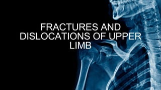
- 1. FRACTURES AND DISLOCATIONS OF UPPER LIMB
- 2. INTRODUCTION Break in the structural continuity of a bone If the underlying skin remain intact - CLOSED (Simple fracture) If skin or one of the body cavities is breeched - OPEN(Compund fracture) FRACTURE
- 7. Click to add text
- 8. COMMON AND UNCOMMON FRACTURES OF UPPER LIMB
- 9. CLAVICLE FRACTURE • Fracture of clavicle is the most common skeletal injury during birth and childhood. • Most common site of clavicle fracture is at the junction of its medial 3/5 and lateral 2/5 junction. • The optimal radiographic assessment include AP veiw with 10-15 degree cephalic tilt. • Incidence of middle 1/3 rd > lateral 1/3rd> medial 1/3rd
- 10. Neurovascular damage Malunion Non union Degenerative arthritis Post traumatic osteoblasts COMPLICATIONS OF CLAVICLE FRACTURES
- 11. SHOULDER DISLOCATION • 1. Anterior - Most common type • Mechanism is abduction + external rotation • 2. Posterior - Mechanism is adduction + internal rotation • More common during seizures and ECT • Empty glenoid and light bulb sign will seen • 3. Inferior - due to hyperabduction 3 TYPES
- 13. Most common complication is recurrence. Recurrent shoulder dislocations may lead to BANKART LESION And HILL SACHS LESION
- 14. PROXIMAL HUMERUS FRACTURE • Most common fracture of humerus. • Higher incidence in elderly. • MECHANISM:- Most commonly fall on outstretched arm.
- 16. HUMERAL SHAFT FRACTURES • MECHANISM : Mainly direct trauma and sometimes indirect trauma such as fall on outstretched hand. • Radiographic evaluation : • AP and Lateral view of Humerus • Traction radiographs may be indicated for hard to classify secondary to severe displacement or a lot of communition.
- 17. HOLSTEIN -LEWIS FRACTURE • Spiral fracture of the Distal 1/3 rd of humerus. • May entrap or lacerate radial nerve.
- 19. SUPRACONDYLAR FRACTURE HUMERUS • Most common humeral fracture in children. • 3 BPR is maintained. • Common complications : • 1. Neurovascular injury (AIN is most common nerve involved) • 2. Malunion • 3. Compartment syndrome • 4. Volkmann’s ischemic contracture. • 5. Myosotis ossificans
- 20. 3 PBR is not preserved in Lateral or Medial Epicondilar fractures of Humerus.
- 21. FRACTURES OF RADIUS AND ULNA • Fracture of proximal 1/3 rd of ulnar shaft with radial head dislocation. • Most common injured nerve is PIN MONTEGGIA FRACTURE
- 22. FRACTURES OF RADIUS AND ULNA • Also known as Reverse Monteggia or Piedmont’s fracture • Fracture of Radius shaft at junction middle and distal 1/3rd with distal radio ulnar joint disruption GALEAZZI’S FRACTURE
- 23. FRACTURES OF RADIUS AND ULNA • Isolated fracture of shaft of ulna. • Usually a defence injury. NIGHTSTICK FRACTURE
- 24. FRACTURES OF RADIUS AND ULNA • Most common fracture in children. • It is an incomplete unicortical fracture seen in forearm bones(more commonly in radius) with concave-cover deformity. GREENSTICK FRACTURE
- 25. FRACTURES OF RADIUS AND ULNA TORUS FRACTURE
- 27. COLLES FRACTURE • Extra articulate fracture of distal end of Radius with dorsal / posterior displacement of distal fragment. • Most common complication is Finger stiffness> Malunion(Dinner fork deformity)
- 29. SMITH’S FRACTURE • AKA Reverse colles fracture. • It is Extra articular fracture of distal end of Radius with anterior/volar displacement of distal fragment. • Most common complication is Malunion(Garden spade deformity)
- 32. CHAUFFER FRACTURE • Intra articular fracture of distal end radius. • Radio-styloid fragment seen with normal radio- scaphoid joint anatomy.
- 34. BARTON’S FRACTURE • Intra articular fracture of distal end of radius. • No Radios-styloid fragment seen. • Radio-scaphoid joint subluxation.
- 36. SCAPHOID FRACTURE • Most common carpal bone fracture. • Most common fracture due to fall on outstretched hand in adults. • Scaphoid forms floor of anatomical snuff box and it has retrograde blood flow. • Most common site of fracture is waist of scaphoid. • Most common complication is Non union >> Avascular Necrosis. • X-Ray is taken in Oblique and PA in 15 degree Ulanar deviation.
- 40. BENNET’S FRACTURE • Intra articular fracture of 1st metacarpal • Oblique fracture line. • More displaced.
- 41. ROLANDO’S FRACTURE • Intra articular fracture of 1st metacarpal. • T / Y shaped comminuted fracture. • Less displaced
- 42. BOXER’S FRACTURE • Fracture of neck of 5th metacarpal. • Commonly seen in boxers.
- 48. THE END