Imaging for Radiotherapy delivery and verification
•
4 j'aime•3,376 vues

This document summarizes the work completed during a medical physics rotation focused on imaging for treatment planning and verification. Key tasks included: - Performing quality assurance tests on the electronic portal imaging device (EPID) including measurements of image uniformity, signal-to-noise ratio, and modulation transfer function at varying imaging parameters. - Analyzing contrast-to-noise ratio, signal-to-noise ratio, and dose dependence using phantoms imaged with the EPID. - Validating calculations of the EPID's modulation transfer function. - Ensuring proper alignment of the EPID with the radiation isocenter using reticule alignment tests at different gantry angles. - Observing clinical treatments for sites

Recommandé
Recommandé
Contenu connexe
Tendances
Tendances (20)
Similaire à Imaging for Radiotherapy delivery and verification
Similaire à Imaging for Radiotherapy delivery and verification (20)
Plus de Miami Cancer Institute
Plus de Miami Cancer Institute (16)
Dernier
Dernier (20)
Imaging for Radiotherapy delivery and verification
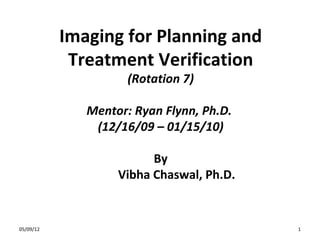
- 1. Imaging for Planning and Treatment Verifica4on (Rota&on 7) Mentor: Ryan Flynn, Ph.D. (12/16/09 – 01/15/10) By Vibha Chaswal, Ph.D. 2/24/14 1
- 3. EPID QA • Uniformity versus thickness (across flat-‐field images) • SNR versus thickness of solid water • MTF • CNR • SNR • EPID’s alignment with gantry isocenter 2/24/14 3
- 4. Addi4onal • • • • • CNR vs Thickness vs Dose Effect of filtraRon on images MTF calculaRon validaRon Contrast calculaRons for Air & Bone in water DidacRc 2/24/14 4
- 5. EPID QA: Image Uniformity vs Thickness 2/24/14 5
- 6. 1 3 5 4 2 Uniformity (Normalized mean PV in an ROI) Image Uniformity vs Solid Water Thickness 1.010 0.980 0.950 ROI_1 (top) ROI_2 (boaom) ROI_3 (right) ROI_4 (leb) ROI_5 (center) 0.920 0.890 0.860 0.830 0.800 0 2/24/14 5 10 15 20 Solid Water Thickness (cm) 25 30 35 6
- 7. EPID QA: Signal-‐to-‐Noise raRo vs Thickness 2/24/14 7
- 8. 2/24/14 8
- 9. Signal-‐to-‐Noise Ra4o vs Thickness of SW slab 300 250 SNR 200 150 100 50 0 0 5 10 15 20 25 30 35 Sold Water Thickness (cm) 2/24/14 9
- 10. EPID QA: Imaging Parameters (SNR, CNR, MTF, Dose dependence) 2/24/14 10
- 11. Material Physical density (g/cm3) PVC 1.00 Al 2.6 Pb 11.34 0.23 lp/mm 5 mm lead 15 mm PVC QC - 3V Phantom s/n 104 0.43 lp/mm 15 mm Al 11 mm Pb 0.76 lp/mm 0.2 lp/mm 0.1 lp/mm 7.5 mm Pb 2/24/14 11
- 12. MTF Calculation Validation 2/24/14 Droege R.T, Morin R. L, ‘A practical method to measure MTF of scanners’, Med. Phys. 9(5), 1982 12
- 13. EPID spatial resolution: MTF Calculation lp/mm 0.1 663.45 0.2 1342.163 0.23 2066.66 0.43 MTF measured using QC-‐3V phantom Std Dev 2315.17 0.76 2825.81 1.20 1.00 MTF 0.80 0.60 0.40 0.20 0.00 0 2/24/14 0.1 0.2 0.3 0.4 0.5 line-‐pairs/mm (lp/mm) 0.6 0.7 0.8 13
- 14. Imaging Parameters: SNR Region Physical density (g/ cm3) Depth (cm) Radiological Path-‐ length (g/cm2) 1 (Pb) 11.34 0.75 8.505 2 (PVC) 1 1.5 1.5 3 (Pb) 11.34 0.5 5.67 4 (Al) 2.6 1.5 3.9 5 (Pb) 11.34 1.1 12.474 6 (Pb) 11.34 0.75 8.505 SNR versus Radiological Path-‐length SNR (standard devia4on) 140 120 100 80 60 40 20 0 0 2/24/14 2 4 6 8 10 Radiological pathlength (g/cm2) 12 14 14
- 15. SNR analysis ROIs 1 & 6 ROI 1 2/24/14 ROI 6 15
- 16. EPID QA: CNR vs Thickness vs Dose 10-QC-10 1MU 2MU 3MU (QC-3V as tumor inside varying solid water thickness patients) 10 cm 20-QC-10 1MU 2MU 3MU 30-QC-10 1MU 2MU 3MU 20 cm 30 cm 2/24/14 16
- 17. CNR of QC-‐3 phantom vs Radiological Thickness (for different doses and solid water thicknesses) 60.00 Contrast-‐to-‐Noise Ra4o w.r.t. 15mm PVC 1 MU 2 MU 50.00 10 cm 3 MU 40.00 30.00 20 cm 20.00 30 cm 10.00 -‐ 0 2 4 6 8 10 12 14 Radiological Thickness (g/cm2) 2/24/14 17
- 18. EDPI QA: EPID Alignment (Oncor A) Gantry 0 degree Gantry 90 degree 2/24/14 18 Gantry 270 degree, Gantry 180 degree
- 19. Fig 4(c): Gantry 180 degree, distance measurements between x-retic and e-retic. 2/24/14 Fig 4(d): Gantry 180 degree, distance measurements between x-retic and e-retic at the central ISIS sphere region that coincides with radiation isocenter; note that distance between x-retic and e-retic is 2.3mm, which is more than the tolerance value of 2mm. Hence, EPID is not aligned within specifications at 19 Gantry angle 180-degrees.
- 20. Fig 4(e): Gantry 180 degree, distance measurements between x-retic and center of pinhole of ISIS. Fig 4(f): Gantry 180 degree, distance measurements between e-retic and center of pinhole of ISIS. 2/24/14 20
- 21. Clinical ObservaRon • • • • • • Whole brain x 1 Head and neck x 2 (MVCBCT) Pelvis x 1 (portal) x 3 (MVCBCT) Thoracic x 1 (portal) x 2 (MVCBCT) Extra-‐cranial lung x 1 (MVCBCT) Supine Breast x 1 (portal) 2/24/14 21
- 22. Treatment Verification: MVCBCT Extra-Cranial Lung Head & Neck 2/24/14 Pelvic 22
- 23. Treatment Verification: Portal Imaging Reference DRR Right Lat Treatment day Right lat portal image Whole Brain DRR Registered DRR and portal image Pelvic DRR Reference Planning PA DRR 2/24/14 Treatment day PA portal image Registered PA images 23
- 24. Thoracic DRR Reference Planning AP setup DRR Reference Planning Left Anterior Oblique field DRR at Gantry 45-deg Reference Planning field DRR Left Lat Gantry 90-deg Reference Planning Right Anterior Oblique field DRR 2/24/14 at Gantry 344.9-deg Treatment day AP double exposure portal image Treatment day Left Anterior Oblique field portal image at Gantry 45-deg Registered AP DRR and portal images Registered Left Anterior Oblique DRR and portal images Treatment day Left Lat field portal image at Gantry 90- Registered Left Lat DRR and portal images deg Treatment day Right Anterior Oblique field portal image at Gantry 344.9-deg Registered Right Anterior Oblique DRR and portal 24 images
- 25. THANK YOU!!!!! 2/24/14 25