SlideShare une entreprise Scribd logo
1  sur  46
Télécharger pour lire hors ligne
Diagnostic Cytopathology
 International CME on                           Hotel O, Pune, India                                      of Effusion Fluids
    Oncopathology                                January 7, 2012                                            Short course


                Diagnostic Cytopathology
                    of Effusion Fluids
                                                   Short course


           Vinod B. Shidham, MD, FRCPath, FIAC
              Vice-chair & Professor
              Director of Cytopathology, Cytotechnology School, Cytopathology fellowship
              Dept of Pathology, Wayne State University Medical School
              Detroit, MI 48201, USA
           Co-editor-in-chief & Executive editor, CytoJournal (www.cytojournal.com)
                                         vshidham@med.wayne.edu

This presentation is available on web for all conference attendees at- http://alturl.com/3ucwx


                                                                                                      Diagnostic Cytopathology
 International CME on                                                                                     of Effusion Fluids
    Oncopathology                                                                                           Short course


                                           Disclosure
              I do not have conflict of any financial interest in the
                         product if cited any in the course.

                                 I am the co-editor of the book *
                                               and
                                      Co-author of th chapter**
                                      C    th     f the h t
                                          on the same topic.

  *Shidham VB and Atkinson BF. Editors and contributors ‘Cytopathologic Diagnosis of Serous Fluids’ Multi-author book with
  15 chapters, Elsevier (W. B. Saunders Company), 2007 (ISBN-13: 9781416001454).
  ** Shidham VB, Falzon M.     Serous effusions: reactive, benign and malignant. In Gray & Kocjan, ed. Diagnostic Cytopathology,
  Elsevier, 3rd edition, Chapter 3.


                                                                                                                                   1
Diagnostic Cytopathology
International CME on                                                         of Effusion Fluids
   Oncopathology               Acknowledgment                                 Short course


                  Many images and tables are based on chapters in:


Shidham VB and Atkinson BF.
Cytopathologic Diagnosis of Serous
Fluids
Elsevier (W. B. Saunders Company) First
edition, 2007.


                                                                         Diagnostic Cytopathology

                                Outline
International CME on                                                         of Effusion Fluids
   Oncopathology                                                              Short course


  Part I :
              Anatomy, histology, cytology, and effusions

              Collection, transportation, and processing

              Factors leading to potential diagnostic pitfalls

              Approach to diagnostic cytopathology of effusions

              The panorama of different faces of mesothelial cells
  Part II :

              Immunocytochemistry of effusion fluids:
                      SCIP (Subtractive Coordinate Immunoreactivity Pattern) approach

              Evaluation of unknown primary sites of origin-
                      Where do they come from?


                                                                                                    2
Diagnostic Cytopathology
    International CME on                                            of Effusion Fluids
       Oncopathology                                                 Short course


              Anatomy, histology, cytology, and effusions


                               a. Peritoneal cavity
                                                  y
                               b. Left & right pleural cavities
                               c. Pericardial cavity


                       Four major serous cavities


                                                                Diagnostic Cytopathology
    International CME on                                            of Effusion Fluids
       Oncopathology                                                 Short course


        Anatomy, histology, cytology, and effusions (continued)
                                         1
                                                       Histology of serous li i
                                                       Hi t l      f           lining
                                                       (inguinal hernia sac). The
                                                       mesothelial cells lining the
                                                       fibrous tissue are flat (1).
                                                       Focal reactive changes are
                                                       seen as hypertrophy of
a                          b                           some cells which assume a
                   2                      3            cuboidal contour (2,3).
                                                                        ( ,3)
                                                       [a-d, HE stain (a, 10X; b-d,
                                                       100X)].


c                          d


                                                                                           3
Diagnostic Cytopathology
     International CME on                                                           of Effusion Fluids
        Oncopathology                                                                Short course


        Anatomy, histology, cytology, and effusions (continued)

                            Peripheral light ectoplasm (1)
                                                              Mesothelial cells (peritoneal
                                                              fluid): show outer faintly stained
                                Inner darker endoplasm (2)
                                                              ectoplasm (1) with inner denser
                                                              endoplasm         (2)     rich     in
                                Slightly off-center nucleus   intermediate      filaments.     The
                                                              nucleus is usually central or near
                                 Nucleolus
                                                              central (a), but may be eccentric
                                                              (b).     Nucleoli     are     readily
                    1                                1        observed.
                                                              observed       The      vacuolation
                                                              generally begins at the periphery
                                                              in ectoplasm (1).
                        2                                     [a,b,     Papanicolaou       stained
                                                          2
                                                              SurePath® Preparation (a,b,
                                                              100XZoomed)]

 a                          b


                                                                                Diagnostic Cytopathology
     International CME on                                                           of Effusion Fluids
        Oncopathology                                                                Short course


Anatomy, histology, cytology, and effusions (continued)

                                                              Peripheral ectoplasm
                                                              Inner endoplasm


                                                              Central to
                                                              slightly eccentric nucleus


                                                              Ruffled cell border
                                                              with blebs

 Mesothelial cell (pleural fluid): shows outer ectoplasm which is denser than the inner
 endoplasm. The nucleus is central to slightly eccentric but not touching the cell
 periphery. The cell margin shows blebs and is ruffled.
 [Diff-Quik stained Cytospin preparation (100XZoomed)]


                                                                                                           4
Diagnostic Cytopathology
  International CME on                                                of Effusion Fluids
     Oncopathology                                                     Short course


Anatomy, histology, cytology, and effusions (continued)
                              Bloody effusions
              (Hemothorax,
              (Hemothorax hemopericardium and hemoperitoneum)
 Homogenously red or dark brown with hemosiderin pigment and the hematocrit of
 the effusion is 10% or more than that of the plasma hematocrit.
 (Occasional blood tinged fluids may be associated with a procedure trauma).
 Bloody effusions are more likely to be associated with an underlying malignancy.

        Reactive conditions associated with Bloody Effusions
 Para pneumonic effusions                         Pancreatitis
 Post traumatic effusions                         Acute aortic dissection

         underlying malignancy
    -Post cardiothoracic procedures / surgeries
    -Thoracic cavity vascular damage
 Pulmonary embolism
                                                  Endometriosis
                                                  Sarcoidosis
                                                  Intralobar pulmonary sequestration
 Asbestos exposure associated pleural effusion    Some Infections –e.g B. Anthrax


                                                                  Diagnostic Cytopathology
  International CME on                                                of Effusion Fluids
     Oncopathology                                                     Short course


  Collection, transportation, and processing
                      Biopsy vs effusion cytology


                                                                                             5
Diagnostic Cytopathology
International CME on                                                                       of Effusion Fluids
   Oncopathology                                                                             Short course


Collection, transportation, and processing (continued)
 To prevent clotting: Collected in anticoagulant such as-
           EDTA (sodium or potasium salts) in final concentration of 4.55+0.85 µmol/ml
          [e.g. di-sodium
          [e g di sodium EDTA dihydrate (EDTA Na2, 2H2O) 1 4 mg per ml of effusion fluid]
                                                             1.4

 Amount of fluid sample: Variable (less than 1 ml to 100 ml or more).
         For optimum cellularity of cytology preparations and cell blocks, it is recommend to submit as
                              much specimen as possible (up to 1000 ml).
         Each laboratory follows its own protocol for determining the amount of sample to be used.

 Submit as a fresh, unfixed sample:
         If delay is expected in transporting to the laboratory-
                     refrigerate at 4oC (with precaution not to freeze the specimen).
                          g             (     p                             p      )
         Alternatively, although not recommended, it may be preserved in a weak fixative


 Fluids collected in fixative- require a wash prior to instrument processing.

 For blood rich specimens- start with smaller aliquots (such as 10 ml) of fresh fluid.
          Concentration of cells and removal of excess erythrocytes in the blood rich specimen
          may be achieved by density gradient centrifugation with Ficoll


International CME on             Collection, transportation,                           Diagnostic Cytopathology
                                                                                           of Effusion Fluids
   Oncopathology                 and processing (continued)                                  Short course


                    Collect fresh effusion fluid                 Concentrate the
                    with or without anticoagulant            effusion fluid specimen

                                                     Concentrate the
                                                     remaining fluid similar to
      Direct smear of              Specimen          specimen without clot
 unconcentrated specimen for       with clot
 semi-quantitative evaluation                               Use homogenized specimen after mixing
                                                            (For paucicelluar fluids: Use cell-rich sediment)


                                                                Concentrate by centrifugation
                            Remove the clot                     (at 600g for 10 minutes)
                            and process for
                            cell-block
                            preparation                          Pour off supernatant and
                                                                 vortex to resuspend cell pellet
Process for paraffin-
embedding after formalin           Cell-block preparation                  Smear preparation
fixation.                          Histogel                     • Direct smears    • SurePrep (Autocyte)
                                   Plasma-Thrombin              • Cytospin         • ThinPrep
                                   Celloidin bag                                   • Other methods


                                                                                                                  6
Diagnostic Cytopathology
International CME on                                                            of Effusion Fluids
   Oncopathology                                                                 Short course


Collection, transportation, and processing (continued)

    Different alternatives available for preparing permanent cytology preparations.
    a. Direct smears
      (Spreading a drop of specimen directly on slide. The specimen may be before
       concentration OR after concentration as sediment)
               i. Wet fixed- Pap stain
               ii. Air-dried-
                           Diff-Quik stain*
                           Pap stain: After rehydration in saline and
                                      post-fixation in 95%ethanol with 5% acetic acid (28)
    b. Cytospin preparations (Shandon EZ Megafunnel™ with Shandon Cytospin®) (31)
               i. Wet fixed- Pap stain
               ii. Air-dried-
               ii Ai d i d
                           Diff-Quik stain
                           Pap stain: After rehydration in saline and
                                      post-fixation in 95%ethanol with 5% acetic acid (28)
    c. Filters
               i. Wet fixed- Pap stain
    d. Liquid based cytology (SurePathTM or ThinPrepTM or other non-proprietary methods)
               i. Wet fixed- Pap stain


                                                                            Diagnostic Cytopathology
International CME on                                                            of Effusion Fluids
   Oncopathology                                                                 Short course


Collection, transportation, and processing (continued)

     Preparation                                          Purpose
                                                          Semiquantitative evaluation of
A    Diff-Quik stained direct smear
     of the undiluted specimen                            cellularity.
     Diff-Quik stained air-dried                          Rapid initial detection of second
B    Shandon EZ Megafunnel™                               population and cytomorphologic
     preparation                                          evaluation of hematopoietic
                                                          cells.
     Pap stained preparation                              Cytomorphologic evaluation
C                                                         especially nuclear details.
                                                                             details


     Cell-block preparation in                            Evaluation of-
D    HistoGelTM                                            a. Immunoprofile,
                                                           b. Other special stains,
                                                           c. Architecture,


                                                                                                       7
Diagnostic Cytopathology
   International CME on                                                 of Effusion Fluids
      Oncopathology                                                      Short course


Approach to diagnostic cytopathology of effusions

                ► GENERAL FEATURES

                ► PROCESSING RELATED

                ► INTERPRETATION STRATEGY

                ► CYTOMORPHOLOGY

                ► IMMUNOCYTOCHEMISTRY


                                                                    Diagnostic Cytopathology
   International CME on                                                 of Effusion Fluids
      Oncopathology                                                      Short course


Approach to diagnostic cytopathology of effusions (continued)

                    ► GENERAL FEATURES
►A general pathology l b
 Any         l     h l   laboratory may receive effusion- all general pathologists and
                                            i    ff i      ll       l    h l i       d
 cytopathologists should be conversant with the diagnostic challenges and pitfalls
 of effusion fluid cytology.
►Finding neoplastic cells in effusion specimens not only reveals that a patient has
 cancer, but it also denotes the advanced nature of the disease which at this stage is
 almost always incurable.
►The morphologic features of most of the cancer cells in effusion smears are
 different f
 diff    t from th
                those seen i exfoliative, b hi
                           in f li ti     brushing, and FNA cytology.
                                                      d       t l
►The standard cytologic criteria of malignancy based on evaluation of single cell
 morphology are not applicable for most of the effusion cytology specimens
 (except in cases with high-grade neoplasms with pleomorphic cells).
►Since cells in a fluid medium ‘round up’ because of surface tension, the native
 shapes of cancer cells cannot be a guiding feature.


                                                                                               8
Diagnostic Cytopathology
  International CME on                                              of Effusion Fluids
     Oncopathology                                                   Short course


Approach to diagnostic cytopathology of effusions (continued)


                  ► PROCESSING RELATED
1. Second population of foreign cells

2. Nuclear details of the ‘second population’.

3. Semiquantitative evaluation

4. Objective confirmation and differential diagnosis of primary neoplasm.


                                                                Diagnostic Cytopathology
  International CME on                                              of Effusion Fluids
     Oncopathology                                                   Short course


Approach to diagnostic cytopathology of effusions (continued)

           ► INTERPRETATION STRATEGY
 a. Reactive mesothelial cells- morphologic overlap with neoplastic cells
 b. A single population- favor mesothelial cells
 c. Second foreign population consistent with metastatic neoplasm
 d. Sarcomas- previous history known
 e. Romanowsky stains highlight the ‘second population’
 f. Immunocytochemistry- objective confirmation of ‘second population’
             y            y
 g. Identical orientation of serial sections for immunocytochemistry
 h. Quantitative and qualitative clues for mesothelioma
 i. Apoptosis


                                                                                           9
Diagnostic Cytopathology
  International CME on                                                                       of Effusion Fluids
     Oncopathology                                                                            Short course


Approach to diagnostic cytopathology of effusions (continued)


                   ► CYTOMORPHOLOGY
  1. Cell groups and intercellular cohesion
          Non-cohesive, good intercellular cohesion, proliferation spheres

  2. Arrangement of neoplastic cells
         Papillary configurations

  3. Cytoplasm of neoplastic cells

  4. Special structures and cytological features

  5. Other features


  International CME on               Algorithm for evaluation of                         Diagnostic Cytopathology
                                                                                             of Effusion Fluids
     Oncopathology                  ‘second foreign population’                               Short course


                                            Reactive-                             4
                                            Usually single cells without large 3-D
                                            groups
        Mesothelial cells       2
                                            Neoplastic- Mesothelioma              5
                                            ♦Quantity- Many cells
                                            ♦Quality- Many large groups
      Cells in effusion fluid 1                                                           Reactive-        6a
                                            Hematopoietic cells                   6       Inflammatory cells
                                            (Non-cohesive cells)
                                                                                          Neoplastic-       6b
      Non mesothelial cells 3
      Non-mesothelial                                                                     Lymphoma
                                                                                          L   h
                                                       7
         Neoplastic-8 (2nd foreign population)              8 ¶Metastatic cancer cells may be the predominant
           Carcinoma (Cohesive cells)                      cells without being seen as a ‘second population’.
           Sarcoma                                         They may be present as scattered solitary cells with
           (Spindle cells may be present. Known            cytomorphology overlapping with floridly reactive
           history of sarcoma is usually crucial for       mesothelial cells. If indicated, immunocytochemistry
           proper interpretation)                          would facilitate confirmation of these cells as non-
           Melanoma (Non-cohesive cells)                   mesothelial.


                                                                                                                    10
Diagnostic Cytopathology
  International CME on                                                      of Effusion Fluids
     Oncopathology                                                           Short course


    The panorama of different faces of mesothelial cells
REACTIVE MESOTHELIAL CELLS
      Binucleation and multinucleation-
                         multinucleation
      Gigantic nuclei
      Phagocytic activity
      Cohesive Clusters and/or Papillary Structures
      ‘The two cell population approach’
      Cell-in-cell configuration
      Cells in sheets
      Groups of reactive mesothelial cells
               I) Hepatomegaly, II) Ischemic conditions such as pulmonary infarction,
               ischemic colitis, and occlusion of mesenteric blood vessels, III)
               Trauma to organs covered with mesothelium such as spleen, liver,
               and lung etc, IV) Large retroperitoneal masses- Slowly growing
               retroperitoneal masses, V) Postoperative- following laparotomy and
               thoracotomy.
‘ATYPICAL’ MESOTHELIAL CELLS


                                                                        Diagnostic Cytopathology
  International CME on                                                      of Effusion Fluids
     Oncopathology                                                           Short course


The panorama of different face of mesothelial cells (continued)


    a                              b                              c
 Mesothelial cells with central to slightly eccentric nuclei (ascitic fluid). The cytoplasm
 shows a two-zone staining pattern.
 [a-c, Diff-Quik stained cytospin preparation (a-c, 100x Zoomed)].


                                                                                                   11
Diagnostic Cytopathology
  International CME on                                                            of Effusion Fluids
     Oncopathology                                                                 Short course


The panorama of different face of mesothelial cells (continued)

                                                                Panorama of mesothelial cells
                                                                (asc t c u d) Ce t a
                                                                (ascitic fluid). Central to near
                                                                                             ea
                                                                central nuclei. Rare mesothelial
  a            b        c           d           e   f           cells may show eccentric nuclei
                                                                touching the cell membrane,
                                                                but usually there is a narrow
                                                                rim of cytoplasm separating the
  g       h        i            j           k           l       nucleus from the cell border
                                                                (arrows).
                                                                [a-x, Diff Q ik stained cytospin
                                                                [     Diff-Quik t i d t        i
                                                                preparations      (a-x,     100x
  m        n        o           p               q   r
                                                                Zoomed)].


  s        t       u        v           w           x


                                                                              Diagnostic Cytopathology
  International CME on                                                            of Effusion Fluids
     Oncopathology                                                                 Short course


The panorama of different face of mesothelial cells (continued)

                                                            1


      a                                 b                            c
 Mesothelial cells with central to slightly eccentric nuclei (ascitic fluid). The cytoplasm
 shows a two-zone staining pattern with peripheral vacuolation (red arrow 1).
 [a-c, Papanicolaou stained ThinPrep preparation (a-c, 100x Zoomed)].


                                                                                                         12
Diagnostic Cytopathology
    International CME on                                                    of Effusion Fluids
       Oncopathology                                                         Short course


The panorama of different face of mesothelial cells (continued)

                                                   Mesothelial cells with central to
                                                   eccentric nuclei (
                                                          ti      l i (ascitic fl id)
                                                                           iti   fluid).
    a    b       c       d           e       f
                                                   Spectrum    of    cytomorphological
                                                   features.
                                                   [a-zc,    Papanicolaou      stained
    g        h       i           j           k     ThinPrep preparation (a-zc, 100x
                                                   Zoomed)]

    l    m       n       o           p       q


    r    s       t           u           v   w


    x    y       z       za          zb      zc


                                                                        Diagnostic Cytopathology
    International CME on                                                    of Effusion Fluids
       Oncopathology                                                         Short course


The panorama of different face of mesothelial cells (continued)


                                                                  Mesothelial cells with
                                                                  eccentric nuclei (ascitic
                                                                  fluid). Careful scrutiny
                                                                  usuallyy    shows       a
                                                                  narrow        rim      of
                                                                  cytoplasm separating
                                                                  the nucleus from the
                                                                  cell border (arrows).
                                                                  [Papanicolaou stained
                                                                  ThinPrep preparation,
l                                    x                            100XZoomed].


                                                                                                   13
Diagnostic Cytopathology
    International CME on                                                              of Effusion Fluids
       Oncopathology                                                                   Short course


The panorama of different face of mesothelial cells (continued)


                                               NC                                         NC
                                                                                            RM
                                 IC
       RM                                                        IC


DQ                                                                                                 Pap


       a                                              b
Metastatic adenocarcinoma (pleural fluid). An example with morphologically identifiable unequivocal
‘second foreign population’ (red arrow NC) other than mesothelial cells (blue arrow RM) and inflammatory
cells (brown arrow ‘IC) in DQ and PAP stained preparations. This ‘second population’ of cells ’ (red
arrow NC) is easy to be identified in DQ stain (a) as compared to that in PAP stain (b).
[a, Diff-Quik stained cytospin preparation; b, Papanicolaou stained ThinPrepTM preparation (a,b, 100x
Zoomed)]


                                                                                  Diagnostic Cytopathology
    International CME on                                                              of Effusion Fluids
       Oncopathology                                                                   Short course


The panorama of different face of mesothelial cells (continued)


a                          b                          c                          d
Pulmonary adenocarcinoma (pleural fluid). The non-cohesive metastatic cancer cells
is the predominant cell population without being seen as a ‘second population’ (a-d).
Some apoptotic tumor cells (arrows in c & d) are present. Differential includes
anaplastic large cell lymphoma
[a-d: PAP stained Cytospin preparation (a, 10X; b, 40X; c,d, 100X)].


                                                                                                             14
Diagnostic Cytopathology
 International CME on                                                                            of Effusion Fluids
    Oncopathology                                                                                  Short course


      Factors leading to potential diagnostic pitfalls
      a. Surface tension related alterations in cytomorphology
      b.
      b Improper specimen processing
      c. Many faces of reactive mesothelial cells
      d. Proliferation related features
               i) Proliferation spheres
               ii) Increased number of mitotic figures
               iii) Prominent nucleoli
      e. Degenerative changes
               Nuclear hyperchromasia
               Cytoplasmic vacuolation
               C       l    i       l i
      f. Presence of some unexpected patterns and unusual structures
               i) Reactive lymphoid population
               ii) Polymorphic lymphocytes
               iii) Single population of cells due to predominance of tumor cells
               iv) Psammoma bodies
               v) Three dimensional benign cell groups
                            Benign papillary inclusions, Gland-like epithelial structures, Mullerian inclusions
               vi) Megakaryocytes


                                                                                             Diagnostic Cytopathology
 International CME on                                                                            of Effusion Fluids
    Oncopathology                                                                                  Short course


 Factors leading to potential diagnostic pitfalls (continued)
TRUE NEGATIVE RESULTS IN EFFUSIONS CAUSED BY CANCER


a. Blockage of lymphatics without exfoliation of neoplastic cells

b. Increased capillary permeability due to VEGF.

c. Lack
c L k off exfoliation b llow-grade sarcomas and spindle cell mesotheliomas.
            f li ti by          d             d i dl      ll     th li

d. Organized thick fibrin serosal covering preventing exfoliation of neoplastic cells.

e. Decrease or total disappearance of neoplastic cells over a time


                                                                                                                        15
Diagnostic Cytopathology
 International CME on                           of Effusion Fluids
    Oncopathology                                Short course


                        Part II


                                            Diagnostic Cytopathology
 International CME on                           of Effusion Fluids
    Oncopathology                                Short course


     Immunocytochemistry of effusion fluids


The most important issue to be considered when applying
immunocytochemistry to effusion fluids is the significant
variation in results due to the many variables incurred
from the time of collection of the specimen to its final
immunostaining.


                                                                       16
Diagnostic Cytopathology
 International CME on                Immunocytochemistry of                             of Effusion Fluids
    Oncopathology                        effusion fluids                                 Short course


 UNIQUENESS OF EFFUSION IMMUNOCYTOCHEMISTRY

    Confirmation of a ‘
    C fi    ti    f ‘second-foreign’ non-inflammatory population of cells other th
                              df i ’         i fl     t         l ti   f ll th than
    mesothelial cells in effusions correlate with metastatic cancer with objectivity.

    Intricacies of finding and locating the cells of interest in cell-block sections may
    adversely affect the final results.

    It is crucial to:
       ►Orient the serial sections identically on all slides
              (to identify more precisely the same cell (or small group of cells) in different sections).
      ►Know the sequence of these serial sections
               (to evaluate their co-ordinate immunoreactivity pattern).


       ►Immunocytochemistry does not have significant role in evaluation of
                            peritoneal washings.


                                                                                    Diagnostic Cytopathology
 International CME on                Immunocytochemistry of                             of Effusion Fluids
    Oncopathology                        effusion fluids                                 Short course


Cell-blocks are the preferred choice.
Formalin-fixed cell-block sections are recommended-
        Other protocols such as the evaluation of various cytology preparations
(direct smears- wet fixed in alcohol or acetone, air-dried fixed with alcohol, air-dried
smears rehydrated and post-fixed in formol alcohol, liquid based cytology
preparations- SurePath or ThinPrep, cytospin preparations, etc) should be
avoided.

For reproducible results a standardized protocol with steps
comparable to the processing of formalin-fixed paraffin-embedded
tissue sections is essential.


                                                                                                               17
Diagnostic Cytopathology
International CME on                                                   of Effusion Fluids
   Oncopathology                                                        Short course


          Critical issues leading to
       the best result with cell blocks
           of cytology specimens
         with singly scattered cells
     Aligning the cells along the cutting surface

     Monitor the depth of section cutting


                                                                   Diagnostic Cytopathology
International CME on                                                   of Effusion Fluids
   Oncopathology                                                        Short course


      Varsegi GM, Shidham V (2009)
      Journal of Visualized Experiments (JoVE) 2009 Jul 21;(29). pii: 1316.
      doi: 10.3791/1316. PMID: 19623160


                                                                                              18
Diagnostic Cytopathology
International CME on                        of Effusion Fluids
   Oncopathology                             Short course


                       From:
                       Varsegi GM, Shidham V (2009)
                       Journal of Visualized Experiments
                       (JoVE) 2009 Jul 21;(29). pii: 1316.
                       doi: 10.3791/1316. PMID: 19623160


                                        Diagnostic Cytopathology
International CME on                        of Effusion Fluids
   Oncopathology                             Short course


                         From:
                         Varsegi GM, Shidham V (2009)
                         Journal of Visualized Experiments
                         (JoVE) 2009 Jul 21;(29). pii: 1316.
                         doi: 10.3791/1316. PMID: 19623160


                                                                   19
Diagnostic Cytopathology
International CME on                          of Effusion Fluids
   Oncopathology                               Short course


                        Modified from:
                        Varsegi GM, Shidham V (2009)
                        Journal of Visualized Experiments
                        (JoVE) 2009 Jul 21;(29). pii: 1316.
                        doi: 10.3791/1316. PMID: 19623160


                                          Diagnostic Cytopathology
International CME on                          of Effusion Fluids
   Oncopathology                               Short course


                       From:
                       Varsegi GM, Shidham V (2009)
                       Journal of Visualized Experiments
                       (JoVE) 2009 Jul 21;(29). pii: 1316.
                       doi: 10.3791/1316. PMID: 19623160


                                                                     20
Diagnostic Cytopathology
International CME on                                                      of Effusion Fluids
   Oncopathology                                                           Short course


The video article is available FREE on the web
            under open access at:
http://www.jove.com/index/Details.stp?ID=1316
    Video f J VE ti l
    Vid of JoVE article (8 minutes 15 sec)
                               i t         )


                         Video of JoVE article (8 minutes 15 sec)


                                                                      Diagnostic Cytopathology
International CME on                                                      of Effusion Fluids
   Oncopathology                                                           Short course


    Immunophenotyping and cell blocks-
      Factors affecting immunoreactivity-
         Loss, reduction, or enhancement of antigen immunoreactivity
      Exposure to different reagents and fixative(s)
      Temperature
      Storage of specimen with or without fixative

      Subtractive Coordinate Immunoreactivity Pattern (SCIP) approach
         Shidham & Atkinson
         Ch 5 Imm noc tochemistr of effusion fl uids: introd ction to SCIP approach
            5. Immunocytochemistry     eff sion    ids introduction        approach.
         ‘Cytopathologic Diagnosis of Serous Fluids’
         Elsevier (W. B. Saunders Company)

         Shidham VB.
         Effusion Fluid Evaluation Made Simple: A brief review of cytomorphologic and SCIP
         approach
         CytoJournal (In press).


                                                                                                 21
Diagnostic Cytopathology
International CME on          Immunocytochemistry of                 of Effusion Fluids
   Oncopathology                  effusion fluids                     Short course


Discrepant results between formalin-fixed paraffin-embedded tissue sections of
surgical pathology material and effusion fluid cell block sections are not uncommon
                                               cell-block                  uncommon.

Reasons for variable reports:
  The variables responsible for such discrepancies include:
          sample size (tiny cell groups or single cells),
          selection of fixatives,
          antigen retrieval methods
                  (i.e., heat-induced
                  (i e heat induced epitope retrieval enzyme digestion etc )
                                              retrieval,     digestion, etc.),
          antibody clones used,
          antibody titer, and
          other variations in immunostaining protocols.
The proteinaceous effusion fluid around suspended cells may contribute to
unexpected nonspecific immunoreactivity.


                                                                 Diagnostic Cytopathology
International CME on          Immunocytochemistry of                 of Effusion Fluids
   Oncopathology                  effusion fluids                     Short course


If findings are equivocal:
  It is prudent to be conservative and recommend to repeat.
  Malignant effusions usually re-accumulate quickly.
  Acquiring a new sample is generally not a problem.


  However, it is not uncommon to submit only a small fraction of a large volume of
  effusion fluid collected.

  To avoid inadequate resubmission, it may be specifically communicated in the
  recommendation as comment :
          “Recommend submission of most of the drained effusion fluid (up to
          1000 mL). Larger volume of specimen facilitates retrieval of adequate
          cellular material in cell-block sections for immunocytochemical
          evaluation”.


                                                                                            22
Diagnostic Cytopathology
International CME on         Immunocytochemistry of                       of Effusion Fluids
   Oncopathology                 effusion fluids                           Short course


Interpretation:
I t     t ti
 All aspects of individual and complimentary immunomarkers
 should be considered collectively rather than applying a
 reflexive positive-negative approach.

 Evaluation of co-ordinate immunoreactivity in different cell
                                          y
 population.


                                                                      Diagnostic Cytopathology
International CME on         Immunocytochemistry of                       of Effusion Fluids
   Oncopathology                 effusion fluids                           Short course


 Immunostaining      None   Nuclear    Nuclear &    Cytoplasmic   Membranous   Microvillous
    pattern                           cytoplasmic

Immunomarker

Calretinin                    X           X

WT-1                          X           X

D2-40 (Podoplanin)                                                    X

Cytokeratin*                                            X

Vimentin                                                X

LCA (CD45)                                              X

PGM1 (CD68)                                             X

EMA                                                   X AdCa                     X meso

HBME-1                                                X AdCa                     X meso


                                                                                                 23
Diagnostic Cytopathology
International CME on      Immunocytochemistry of                       of Effusion Fluids
   Oncopathology              effusion fluids                           Short course


 Immunostaining   None   Nuclear    Nuclear &    Cytoplasmic   Membranous   Microvillous
    pattern                        cytoplasmic

Immunomarker

B72.3                                                 X

BerEP4                                                             X

Cadherins                                                          X

MOC-31                                                             X

CD44S                                                              X

Mesothelin                                                         X

mCEA                                                  X

CK 5/6                                                X

CD15 (Lue-M1)                                         X

CA19.9                                                X

Ttf-1                       X


                                                                   Diagnostic Cytopathology
International CME on      Immunocytochemistry of                       of Effusion Fluids
   Oncopathology              effusion fluids                           Short course


                                                       Immunoreactivity
                                                       pattern with EMA
                                                       (epithelioid mesothelioma,
                                                       pleural fluid).
                                        EMA            Mesothelioma cells with
                                                       membranous (arrow) and
                                                       cytoplasmic
                                                       immunostaining. Note the
                                                       microvilli (arrowhead).
                                                       [Immunostained     cell-block
                                                       section (100XZoomed)].


                                                                                              24
Diagnostic Cytopathology
  International CME on        Immunocytochemistry of              of Effusion Fluids
     Oncopathology                effusion fluids                  Short course


   a                         HBME-1     b                             HBME-1

HBME-1 immunoreactivity pattern (epithelioid mesothelioma, pleural fluid).
Mesothelioma cells with membranous (arrow in a) and cytoplasmic immunostaining.
Note the microvilli (arrowhead in b).


                                                              Diagnostic Cytopathology
  International CME on        Immunocytochemistry of              of Effusion Fluids
     Oncopathology                effusion fluids                  Short course


                                                       Pan-cytokeratin
                                                       immunoreactivity pattern
                                                       (pleural fluid).
                                                       Reactive mesothelial cells
                                                       with             cytoplasmic
                                                       immunostaining (arrow in
                                                       inset).     Some     reactive
                                                       mesothelial cells may show
                                                       a                 concentric
                                                       immunostaining
                                                       i          t i i      pattern
                                                                               tt
                                                       around the nucleus better
                                                       appreciated by adjusting
                                                       fine focus.


                                   Pan-cytokeratin


                                                                                         25
Diagnostic Cytopathology
    International CME on         Immunocytochemistry of                    of Effusion Fluids
       Oncopathology                 effusion fluids                        Short course


                                                 Cytokeratin 7
                                                 immunoreactivity
                                                 pattern
                                                 (epithelioid mesothelioma,
                                                 pleural fluid).
                                                 Neoplastic mesothelial
                                                 cells with cytoplasmic
                                                 immunostaining. Note the
                                                 bushy microvilli
                                                 (arrowhead).
                            Cytokeratin 7


                                                                       Diagnostic Cytopathology
    International CME on         Immunocytochemistry of                    of Effusion Fluids
       Oncopathology                 effusion fluids                        Short course


                                                                       2
                                                                                      1

a                             Calretinin    b             Calretinin
Calretinin immunoreactivity pattern (epithelioid mesothelioma, pleural fluid).
Mesothelioma cells (arrow in a) show nuclear (arrowhead 1) immunoreactivity usually with
cytoplasmic immunostaining (arrowhead 2) imparting the so called ‘fried-egg’ appearance.


                                                                                                  26
Diagnostic Cytopathology
International CME on   Immunocytochemistry of               of Effusion Fluids
   Oncopathology           effusion fluids                   Short course


                                                 Calretinin
                                                 immunoreactivity
                                                 pattern (pleural fluid).
                                                 Reactive mesothelial
                                                 cells (blue arrows).
                                                 The effusion also
                                                 contains metastatic
   RM
                                                 mammary   y
                                                 carcinoma cells (red
                                                 arrow NC).

           NC
                                     Calretnin


                                                        Diagnostic Cytopathology
International CME on   Immunocytochemistry of               of Effusion Fluids
   Oncopathology           effusion fluids                   Short course


                                                 WT-1 immunoreactivity
                                                 p
                                                 pattern
                                                 (Metastatic colonic
                                                 adenocarcinoma,
                                                 peritoneal fluid).
                                                 Reactive mesothelial cells
                                                 (arrow RM) show nuclear
                                                 immunoreactivity (arrowhead
                                                 in    inset)  with     some
                                                 cytoplasmic immunostaining.
                                                   y p                     g
                                                 Rare adenocarcinoma cells
                                                 demonstrating        nuclear
                                                 immunoreactivity for CDX2
                         RM                      were also seen in other
                                                 section.
                                         WT-1


                                                                                   27
Diagnostic Cytopathology
International CME on   Immunocytochemistry of                  of Effusion Fluids
   Oncopathology           effusion fluids                      Short course


                                                B72.3
                                                immunoreactivity
                                                pattern
                                                (Metastatic mammary
                                                adenocarcinoma, pleural
                                                fluid).
                                                Metastatic adenocarcinoma
                                                cells (red arrow NC) show a
                                                cytoplasmic immunoreactivity
                                                pattern.
         NC

                                     B72.3


                                                           Diagnostic Cytopathology
International CME on   Immunocytochemistry of                  of Effusion Fluids
   Oncopathology           effusion fluids                      Short course


                                                Vimentin
                                                immunoreactivity
                                                pattern
                                                (peritoneal wash).
                                                Reactive mesothelial cells
                                                (arrow RM) show cytoplasmic
                                                immunoreactivity      pattern
                                                (arrowhead in inset)
                                                              inset).
                             RM


                                vimentin


                                                                                      28
Diagnostic Cytopathology
    International CME on    Immunocytochemistry of                   of Effusion Fluids
       Oncopathology            effusion fluids                       Short course


                                                     LCA (CD45 )
                                                     immunoreactivity
                                                     pattern
                                                     (pleural fluid).
                                                     Reactive mesothelial cells
                                                     (blue arrow RM) with chronic
                              RM                     inflammatory cells (red
                                                     arrows). The inflammatory
                                                     cells show a strong
                                                     cytoplasmic immunoreactivity
                                                     pattern obscuring the nucleus
                                                     (arrowhead in inset).
                           LCA


                                                                 Diagnostic Cytopathology
    International CME on    Immunocytochemistry of                   of Effusion Fluids
       Oncopathology            effusion fluids                       Short course


                                                        CD 68 (PGM1 )
                                                        immunoreactivity
                                                        pattern (M
                                                          tt    (Metastatic
                                                                         i
                                                        mammary adenocarcinoma
    H                                                   with proliferation spheres
                                 H                      (red arrow NC), pleural
                                                        fluid).
                                                        Histiocytes show CD68
H
                                                        immunoreactivity (blue
                                                        arrows H). In our experience,
                                                        PGM1 does not show non  non-
                                                        specific immunostaining
                                                        usually associated with KP1.
                            NC                          Inset- Histiocytes (blue arrow
                                                        H) with cytoplasmic
             H                                          immunoreactivity pattern
                                             CD68       around the nucleus.


                                                                                            29
Diagnostic Cytopathology
    International CME on            Immunocytochemistry of                  of Effusion Fluids
       Oncopathology                    effusion fluids                      Short course


                                       NC
                                                       NC


                                                                              NC
a                                   BerEP4    b                                     BerEP4

BerEP4 immunoreactivity pattern (Metastatic mammary adenocarcinoma, pleural fluid).
a. The neoplastic cells in proliferation spheres (red arrow NC)- membranous immunostaining
with a honey comb-like pattern. b. Solitary adenocarcinoma cells (red arrow NC)- membranous
immunostaining pattern along the cell membrane (arrowhead in inset).


                                                                        Diagnostic Cytopathology
    International CME on            Immunocytochemistry of                  of Effusion Fluids
       Oncopathology                    effusion fluids                      Short course


                     NC                                 NC        Comparison of
                                                                  immunoreactivity with
            NC                                                    BerEP4 and B72.3
                                                                  (Metastatic mammary
                                                                  adenocarcinoma, pleural
                                                                  fluid).
                                                                  As Compared to B72.3,
a                      BerEP4   b                 NC   BerEP4     most of the
                                                                  adenocarcinoma cells (red
                      NC                                          arrows NC) show strong
                                                                             )          g
                                                             NC   membranous BerEP4
                                                                  immunoreactivity.


c                       B72.3   d                       B72.3


                                                                                                   30
Diagnostic Cytopathology
International CME on   Immunocytochemistry of               of Effusion Fluids
   Oncopathology           effusion fluids                   Short course


                                                Monoclonal CEA
                                                (mCEA)
                        m                       immunoreactivity
                                                pattern
                                                (Metastatic ovarian
                                                carcinoma, peritoneal
                                                fluid).
                                                Metastatic adenocarcinoma
                                                cells show cytoplasmic (c)
                                                and membranous (m)
                   c                            immunostaining.


                                      mCEA


                                                        Diagnostic Cytopathology
International CME on   Immunocytochemistry of               of Effusion Fluids
   Oncopathology           effusion fluids                   Short course


                                                CDX2
                                                               y
                                                immunoreactivity
                                                pattern
                           NC
                                                (Metastatic colonic
                                                adenocarcinoma,
                                                peritoneal fluid).
                                                The adenocarcinoma cells
                                                show nuclear
                                                immunoreactivity (arrow
                                                NC). Compare with non-
                                                immunoreactive nuclei with
                                                blue hematoxylin
                                                counterstain (arrowhead).

                                         CDX2


                                                                                   31
Diagnostic Cytopathology
International CME on        Immunocytochemistry of                   of Effusion Fluids
   Oncopathology                effusion fluids                       Short course


                                                     TTF-1 immunoreactivity
                                                                          y
                                                     pattern
                                                     (Metastatic pulmonary
                                                     carcinoma, pleural fluid).
                                                     The solitary adenocarcinoma
                                                     cells as the predominant
                                                     population (arrows NC) show
     NC                                              nuclear immunoreactivity
                                                     (arrowheads in inset).


                       NC
                                         TTF-1


                                                                 Diagnostic Cytopathology
International CME on        Immunocytochemistry of                   of Effusion Fluids
   Oncopathology                effusion fluids                       Short course


                                                       MOC-31 immunoreactivity
                                     m                 pattern
                                                       (Metastatic mammary
                                                       carcinoma, pleural fluid).
                                                       The adenocarcinoma cells
                                                       show predominantly
                                                       membranous (m) with
                                                       cytoplasmic (c)
                                                       immunoreactivity.
                               c
                                         MOC-31


                                                                                            32
Diagnostic Cytopathology
 International CME on          Immunocytochemistry of                of Effusion Fluids
    Oncopathology                  effusion fluids                    Short course


                                                        Diffuse malignant
                                                        mesothelioma of epithelial
                                                        type,
     EMA


                                                        (pleural fluid).
                                                        Neoplastic cells are
                                                        immunoreactive for EMA (a &
                                                        b) and HBME-1 (c, d, & e)
              a                b                        with a membranous
                                                        immunostaining pattern
                                                        (arrows) highlighting long,
                                                        slender, microvilli
     HBME-1


                                                        (arrowheads).


              c         d              e


                                                                 Diagnostic Cytopathology
 International CME on          Immunocytochemistry of                of Effusion Fluids
    Oncopathology                  effusion fluids                    Short course


    a                                EMA     b                           HBME-1

Adenocarcinoma, peritoneal fluid.
Cytoplasmic immunostaining pattern (arrows) with focal blotchy immunostaining for
EMA (a) and HBME-1 (b) along the cell membrane .


                                                                                            33
Diagnostic Cytopathology
 International CME on              Immunocytochemistry of                     of Effusion Fluids
    Oncopathology                      effusion fluids                         Short course


    For reproducible results, it is important to select any
    immunopanel which will fundamentally
    identify most of the mesothelial and inflammatory cells
    to create the basic map
    for confirmation of a ‘second-foreign’ population by ‘Subtractive
    Coordinate Immunoreactivity Pattern’ (SCIP) approach


*  Shidham VB, Atkinson BF. Immunocytochemistry of effusion fluids: Introduction to the SCIP
approach. In: Shidham VB and Atkinson BF. Editors ‘Cytopathologic Diagnosis of Serous Fluids’
First edition, Elsevier (W. B. Saunders Company); 2007. Ch 5, pp. 55-78.
*Shidham VB. Diagnostic Cytopathology of Serous Fluids- A brief review of cytomorphologic and
SCIP approach. CytoJournal (In press).


                                                                          Diagnostic Cytopathology
 International CME on              Immunocytochemistry of                     of Effusion Fluids
    Oncopathology                      effusion fluids                         Short course


                      Evaluation of
    ‘Subtractive Coordinate Immunoreactivity Pattern’
                         (SCIP)


                                                                                                     34
Mesothelial & X                Metastasis       Y          Metastasis     Z
                                                                                          ry
                                                                                inflammator cells               (carcinoma)               (non-carcinoma)

                                                               vimentin         1               2           1                 2       1             2
                                                          A
                                                                                            3                                                   3
                                                                                4                       3                             4
                                                                                                    6                         5                         6
                                                                                        5                           4

                                                                                7                       6
                                                                                                    8                         7       5
                                                                                                                                                        7


       SCIP
                                                                                1               2       1                     2                     2
                                                          B    Pan CK                                                                 1


     approach
                                                          (Mixture of AE1/AE3               3
                                                                                                                                                3
                                                               & CAM5.2)        4                       3
                                                                                                    6                         5       4                 6
                                                                                                                    4


                     Oncopathology
                                                                                                                                                                     Oncopathology


                                                                                        5
                                                                                7                       6


                  International CME on
                                                                                                                                                                  International CME on


                                                                                                    8                         7       5
                                                                                                                                                        7

                                                                                1               2       1                     2                     2
                                                          C    LCA(CD45)                                                              1
                                                          [or PGM1(CD68)
                                                                                            3
                                                                                                                                                3
                                                          or mixture of LCA     4                       3
                                                                                                    6                         5       4                 6
                                                          & PGM1]                                                   4
                                                                                        5
                                                                                7                       6
                                                                                                    8                         7       5
                                                                                                                                                        7

                                                                                1               2       1                     2                     2
                                                           D   Calretinin                                                             1
                                                                                            3
                                                                                                                                                3
                                                                                4                       3
                                                                                                    6                         5       4                 6
                                                                                        5                           4


                        effusion fluids
                                                                                                                                                                        effusion fluids


                                                                                7                       6
                                                                                                    8                         7       5
                                                                                                                                                        7


                    Immunocytochemistry of
                                                                                                                                                                    Immunocytochemistry of


                                                                                    1           2       1                     2
                                                          E                                                                           1
                                                               WT-1                         3                                                   3
                                                                                4                       3
                                                                                                    6                         5       4                 6
                                                                                        5                           4

                                                                                7                       6
                                                                                                    8                         7       5
                                                                                                                                                        7
                                                                                                          SCIP


                Short course
                                                                                                                                                                Short course


                                   of Effusion Fluids
                                                                                                                                                                                   of Effusion Fluids


                                                                                                        approach


                               Diagnostic Cytopathology
                                                                                                                                                                               Diagnostic Cytopathology


35
Diagnostic Cytopathology
               International CME on                                                                                                    Immunocytochemistry of                            of Effusion Fluids
                  Oncopathology                                                                                                            effusion fluids                                 Short course


  SCIP
approach


                                                                                                                   a                       b


                                                                                                                                                                                     Diagnostic Cytopathology
               International CME on                                                                                                    Immunocytochemistry of
                                                                                                                                                                                         of Effusion Fluids
                  Oncopathology                                                                                                            effusion fluids
                                                                                                                                                                                           Short course

                                                                                                                                                                                 Immunoreactive for ‘negative’
                                                                                      lretinin, D2-40)


                                                                                                                                                                                 mesothelial markers such as-
                                                                                                                                                                                 BerEP4, B72.3, MOC-31, mCEA, .
                                                                                                                                                                                   Proceed with:
                                                                                                                                           CK+,vim –/+         Carcinoma         Immunopanel for unknown primary
                               CIP


                                                                                                                                                                                 OR
Basic panel for evaluation by SC
                                     (vimentin, PanCK,CK CK20, Ber/EP4, B72.3, MOC31Cal


                                                                                                                                                                                 Restricted panel for known primary
                                                                                                                    With
                                                                                                         ‘second-foreign’ population                         LCA+
                                                                                                                                                                                                 Lymphoma panel
                                                                                                                                                                            Lymphoma               Cytogenetics
                                                                                                                                                                                                Gene rearrangement
                                                                                                                                            CK-,vim+
                                                                                                                                                                            Melanoma/             S-100 protein &
                                                                                                                                                             LCA–            Sarcoma             Melanoma markers


                                                                                                                 Without                    Qualitative & quantitative                            –          +
                                                       K7,


                                                                                                         ‘second foreign’ population        features of mesothelioma
      p


                                                                                                          second-foreign
                                                                                                                                                                                                            Melanoma
                                                                                                                                                                                                 Sarcoma


                                                                                                                                           Absent             Present


                                                                                                                                        Negative for        Malignant
                                                                                                                                        malignancy         mesothelioma
                                                                                                                                                         EMA/HBME-1:             Immunopanel for sarcoma
                                                                                                                                                          Microvillous pattern   OR
                                                                                                                                                         B72.3–, BerEP4–         Restricted panel for known primary


                                                                                                                                                                                                                       36
‘Subtractive coordinate immunoreactivity pattern’
                                                           (SCIP) in cell block sections
                                                                                                                   Diagnostic Cytopathology
International CME on                                                                                                   of Effusion Fluids
   Oncopathology A. Vimentin                                                                                            Short course
                              Non-immunoreactive                       NC
                                                                                            NC
                                                                            10X                     40X


   SCIP                       B. Pan-cytokeratin
                              Immunoreactive

 approach                                                              NC

                                                                            10X
                                                                                            NC
                                                                                                    40X


                              C. LCA (CD45)
                              Non-immunoreactive                      NC
                                                                                                           Metastatic colonic
                                                                                            NC
                                                                            10X                     40X    adenocarcinoma,
                              D. Calretinin
                              Non-immunoreactive
                                                                                                           (peritoneal fluid).
                              (Inset {2}-          RM
                              Mesothelial cell                        NC
                              immunoreactive
                              nuclear-cytoplasmic) RM
                                        y p      )                                          NC
                                                                            10X                     40X

                              E. WT-1
                              Non-immunoreactive             RM
        HE                    (Arrow 2 with inset:
        stained               Mesothelial cell-                       NC
        cell block            immunoreactive       RM                                        NC
        section               nuclear-cytoplasmic)                          10X                     40X

              NC
                              F. CDX2
                              Immunoreactive
                              nuclear                                 NC
                                                                                            NC
                       40X                                                  10X           40X       100X


                                                  ‘Subtractive coordinate immunoreactivity pattern’
                                                             (SCIP) in cell block sections
                                                                                                                   Diagnostic Cytopathology
International CME on                                                                                RM
                                                                                                                       of Effusion Fluids
   Oncopathology A. Vimentin                                                                                            Short course
                              Non-immunoreactive
                                                                                            NC
                                                                            10X                 Zoomed


   SCIP                       B. Pan-cytokeratin
                              Immunoreactive

 approach                                                                   10X

                                                                                    RM
                              C. Calretinin                                                           NC
                              Non-immunoreactive
                              [Mesothelial cells (RM)
                              immunoreactive
                              nuclear-cytoplasmic]
                                                                            10X                 Zoomed

                                                                                    RM

        HE                    D. BerEP4
        stained               Immunoreactive                                                NC
        cell block
        sect o
        section                                                             10X                 Zoomed


                              E. Cytokeratin 7
                              Immunoreactive
                                                                                                 Metastatic ovarian carcinoma,
                                                                                                 (peritoneal fluid).
                        10X                                                 10X
              NC


                        RM    F. Cytokeratin 20
                              Non-immunoreactive

                     Zoomed                                                 10X


                                                                                                                                              37
‘Subtractive coordinate immunoreactivity pattern’
                                                              (SCIP) in cell block sections
                                                                                                                     Diagnostic Cytopathology
  International CME on                                                                                                   of Effusion Fluids
     Oncopathology                                                                                                        Short course
                                            A. Vimentin
                                            Non-immunoreactive


                                                                                                 40X

    SCIP
  approach                                  B. CD68 (PGM1)
                                            Non-immunoreactive


                                                                                                 40X

                                                                            RM
                                            C. Calretinin
                                            Non-immunoreactive
                                            Mesothelial cell (RM)
                                            immunoreactive                                              Metastatic mammary
                                            nuclear-cytoplasmic)
                                                                                                 40X
                                                                                                        adenocarcinoma,
                                                                                                        (pleural effusion).
                                                                            RM

                                            D. Cytokeratin 7
            HE
                                            Immunoreactive
            stained
            cell block                                                                            NC
            section
                                                                                                 40X

                                                                           RM
                         NC

                                            E. BerEP4
                                            Immunoreactive
                                                                                                 NC
                                      40X                                                        40X


                                                  ‘Subtractive coordinate immunoreactivity pattern’
                                                             (SCIP) in cell block sections
                                                                                                                     Diagnostic Cytopathology
  International CME on                                                                                                   of Effusion Fluids
     Oncopathology
             A. Vimentin                                                                                                  Short course
             Non-immunoreactive
             (Mesothelial &
             inflammatory cells are
             immunoreactive)
                                                                    20X                           40X

  SCIP                                                                                                  Metastatic
             B. CD68 (PGM1)
approach     Non-immunoreactive
             (inflammatory cells are
                                                                                                        mammary adenocarcinoma,
             immunoreactive)                                                                            (pleural effusion).
                                                                    20X                           40X

                                                                                                           RM
             C. Calretinin
             Non-immunoreactive
             (Rare mesothelial
             cell [blue arrow] is
             immunoreactive
             nuclear-cytoplasmic)
                                                                    20X                           40X


             D. BerEP4
             Immunoreactive                                                                                     NC


                                                                    20X                           40X

                                                                                                           NC

             E. Estrogen
             receptors
                                                                                                                        NC
             Immunoreactive


                                                                    20X                           40X


                                                                                                                                                38
Ser fl cytol ico  pune jan 7, 2012 (handout)

Contenu connexe

Tendances

Polymerase Chain Reaction (PCR)-Based Sex Determination Using Unembalmed Huma...
Polymerase Chain Reaction (PCR)-Based Sex Determination Using Unembalmed Huma...Polymerase Chain Reaction (PCR)-Based Sex Determination Using Unembalmed Huma...
Polymerase Chain Reaction (PCR)-Based Sex Determination Using Unembalmed Huma...IOSR Journals
 
Genomic Cytometry: Using Multi-Omic Approaches to Increase Dimensionality in ...
Genomic Cytometry: Using Multi-Omic Approaches to Increase Dimensionality in ...Genomic Cytometry: Using Multi-Omic Approaches to Increase Dimensionality in ...
Genomic Cytometry: Using Multi-Omic Approaches to Increase Dimensionality in ...Robert (Rob) Salomon
 
lab diagnosis of neoplasm or tumours
lab diagnosis of neoplasm or tumourslab diagnosis of neoplasm or tumours
lab diagnosis of neoplasm or tumoursUday Shanker Pandey
 
3 d biomatrix-white-paper-3d-cell-culture-101
3 d biomatrix-white-paper-3d-cell-culture-1013 d biomatrix-white-paper-3d-cell-culture-101
3 d biomatrix-white-paper-3d-cell-culture-101ratna azizah
 
3D cell culture techniques for the tumor models
3D cell culture techniques for the tumor models3D cell culture techniques for the tumor models
3D cell culture techniques for the tumor modelsDurgesh Jha
 
biochem of cancer modified dialysis treatment
biochem of cancer modified dialysis treatmentbiochem of cancer modified dialysis treatment
biochem of cancer modified dialysis treatmentThomas Brinkman
 
Cfri presentation 2014
Cfri presentation 2014Cfri presentation 2014
Cfri presentation 2014ice1n36
 
Sigma Xi - A Living Band-Aid
Sigma Xi - A Living Band-AidSigma Xi - A Living Band-Aid
Sigma Xi - A Living Band-Aidalivingbandaid
 
Colitis in rats_Dig Dis Sci
Colitis in rats_Dig Dis SciColitis in rats_Dig Dis Sci
Colitis in rats_Dig Dis SciWendy McLaren
 
Sigma Xi - A Living Band-Aid
Sigma Xi - A Living Band-AidSigma Xi - A Living Band-Aid
Sigma Xi - A Living Band-Aidalivingbandaid
 
3D In Vitro Models for Drug Efficiency Testing
3D In Vitro Models for Drug Efficiency Testing3D In Vitro Models for Drug Efficiency Testing
3D In Vitro Models for Drug Efficiency TestingTiffany Ho
 

Tendances (20)

Polymerase Chain Reaction (PCR)-Based Sex Determination Using Unembalmed Huma...
Polymerase Chain Reaction (PCR)-Based Sex Determination Using Unembalmed Huma...Polymerase Chain Reaction (PCR)-Based Sex Determination Using Unembalmed Huma...
Polymerase Chain Reaction (PCR)-Based Sex Determination Using Unembalmed Huma...
 
Genomic Cytometry: Using Multi-Omic Approaches to Increase Dimensionality in ...
Genomic Cytometry: Using Multi-Omic Approaches to Increase Dimensionality in ...Genomic Cytometry: Using Multi-Omic Approaches to Increase Dimensionality in ...
Genomic Cytometry: Using Multi-Omic Approaches to Increase Dimensionality in ...
 
Vanina keloid explant culture
Vanina keloid explant cultureVanina keloid explant culture
Vanina keloid explant culture
 
lab diagnosis of neoplasm or tumours
lab diagnosis of neoplasm or tumourslab diagnosis of neoplasm or tumours
lab diagnosis of neoplasm or tumours
 
Regulation concerned about stem cell research
Regulation concerned about stem cell researchRegulation concerned about stem cell research
Regulation concerned about stem cell research
 
Aeha muller 2
Aeha muller 2Aeha muller 2
Aeha muller 2
 
Reza Khorramirouz
Reza KhorramirouzReza Khorramirouz
Reza Khorramirouz
 
Eleanor Gallo_Hendrikx CV
Eleanor Gallo_Hendrikx CVEleanor Gallo_Hendrikx CV
Eleanor Gallo_Hendrikx CV
 
JoU
JoUJoU
JoU
 
3 d biomatrix-white-paper-3d-cell-culture-101
3 d biomatrix-white-paper-3d-cell-culture-1013 d biomatrix-white-paper-3d-cell-culture-101
3 d biomatrix-white-paper-3d-cell-culture-101
 
3D cell culture techniques for the tumor models
3D cell culture techniques for the tumor models3D cell culture techniques for the tumor models
3D cell culture techniques for the tumor models
 
Bioprinting
BioprintingBioprinting
Bioprinting
 
biochem of cancer modified dialysis treatment
biochem of cancer modified dialysis treatmentbiochem of cancer modified dialysis treatment
biochem of cancer modified dialysis treatment
 
Cfri presentation 2014
Cfri presentation 2014Cfri presentation 2014
Cfri presentation 2014
 
Sigma Xi - A Living Band-Aid
Sigma Xi - A Living Band-AidSigma Xi - A Living Band-Aid
Sigma Xi - A Living Band-Aid
 
สัมมนา
สัมมนาสัมมนา
สัมมนา
 
Colitis in rats_Dig Dis Sci
Colitis in rats_Dig Dis SciColitis in rats_Dig Dis Sci
Colitis in rats_Dig Dis Sci
 
3D bioprinting
3D bioprinting3D bioprinting
3D bioprinting
 
Sigma Xi - A Living Band-Aid
Sigma Xi - A Living Band-AidSigma Xi - A Living Band-Aid
Sigma Xi - A Living Band-Aid
 
3D In Vitro Models for Drug Efficiency Testing
3D In Vitro Models for Drug Efficiency Testing3D In Vitro Models for Drug Efficiency Testing
3D In Vitro Models for Drug Efficiency Testing
 

En vedette

Cell blocks in cytol (iac wrkshp i) 5 11-10
Cell blocks in cytol (iac wrkshp i) 5 11-10Cell blocks in cytol (iac wrkshp i) 5 11-10
Cell blocks in cytol (iac wrkshp i) 5 11-10vshidham
 
Cytotechniques
Cytotechniques  Cytotechniques
Cytotechniques drtousif
 
Fixatives used in histopathology
Fixatives used in histopathologyFixatives used in histopathology
Fixatives used in histopathologyHitendra Prajapati
 
Cell block and liquid based cytology
Cell block and liquid based cytologyCell block and liquid based cytology
Cell block and liquid based cytologyDr Neha Mahajan
 

En vedette (8)

Cell blocks in cytol (iac wrkshp i) 5 11-10
Cell blocks in cytol (iac wrkshp i) 5 11-10Cell blocks in cytol (iac wrkshp i) 5 11-10
Cell blocks in cytol (iac wrkshp i) 5 11-10
 
Cell block and its utility
Cell block and its utilityCell block and its utility
Cell block and its utility
 
Cytotechniques
Cytotechniques  Cytotechniques
Cytotechniques
 
Fixatives used in histopathology
Fixatives used in histopathologyFixatives used in histopathology
Fixatives used in histopathology
 
cytopreparation techniques part 1
cytopreparation techniques part 1cytopreparation techniques part 1
cytopreparation techniques part 1
 
cytopreparation techniques part 2
cytopreparation techniques part 2cytopreparation techniques part 2
cytopreparation techniques part 2
 
Exfoliative cytology
Exfoliative cytologyExfoliative cytology
Exfoliative cytology
 
Cell block and liquid based cytology
Cell block and liquid based cytologyCell block and liquid based cytology
Cell block and liquid based cytology
 

Similaire à Ser fl cytol ico pune jan 7, 2012 (handout)

00 Initial Pages A Through C (10 26 07) Vapm
00 Initial Pages A Through C (10 26 07) Vapm00 Initial Pages A Through C (10 26 07) Vapm
00 Initial Pages A Through C (10 26 07) Vapmguestacce40
 
01 Presentation I VS (8-55MB)- (3-28-08).pps
01 Presentation I VS (8-55MB)-  (3-28-08).pps01 Presentation I VS (8-55MB)-  (3-28-08).pps
01 Presentation I VS (8-55MB)- (3-28-08).ppsvshidham
 
NMunce uCT for CTOs
NMunce uCT for CTOsNMunce uCT for CTOs
NMunce uCT for CTOsNigel Munce
 
The Pathology Of Endometriosis
The Pathology Of EndometriosisThe Pathology Of Endometriosis
The Pathology Of EndometriosisCelso Silva
 
Internship - FMRI, Gurgaon (Dec '18 - Jan '19)
Internship - FMRI, Gurgaon (Dec '18 - Jan '19) Internship - FMRI, Gurgaon (Dec '18 - Jan '19)
Internship - FMRI, Gurgaon (Dec '18 - Jan '19) AryanDugar
 
HistoPATHOLOGY
HistoPATHOLOGYHistoPATHOLOGY
HistoPATHOLOGYMusa Khan
 
histo-130626122815-phpapp01.pdf
histo-130626122815-phpapp01.pdfhisto-130626122815-phpapp01.pdf
histo-130626122815-phpapp01.pdfRaniaSaad31
 
04 Presentations IV VS (8MB)- (3-28-08) .pps
04 Presentations IV VS (8MB)- (3-28-08) .pps04 Presentations IV VS (8MB)- (3-28-08) .pps
04 Presentations IV VS (8MB)- (3-28-08) .ppsvshidham
 
Lymphedema & Latest Research Initiatives, Dr. Eva Sevick - 7th Annual Breast ...
Lymphedema & Latest Research Initiatives, Dr. Eva Sevick - 7th Annual Breast ...Lymphedema & Latest Research Initiatives, Dr. Eva Sevick - 7th Annual Breast ...
Lymphedema & Latest Research Initiatives, Dr. Eva Sevick - 7th Annual Breast ...Breast Health Collaborative of Texas
 
Van schie2010
Van schie2010Van schie2010
Van schie2010physiovj
 
Motion-Based Angiogenesis Analysis_A Simple Method to Quanitfy Blood Vessel G...
Motion-Based Angiogenesis Analysis_A Simple Method to Quanitfy Blood Vessel G...Motion-Based Angiogenesis Analysis_A Simple Method to Quanitfy Blood Vessel G...
Motion-Based Angiogenesis Analysis_A Simple Method to Quanitfy Blood Vessel G...Joe Lee
 
Blood investigations in clinical dentistry
Blood investigations in clinical dentistryBlood investigations in clinical dentistry
Blood investigations in clinical dentistryPiyushPandey147
 
Non invasive high resolution
Non invasive high resolutionNon invasive high resolution
Non invasive high resolutionronchardman
 
Bohomolets 3rd year Surgery Peritonitis
Bohomolets 3rd year Surgery PeritonitisBohomolets 3rd year Surgery Peritonitis
Bohomolets 3rd year Surgery PeritonitisDr. Rubz
 

Similaire à Ser fl cytol ico pune jan 7, 2012 (handout) (20)

00 Initial Pages A Through C (10 26 07) Vapm
00 Initial Pages A Through C (10 26 07) Vapm00 Initial Pages A Through C (10 26 07) Vapm
00 Initial Pages A Through C (10 26 07) Vapm
 
01 Presentation I VS (8-55MB)- (3-28-08).pps
01 Presentation I VS (8-55MB)-  (3-28-08).pps01 Presentation I VS (8-55MB)-  (3-28-08).pps
01 Presentation I VS (8-55MB)- (3-28-08).pps
 
Dr. Obumneke Amadi-Onuoha scripts-30
Dr. Obumneke Amadi-Onuoha scripts-30Dr. Obumneke Amadi-Onuoha scripts-30
Dr. Obumneke Amadi-Onuoha scripts-30
 
NMunce uCT for CTOs
NMunce uCT for CTOsNMunce uCT for CTOs
NMunce uCT for CTOs
 
The Pathology Of Endometriosis
The Pathology Of EndometriosisThe Pathology Of Endometriosis
The Pathology Of Endometriosis
 
Internship - FMRI, Gurgaon (Dec '18 - Jan '19)
Internship - FMRI, Gurgaon (Dec '18 - Jan '19) Internship - FMRI, Gurgaon (Dec '18 - Jan '19)
Internship - FMRI, Gurgaon (Dec '18 - Jan '19)
 
HistoPATHOLOGY
HistoPATHOLOGYHistoPATHOLOGY
HistoPATHOLOGY
 
histo-130626122815-phpapp01.pdf
histo-130626122815-phpapp01.pdfhisto-130626122815-phpapp01.pdf
histo-130626122815-phpapp01.pdf
 
Poster v4
Poster v4Poster v4
Poster v4
 
E0351026032
E0351026032E0351026032
E0351026032
 
04 Presentations IV VS (8MB)- (3-28-08) .pps
04 Presentations IV VS (8MB)- (3-28-08) .pps04 Presentations IV VS (8MB)- (3-28-08) .pps
04 Presentations IV VS (8MB)- (3-28-08) .pps
 
Lymphedema & Latest Research Initiatives, Dr. Eva Sevick - 7th Annual Breast ...
Lymphedema & Latest Research Initiatives, Dr. Eva Sevick - 7th Annual Breast ...Lymphedema & Latest Research Initiatives, Dr. Eva Sevick - 7th Annual Breast ...
Lymphedema & Latest Research Initiatives, Dr. Eva Sevick - 7th Annual Breast ...
 
Van schie2010
Van schie2010Van schie2010
Van schie2010
 
Motion-Based Angiogenesis Analysis_A Simple Method to Quanitfy Blood Vessel G...
Motion-Based Angiogenesis Analysis_A Simple Method to Quanitfy Blood Vessel G...Motion-Based Angiogenesis Analysis_A Simple Method to Quanitfy Blood Vessel G...
Motion-Based Angiogenesis Analysis_A Simple Method to Quanitfy Blood Vessel G...
 
Blood investigations in clinical dentistry
Blood investigations in clinical dentistryBlood investigations in clinical dentistry
Blood investigations in clinical dentistry
 
Non invasive high resolution
Non invasive high resolutionNon invasive high resolution
Non invasive high resolution
 
bone marrow
bone marrowbone marrow
bone marrow
 
Can Ince - Capturing capillary leak - IFAD 2012
Can Ince - Capturing capillary leak - IFAD 2012Can Ince - Capturing capillary leak - IFAD 2012
Can Ince - Capturing capillary leak - IFAD 2012
 
Bohomolets 3rd year Surgery Peritonitis
Bohomolets 3rd year Surgery PeritonitisBohomolets 3rd year Surgery Peritonitis
Bohomolets 3rd year Surgery Peritonitis
 
Koutsaris Letter 2016
Koutsaris Letter 2016Koutsaris Letter 2016
Koutsaris Letter 2016
 

Plus de vshidham

Vshidham how to write article 9-25-2011
Vshidham  how to write article 9-25-2011Vshidham  how to write article 9-25-2011
Vshidham how to write article 9-25-2011vshidham
 
Dr Shidham brief bio-sketch-2010
Dr Shidham  brief bio-sketch-2010Dr Shidham  brief bio-sketch-2010
Dr Shidham brief bio-sketch-2010vshidham
 
Teaching Pathology To Medical Students 10 25 09
Teaching Pathology To Medical Students 10 25 09Teaching Pathology To Medical Students 10 25 09
Teaching Pathology To Medical Students 10 25 09vshidham
 
CytoJournal- Open Access & CMAS on EUS FNA of Pancreas
CytoJournal- Open Access & CMAS on EUS FNA of PancreasCytoJournal- Open Access & CMAS on EUS FNA of Pancreas
CytoJournal- Open Access & CMAS on EUS FNA of Pancreasvshidham
 
Recent Advances in Pathologic Evaluation of Melanoma Sentinel Lymph Nodes. Sl...
Recent Advances in Pathologic Evaluation of Melanoma Sentinel Lymph Nodes. Sl...Recent Advances in Pathologic Evaluation of Melanoma Sentinel Lymph Nodes. Sl...
Recent Advances in Pathologic Evaluation of Melanoma Sentinel Lymph Nodes. Sl...vshidham
 
Potpouri Of Asc H Shidham Et Al (Wk#13 Asc Annual Meeting 2008)
Potpouri Of Asc H  Shidham Et Al (Wk#13 Asc Annual Meeting 2008)Potpouri Of Asc H  Shidham Et Al (Wk#13 Asc Annual Meeting 2008)
Potpouri Of Asc H Shidham Et Al (Wk#13 Asc Annual Meeting 2008)vshidham
 
03 Asc H Medicolegal Austin
03 Asc H  Medicolegal  Austin03 Asc H  Medicolegal  Austin
03 Asc H Medicolegal Austinvshidham
 
02 Asc H Continued Chivkula
02 Asc H Continued  Chivkula02 Asc H Continued  Chivkula
02 Asc H Continued Chivkulavshidham
 
01 Potpouri Of Asc H Shidham
01 Potpouri Of Asc H  Shidham01 Potpouri Of Asc H  Shidham
01 Potpouri Of Asc H Shidhamvshidham
 
00 Potpouri Of Asc H
00 Potpouri Of Asc H00 Potpouri Of Asc H
00 Potpouri Of Asc Hvshidham
 
02 Presentations Ii Vs (14 4 Mb) (3 30 08)
02 Presentations Ii Vs (14 4 Mb)  (3 30 08)02 Presentations Ii Vs (14 4 Mb)  (3 30 08)
02 Presentations Ii Vs (14 4 Mb) (3 30 08)vshidham
 

Plus de vshidham (11)

Vshidham how to write article 9-25-2011
Vshidham  how to write article 9-25-2011Vshidham  how to write article 9-25-2011
Vshidham how to write article 9-25-2011
 
Dr Shidham brief bio-sketch-2010
Dr Shidham  brief bio-sketch-2010Dr Shidham  brief bio-sketch-2010
Dr Shidham brief bio-sketch-2010
 
Teaching Pathology To Medical Students 10 25 09
Teaching Pathology To Medical Students 10 25 09Teaching Pathology To Medical Students 10 25 09
Teaching Pathology To Medical Students 10 25 09
 
CytoJournal- Open Access & CMAS on EUS FNA of Pancreas
CytoJournal- Open Access & CMAS on EUS FNA of PancreasCytoJournal- Open Access & CMAS on EUS FNA of Pancreas
CytoJournal- Open Access & CMAS on EUS FNA of Pancreas
 
Recent Advances in Pathologic Evaluation of Melanoma Sentinel Lymph Nodes. Sl...
Recent Advances in Pathologic Evaluation of Melanoma Sentinel Lymph Nodes. Sl...Recent Advances in Pathologic Evaluation of Melanoma Sentinel Lymph Nodes. Sl...
Recent Advances in Pathologic Evaluation of Melanoma Sentinel Lymph Nodes. Sl...
 
Potpouri Of Asc H Shidham Et Al (Wk#13 Asc Annual Meeting 2008)
Potpouri Of Asc H  Shidham Et Al (Wk#13 Asc Annual Meeting 2008)Potpouri Of Asc H  Shidham Et Al (Wk#13 Asc Annual Meeting 2008)
Potpouri Of Asc H Shidham Et Al (Wk#13 Asc Annual Meeting 2008)
 
03 Asc H Medicolegal Austin
03 Asc H  Medicolegal  Austin03 Asc H  Medicolegal  Austin
03 Asc H Medicolegal Austin
 
02 Asc H Continued Chivkula
02 Asc H Continued  Chivkula02 Asc H Continued  Chivkula
02 Asc H Continued Chivkula
 
01 Potpouri Of Asc H Shidham
01 Potpouri Of Asc H  Shidham01 Potpouri Of Asc H  Shidham
01 Potpouri Of Asc H Shidham
 
00 Potpouri Of Asc H
00 Potpouri Of Asc H00 Potpouri Of Asc H
00 Potpouri Of Asc H
 
02 Presentations Ii Vs (14 4 Mb) (3 30 08)
02 Presentations Ii Vs (14 4 Mb)  (3 30 08)02 Presentations Ii Vs (14 4 Mb)  (3 30 08)
02 Presentations Ii Vs (14 4 Mb) (3 30 08)
 

Dernier

(Rocky) Jaipur Call Girl - 09521753030 Escorts Service 50% Off with Cash ON D...
(Rocky) Jaipur Call Girl - 09521753030 Escorts Service 50% Off with Cash ON D...(Rocky) Jaipur Call Girl - 09521753030 Escorts Service 50% Off with Cash ON D...
(Rocky) Jaipur Call Girl - 09521753030 Escorts Service 50% Off with Cash ON D...indiancallgirl4rent
 
Pondicherry Call Girls Book Now 9630942363 Top Class Pondicherry Escort Servi...
Pondicherry Call Girls Book Now 9630942363 Top Class Pondicherry Escort Servi...Pondicherry Call Girls Book Now 9630942363 Top Class Pondicherry Escort Servi...
Pondicherry Call Girls Book Now 9630942363 Top Class Pondicherry Escort Servi...Genuine Call Girls
 
Top Rated Bangalore Call Girls Mg Road ⟟ 9332606886 ⟟ Call Me For Genuine Se...
Top Rated Bangalore Call Girls Mg Road ⟟  9332606886 ⟟ Call Me For Genuine Se...Top Rated Bangalore Call Girls Mg Road ⟟  9332606886 ⟟ Call Me For Genuine Se...
Top Rated Bangalore Call Girls Mg Road ⟟ 9332606886 ⟟ Call Me For Genuine Se...narwatsonia7
 
Call Girls Faridabad Just Call 9907093804 Top Class Call Girl Service Available
Call Girls Faridabad Just Call 9907093804 Top Class Call Girl Service AvailableCall Girls Faridabad Just Call 9907093804 Top Class Call Girl Service Available
Call Girls Faridabad Just Call 9907093804 Top Class Call Girl Service AvailableDipal Arora
 
Call Girls Varanasi Just Call 9907093804 Top Class Call Girl Service Available
Call Girls Varanasi Just Call 9907093804 Top Class Call Girl Service AvailableCall Girls Varanasi Just Call 9907093804 Top Class Call Girl Service Available
Call Girls Varanasi Just Call 9907093804 Top Class Call Girl Service AvailableDipal Arora
 
Manyata Tech Park ( Call Girls ) Bangalore ✔ 6297143586 ✔ Hot Model With Sexy...
Manyata Tech Park ( Call Girls ) Bangalore ✔ 6297143586 ✔ Hot Model With Sexy...Manyata Tech Park ( Call Girls ) Bangalore ✔ 6297143586 ✔ Hot Model With Sexy...
Manyata Tech Park ( Call Girls ) Bangalore ✔ 6297143586 ✔ Hot Model With Sexy...vidya singh
 
Call Girls Tirupati Just Call 9907093804 Top Class Call Girl Service Available
Call Girls Tirupati Just Call 9907093804 Top Class Call Girl Service AvailableCall Girls Tirupati Just Call 9907093804 Top Class Call Girl Service Available
Call Girls Tirupati Just Call 9907093804 Top Class Call Girl Service AvailableDipal Arora
 
College Call Girls in Haridwar 9667172968 Short 4000 Night 10000 Best call gi...
College Call Girls in Haridwar 9667172968 Short 4000 Night 10000 Best call gi...College Call Girls in Haridwar 9667172968 Short 4000 Night 10000 Best call gi...
College Call Girls in Haridwar 9667172968 Short 4000 Night 10000 Best call gi...perfect solution
 
💎VVIP Kolkata Call Girls Parganas🩱7001035870🩱Independent Girl ( Ac Rooms Avai...
💎VVIP Kolkata Call Girls Parganas🩱7001035870🩱Independent Girl ( Ac Rooms Avai...💎VVIP Kolkata Call Girls Parganas🩱7001035870🩱Independent Girl ( Ac Rooms Avai...
💎VVIP Kolkata Call Girls Parganas🩱7001035870🩱Independent Girl ( Ac Rooms Avai...Taniya Sharma
 
Call Girls Bareilly Just Call 9907093804 Top Class Call Girl Service Available
Call Girls Bareilly Just Call 9907093804 Top Class Call Girl Service AvailableCall Girls Bareilly Just Call 9907093804 Top Class Call Girl Service Available
Call Girls Bareilly Just Call 9907093804 Top Class Call Girl Service AvailableDipal Arora
 
Premium Bangalore Call Girls Jigani Dail 6378878445 Escort Service For Hot Ma...
Premium Bangalore Call Girls Jigani Dail 6378878445 Escort Service For Hot Ma...Premium Bangalore Call Girls Jigani Dail 6378878445 Escort Service For Hot Ma...
Premium Bangalore Call Girls Jigani Dail 6378878445 Escort Service For Hot Ma...tanya dube
 
Call Girls Gwalior Just Call 9907093804 Top Class Call Girl Service Available
Call Girls Gwalior Just Call 9907093804 Top Class Call Girl Service AvailableCall Girls Gwalior Just Call 9907093804 Top Class Call Girl Service Available
Call Girls Gwalior Just Call 9907093804 Top Class Call Girl Service AvailableDipal Arora
 
Call Girls Visakhapatnam Just Call 9907093804 Top Class Call Girl Service Ava...
Call Girls Visakhapatnam Just Call 9907093804 Top Class Call Girl Service Ava...Call Girls Visakhapatnam Just Call 9907093804 Top Class Call Girl Service Ava...
Call Girls Visakhapatnam Just Call 9907093804 Top Class Call Girl Service Ava...Dipal Arora
 
VIP Call Girls Indore Kirti 💚😋 9256729539 🚀 Indore Escorts
VIP Call Girls Indore Kirti 💚😋  9256729539 🚀 Indore EscortsVIP Call Girls Indore Kirti 💚😋  9256729539 🚀 Indore Escorts
VIP Call Girls Indore Kirti 💚😋 9256729539 🚀 Indore Escortsaditipandeya
 
Call Girls Dehradun Just Call 9907093804 Top Class Call Girl Service Available
Call Girls Dehradun Just Call 9907093804 Top Class Call Girl Service AvailableCall Girls Dehradun Just Call 9907093804 Top Class Call Girl Service Available
Call Girls Dehradun Just Call 9907093804 Top Class Call Girl Service AvailableDipal Arora
 
♛VVIP Hyderabad Call Girls Chintalkunta🖕7001035870🖕Riya Kappor Top Call Girl ...
♛VVIP Hyderabad Call Girls Chintalkunta🖕7001035870🖕Riya Kappor Top Call Girl ...♛VVIP Hyderabad Call Girls Chintalkunta🖕7001035870🖕Riya Kappor Top Call Girl ...
♛VVIP Hyderabad Call Girls Chintalkunta🖕7001035870🖕Riya Kappor Top Call Girl ...astropune
 
Call Girls Kochi Just Call 9907093804 Top Class Call Girl Service Available
Call Girls Kochi Just Call 9907093804 Top Class Call Girl Service AvailableCall Girls Kochi Just Call 9907093804 Top Class Call Girl Service Available
Call Girls Kochi Just Call 9907093804 Top Class Call Girl Service AvailableDipal Arora
 
Top Quality Call Girl Service Kalyanpur 6378878445 Available Call Girls Any Time
Top Quality Call Girl Service Kalyanpur 6378878445 Available Call Girls Any TimeTop Quality Call Girl Service Kalyanpur 6378878445 Available Call Girls Any Time
Top Quality Call Girl Service Kalyanpur 6378878445 Available Call Girls Any TimeCall Girls Delhi
 
Call Girls Jabalpur Just Call 9907093804 Top Class Call Girl Service Available
Call Girls Jabalpur Just Call 9907093804 Top Class Call Girl Service AvailableCall Girls Jabalpur Just Call 9907093804 Top Class Call Girl Service Available
Call Girls Jabalpur Just Call 9907093804 Top Class Call Girl Service AvailableDipal Arora
 
Call Girls Bangalore Just Call 9907093804 Top Class Call Girl Service Available
Call Girls Bangalore Just Call 9907093804 Top Class Call Girl Service AvailableCall Girls Bangalore Just Call 9907093804 Top Class Call Girl Service Available
Call Girls Bangalore Just Call 9907093804 Top Class Call Girl Service AvailableDipal Arora
 

Dernier (20)

(Rocky) Jaipur Call Girl - 09521753030 Escorts Service 50% Off with Cash ON D...
(Rocky) Jaipur Call Girl - 09521753030 Escorts Service 50% Off with Cash ON D...(Rocky) Jaipur Call Girl - 09521753030 Escorts Service 50% Off with Cash ON D...
(Rocky) Jaipur Call Girl - 09521753030 Escorts Service 50% Off with Cash ON D...
 
Pondicherry Call Girls Book Now 9630942363 Top Class Pondicherry Escort Servi...
Pondicherry Call Girls Book Now 9630942363 Top Class Pondicherry Escort Servi...Pondicherry Call Girls Book Now 9630942363 Top Class Pondicherry Escort Servi...
Pondicherry Call Girls Book Now 9630942363 Top Class Pondicherry Escort Servi...
 
Top Rated Bangalore Call Girls Mg Road ⟟ 9332606886 ⟟ Call Me For Genuine Se...
Top Rated Bangalore Call Girls Mg Road ⟟  9332606886 ⟟ Call Me For Genuine Se...Top Rated Bangalore Call Girls Mg Road ⟟  9332606886 ⟟ Call Me For Genuine Se...
Top Rated Bangalore Call Girls Mg Road ⟟ 9332606886 ⟟ Call Me For Genuine Se...
 
Call Girls Faridabad Just Call 9907093804 Top Class Call Girl Service Available
Call Girls Faridabad Just Call 9907093804 Top Class Call Girl Service AvailableCall Girls Faridabad Just Call 9907093804 Top Class Call Girl Service Available
Call Girls Faridabad Just Call 9907093804 Top Class Call Girl Service Available
 
Call Girls Varanasi Just Call 9907093804 Top Class Call Girl Service Available
Call Girls Varanasi Just Call 9907093804 Top Class Call Girl Service AvailableCall Girls Varanasi Just Call 9907093804 Top Class Call Girl Service Available
Call Girls Varanasi Just Call 9907093804 Top Class Call Girl Service Available
 
Manyata Tech Park ( Call Girls ) Bangalore ✔ 6297143586 ✔ Hot Model With Sexy...
Manyata Tech Park ( Call Girls ) Bangalore ✔ 6297143586 ✔ Hot Model With Sexy...Manyata Tech Park ( Call Girls ) Bangalore ✔ 6297143586 ✔ Hot Model With Sexy...
Manyata Tech Park ( Call Girls ) Bangalore ✔ 6297143586 ✔ Hot Model With Sexy...
 
Call Girls Tirupati Just Call 9907093804 Top Class Call Girl Service Available
Call Girls Tirupati Just Call 9907093804 Top Class Call Girl Service AvailableCall Girls Tirupati Just Call 9907093804 Top Class Call Girl Service Available
Call Girls Tirupati Just Call 9907093804 Top Class Call Girl Service Available
 
College Call Girls in Haridwar 9667172968 Short 4000 Night 10000 Best call gi...
College Call Girls in Haridwar 9667172968 Short 4000 Night 10000 Best call gi...College Call Girls in Haridwar 9667172968 Short 4000 Night 10000 Best call gi...
College Call Girls in Haridwar 9667172968 Short 4000 Night 10000 Best call gi...
 
💎VVIP Kolkata Call Girls Parganas🩱7001035870🩱Independent Girl ( Ac Rooms Avai...
💎VVIP Kolkata Call Girls Parganas🩱7001035870🩱Independent Girl ( Ac Rooms Avai...💎VVIP Kolkata Call Girls Parganas🩱7001035870🩱Independent Girl ( Ac Rooms Avai...
💎VVIP Kolkata Call Girls Parganas🩱7001035870🩱Independent Girl ( Ac Rooms Avai...
 
Call Girls Bareilly Just Call 9907093804 Top Class Call Girl Service Available
Call Girls Bareilly Just Call 9907093804 Top Class Call Girl Service AvailableCall Girls Bareilly Just Call 9907093804 Top Class Call Girl Service Available
Call Girls Bareilly Just Call 9907093804 Top Class Call Girl Service Available
 
Premium Bangalore Call Girls Jigani Dail 6378878445 Escort Service For Hot Ma...
Premium Bangalore Call Girls Jigani Dail 6378878445 Escort Service For Hot Ma...Premium Bangalore Call Girls Jigani Dail 6378878445 Escort Service For Hot Ma...
Premium Bangalore Call Girls Jigani Dail 6378878445 Escort Service For Hot Ma...
 
Call Girls Gwalior Just Call 9907093804 Top Class Call Girl Service Available
Call Girls Gwalior Just Call 9907093804 Top Class Call Girl Service AvailableCall Girls Gwalior Just Call 9907093804 Top Class Call Girl Service Available
Call Girls Gwalior Just Call 9907093804 Top Class Call Girl Service Available
 
Call Girls Visakhapatnam Just Call 9907093804 Top Class Call Girl Service Ava...
Call Girls Visakhapatnam Just Call 9907093804 Top Class Call Girl Service Ava...Call Girls Visakhapatnam Just Call 9907093804 Top Class Call Girl Service Ava...
Call Girls Visakhapatnam Just Call 9907093804 Top Class Call Girl Service Ava...
 
VIP Call Girls Indore Kirti 💚😋 9256729539 🚀 Indore Escorts
VIP Call Girls Indore Kirti 💚😋  9256729539 🚀 Indore EscortsVIP Call Girls Indore Kirti 💚😋  9256729539 🚀 Indore Escorts
VIP Call Girls Indore Kirti 💚😋 9256729539 🚀 Indore Escorts
 
Call Girls Dehradun Just Call 9907093804 Top Class Call Girl Service Available
Call Girls Dehradun Just Call 9907093804 Top Class Call Girl Service AvailableCall Girls Dehradun Just Call 9907093804 Top Class Call Girl Service Available
Call Girls Dehradun Just Call 9907093804 Top Class Call Girl Service Available
 
♛VVIP Hyderabad Call Girls Chintalkunta🖕7001035870🖕Riya Kappor Top Call Girl ...
♛VVIP Hyderabad Call Girls Chintalkunta🖕7001035870🖕Riya Kappor Top Call Girl ...♛VVIP Hyderabad Call Girls Chintalkunta🖕7001035870🖕Riya Kappor Top Call Girl ...
♛VVIP Hyderabad Call Girls Chintalkunta🖕7001035870🖕Riya Kappor Top Call Girl ...
 
Call Girls Kochi Just Call 9907093804 Top Class Call Girl Service Available
Call Girls Kochi Just Call 9907093804 Top Class Call Girl Service AvailableCall Girls Kochi Just Call 9907093804 Top Class Call Girl Service Available
Call Girls Kochi Just Call 9907093804 Top Class Call Girl Service Available
 
Top Quality Call Girl Service Kalyanpur 6378878445 Available Call Girls Any Time
Top Quality Call Girl Service Kalyanpur 6378878445 Available Call Girls Any TimeTop Quality Call Girl Service Kalyanpur 6378878445 Available Call Girls Any Time
Top Quality Call Girl Service Kalyanpur 6378878445 Available Call Girls Any Time
 
Call Girls Jabalpur Just Call 9907093804 Top Class Call Girl Service Available
Call Girls Jabalpur Just Call 9907093804 Top Class Call Girl Service AvailableCall Girls Jabalpur Just Call 9907093804 Top Class Call Girl Service Available
Call Girls Jabalpur Just Call 9907093804 Top Class Call Girl Service Available
 
Call Girls Bangalore Just Call 9907093804 Top Class Call Girl Service Available
Call Girls Bangalore Just Call 9907093804 Top Class Call Girl Service AvailableCall Girls Bangalore Just Call 9907093804 Top Class Call Girl Service Available
Call Girls Bangalore Just Call 9907093804 Top Class Call Girl Service Available
 

Ser fl cytol ico pune jan 7, 2012 (handout)

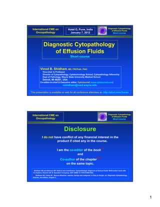
  • 1. Diagnostic Cytopathology International CME on Hotel O, Pune, India of Effusion Fluids Oncopathology January 7, 2012 Short course Diagnostic Cytopathology of Effusion Fluids Short course Vinod B. Shidham, MD, FRCPath, FIAC Vice-chair & Professor Director of Cytopathology, Cytotechnology School, Cytopathology fellowship Dept of Pathology, Wayne State University Medical School Detroit, MI 48201, USA Co-editor-in-chief & Executive editor, CytoJournal (www.cytojournal.com) vshidham@med.wayne.edu This presentation is available on web for all conference attendees at- http://alturl.com/3ucwx Diagnostic Cytopathology International CME on of Effusion Fluids Oncopathology Short course Disclosure I do not have conflict of any financial interest in the product if cited any in the course. I am the co-editor of the book * and Co-author of th chapter** C th f the h t on the same topic. *Shidham VB and Atkinson BF. Editors and contributors ‘Cytopathologic Diagnosis of Serous Fluids’ Multi-author book with 15 chapters, Elsevier (W. B. Saunders Company), 2007 (ISBN-13: 9781416001454). ** Shidham VB, Falzon M. Serous effusions: reactive, benign and malignant. In Gray & Kocjan, ed. Diagnostic Cytopathology, Elsevier, 3rd edition, Chapter 3. 1
  • 2. Diagnostic Cytopathology International CME on of Effusion Fluids Oncopathology Acknowledgment Short course Many images and tables are based on chapters in: Shidham VB and Atkinson BF. Cytopathologic Diagnosis of Serous Fluids Elsevier (W. B. Saunders Company) First edition, 2007. Diagnostic Cytopathology Outline International CME on of Effusion Fluids Oncopathology Short course Part I : Anatomy, histology, cytology, and effusions Collection, transportation, and processing Factors leading to potential diagnostic pitfalls Approach to diagnostic cytopathology of effusions The panorama of different faces of mesothelial cells Part II : Immunocytochemistry of effusion fluids: SCIP (Subtractive Coordinate Immunoreactivity Pattern) approach Evaluation of unknown primary sites of origin- Where do they come from? 2
  • 3. Diagnostic Cytopathology International CME on of Effusion Fluids Oncopathology Short course Anatomy, histology, cytology, and effusions a. Peritoneal cavity y b. Left & right pleural cavities c. Pericardial cavity Four major serous cavities Diagnostic Cytopathology International CME on of Effusion Fluids Oncopathology Short course Anatomy, histology, cytology, and effusions (continued) 1 Histology of serous li i Hi t l f lining (inguinal hernia sac). The mesothelial cells lining the fibrous tissue are flat (1). Focal reactive changes are seen as hypertrophy of a b some cells which assume a 2 3 cuboidal contour (2,3). ( ,3) [a-d, HE stain (a, 10X; b-d, 100X)]. c d 3
  • 4. Diagnostic Cytopathology International CME on of Effusion Fluids Oncopathology Short course Anatomy, histology, cytology, and effusions (continued) Peripheral light ectoplasm (1) Mesothelial cells (peritoneal fluid): show outer faintly stained Inner darker endoplasm (2) ectoplasm (1) with inner denser endoplasm (2) rich in Slightly off-center nucleus intermediate filaments. The nucleus is usually central or near Nucleolus central (a), but may be eccentric (b). Nucleoli are readily 1 1 observed. observed The vacuolation generally begins at the periphery in ectoplasm (1). 2 [a,b, Papanicolaou stained 2 SurePath® Preparation (a,b, 100XZoomed)] a b Diagnostic Cytopathology International CME on of Effusion Fluids Oncopathology Short course Anatomy, histology, cytology, and effusions (continued) Peripheral ectoplasm Inner endoplasm Central to slightly eccentric nucleus Ruffled cell border with blebs Mesothelial cell (pleural fluid): shows outer ectoplasm which is denser than the inner endoplasm. The nucleus is central to slightly eccentric but not touching the cell periphery. The cell margin shows blebs and is ruffled. [Diff-Quik stained Cytospin preparation (100XZoomed)] 4
  • 5. Diagnostic Cytopathology International CME on of Effusion Fluids Oncopathology Short course Anatomy, histology, cytology, and effusions (continued) Bloody effusions (Hemothorax, (Hemothorax hemopericardium and hemoperitoneum) Homogenously red or dark brown with hemosiderin pigment and the hematocrit of the effusion is 10% or more than that of the plasma hematocrit. (Occasional blood tinged fluids may be associated with a procedure trauma). Bloody effusions are more likely to be associated with an underlying malignancy. Reactive conditions associated with Bloody Effusions Para pneumonic effusions Pancreatitis Post traumatic effusions Acute aortic dissection underlying malignancy -Post cardiothoracic procedures / surgeries -Thoracic cavity vascular damage Pulmonary embolism Endometriosis Sarcoidosis Intralobar pulmonary sequestration Asbestos exposure associated pleural effusion Some Infections –e.g B. Anthrax Diagnostic Cytopathology International CME on of Effusion Fluids Oncopathology Short course Collection, transportation, and processing Biopsy vs effusion cytology 5
  • 6. Diagnostic Cytopathology International CME on of Effusion Fluids Oncopathology Short course Collection, transportation, and processing (continued) To prevent clotting: Collected in anticoagulant such as- EDTA (sodium or potasium salts) in final concentration of 4.55+0.85 µmol/ml [e.g. di-sodium [e g di sodium EDTA dihydrate (EDTA Na2, 2H2O) 1 4 mg per ml of effusion fluid] 1.4 Amount of fluid sample: Variable (less than 1 ml to 100 ml or more). For optimum cellularity of cytology preparations and cell blocks, it is recommend to submit as much specimen as possible (up to 1000 ml). Each laboratory follows its own protocol for determining the amount of sample to be used. Submit as a fresh, unfixed sample: If delay is expected in transporting to the laboratory- refrigerate at 4oC (with precaution not to freeze the specimen). g ( p p ) Alternatively, although not recommended, it may be preserved in a weak fixative Fluids collected in fixative- require a wash prior to instrument processing. For blood rich specimens- start with smaller aliquots (such as 10 ml) of fresh fluid. Concentration of cells and removal of excess erythrocytes in the blood rich specimen may be achieved by density gradient centrifugation with Ficoll International CME on Collection, transportation, Diagnostic Cytopathology of Effusion Fluids Oncopathology and processing (continued) Short course Collect fresh effusion fluid Concentrate the with or without anticoagulant effusion fluid specimen Concentrate the remaining fluid similar to Direct smear of Specimen specimen without clot unconcentrated specimen for with clot semi-quantitative evaluation Use homogenized specimen after mixing (For paucicelluar fluids: Use cell-rich sediment) Concentrate by centrifugation Remove the clot (at 600g for 10 minutes) and process for cell-block preparation Pour off supernatant and vortex to resuspend cell pellet Process for paraffin- embedding after formalin Cell-block preparation Smear preparation fixation. Histogel • Direct smears • SurePrep (Autocyte) Plasma-Thrombin • Cytospin • ThinPrep Celloidin bag • Other methods 6
  • 7. Diagnostic Cytopathology International CME on of Effusion Fluids Oncopathology Short course Collection, transportation, and processing (continued) Different alternatives available for preparing permanent cytology preparations. a. Direct smears (Spreading a drop of specimen directly on slide. The specimen may be before concentration OR after concentration as sediment) i. Wet fixed- Pap stain ii. Air-dried- Diff-Quik stain* Pap stain: After rehydration in saline and post-fixation in 95%ethanol with 5% acetic acid (28) b. Cytospin preparations (Shandon EZ Megafunnel™ with Shandon Cytospin®) (31) i. Wet fixed- Pap stain ii. Air-dried- ii Ai d i d Diff-Quik stain Pap stain: After rehydration in saline and post-fixation in 95%ethanol with 5% acetic acid (28) c. Filters i. Wet fixed- Pap stain d. Liquid based cytology (SurePathTM or ThinPrepTM or other non-proprietary methods) i. Wet fixed- Pap stain Diagnostic Cytopathology International CME on of Effusion Fluids Oncopathology Short course Collection, transportation, and processing (continued) Preparation Purpose Semiquantitative evaluation of A Diff-Quik stained direct smear of the undiluted specimen cellularity. Diff-Quik stained air-dried Rapid initial detection of second B Shandon EZ Megafunnel™ population and cytomorphologic preparation evaluation of hematopoietic cells. Pap stained preparation Cytomorphologic evaluation C especially nuclear details. details Cell-block preparation in Evaluation of- D HistoGelTM a. Immunoprofile, b. Other special stains, c. Architecture, 7
  • 8. Diagnostic Cytopathology International CME on of Effusion Fluids Oncopathology Short course Approach to diagnostic cytopathology of effusions ► GENERAL FEATURES ► PROCESSING RELATED ► INTERPRETATION STRATEGY ► CYTOMORPHOLOGY ► IMMUNOCYTOCHEMISTRY Diagnostic Cytopathology International CME on of Effusion Fluids Oncopathology Short course Approach to diagnostic cytopathology of effusions (continued) ► GENERAL FEATURES ►A general pathology l b Any l h l laboratory may receive effusion- all general pathologists and i ff i ll l h l i d cytopathologists should be conversant with the diagnostic challenges and pitfalls of effusion fluid cytology. ►Finding neoplastic cells in effusion specimens not only reveals that a patient has cancer, but it also denotes the advanced nature of the disease which at this stage is almost always incurable. ►The morphologic features of most of the cancer cells in effusion smears are different f diff t from th those seen i exfoliative, b hi in f li ti brushing, and FNA cytology. d t l ►The standard cytologic criteria of malignancy based on evaluation of single cell morphology are not applicable for most of the effusion cytology specimens (except in cases with high-grade neoplasms with pleomorphic cells). ►Since cells in a fluid medium ‘round up’ because of surface tension, the native shapes of cancer cells cannot be a guiding feature. 8
  • 9. Diagnostic Cytopathology International CME on of Effusion Fluids Oncopathology Short course Approach to diagnostic cytopathology of effusions (continued) ► PROCESSING RELATED 1. Second population of foreign cells 2. Nuclear details of the ‘second population’. 3. Semiquantitative evaluation 4. Objective confirmation and differential diagnosis of primary neoplasm. Diagnostic Cytopathology International CME on of Effusion Fluids Oncopathology Short course Approach to diagnostic cytopathology of effusions (continued) ► INTERPRETATION STRATEGY a. Reactive mesothelial cells- morphologic overlap with neoplastic cells b. A single population- favor mesothelial cells c. Second foreign population consistent with metastatic neoplasm d. Sarcomas- previous history known e. Romanowsky stains highlight the ‘second population’ f. Immunocytochemistry- objective confirmation of ‘second population’ y y g. Identical orientation of serial sections for immunocytochemistry h. Quantitative and qualitative clues for mesothelioma i. Apoptosis 9
  • 10. Diagnostic Cytopathology International CME on of Effusion Fluids Oncopathology Short course Approach to diagnostic cytopathology of effusions (continued) ► CYTOMORPHOLOGY 1. Cell groups and intercellular cohesion Non-cohesive, good intercellular cohesion, proliferation spheres 2. Arrangement of neoplastic cells Papillary configurations 3. Cytoplasm of neoplastic cells 4. Special structures and cytological features 5. Other features International CME on Algorithm for evaluation of Diagnostic Cytopathology of Effusion Fluids Oncopathology ‘second foreign population’ Short course Reactive- 4 Usually single cells without large 3-D groups Mesothelial cells 2 Neoplastic- Mesothelioma 5 ♦Quantity- Many cells ♦Quality- Many large groups Cells in effusion fluid 1 Reactive- 6a Hematopoietic cells 6 Inflammatory cells (Non-cohesive cells) Neoplastic- 6b Non mesothelial cells 3 Non-mesothelial Lymphoma L h 7 Neoplastic-8 (2nd foreign population) 8 ¶Metastatic cancer cells may be the predominant Carcinoma (Cohesive cells) cells without being seen as a ‘second population’. Sarcoma They may be present as scattered solitary cells with (Spindle cells may be present. Known cytomorphology overlapping with floridly reactive history of sarcoma is usually crucial for mesothelial cells. If indicated, immunocytochemistry proper interpretation) would facilitate confirmation of these cells as non- Melanoma (Non-cohesive cells) mesothelial. 10
  • 11. Diagnostic Cytopathology International CME on of Effusion Fluids Oncopathology Short course The panorama of different faces of mesothelial cells REACTIVE MESOTHELIAL CELLS Binucleation and multinucleation- multinucleation Gigantic nuclei Phagocytic activity Cohesive Clusters and/or Papillary Structures ‘The two cell population approach’ Cell-in-cell configuration Cells in sheets Groups of reactive mesothelial cells I) Hepatomegaly, II) Ischemic conditions such as pulmonary infarction, ischemic colitis, and occlusion of mesenteric blood vessels, III) Trauma to organs covered with mesothelium such as spleen, liver, and lung etc, IV) Large retroperitoneal masses- Slowly growing retroperitoneal masses, V) Postoperative- following laparotomy and thoracotomy. ‘ATYPICAL’ MESOTHELIAL CELLS Diagnostic Cytopathology International CME on of Effusion Fluids Oncopathology Short course The panorama of different face of mesothelial cells (continued) a b c Mesothelial cells with central to slightly eccentric nuclei (ascitic fluid). The cytoplasm shows a two-zone staining pattern. [a-c, Diff-Quik stained cytospin preparation (a-c, 100x Zoomed)]. 11
  • 12. Diagnostic Cytopathology International CME on of Effusion Fluids Oncopathology Short course The panorama of different face of mesothelial cells (continued) Panorama of mesothelial cells (asc t c u d) Ce t a (ascitic fluid). Central to near ea central nuclei. Rare mesothelial a b c d e f cells may show eccentric nuclei touching the cell membrane, but usually there is a narrow rim of cytoplasm separating the g h i j k l nucleus from the cell border (arrows). [a-x, Diff Q ik stained cytospin [ Diff-Quik t i d t i preparations (a-x, 100x m n o p q r Zoomed)]. s t u v w x Diagnostic Cytopathology International CME on of Effusion Fluids Oncopathology Short course The panorama of different face of mesothelial cells (continued) 1 a b c Mesothelial cells with central to slightly eccentric nuclei (ascitic fluid). The cytoplasm shows a two-zone staining pattern with peripheral vacuolation (red arrow 1). [a-c, Papanicolaou stained ThinPrep preparation (a-c, 100x Zoomed)]. 12
  • 13. Diagnostic Cytopathology International CME on of Effusion Fluids Oncopathology Short course The panorama of different face of mesothelial cells (continued) Mesothelial cells with central to eccentric nuclei ( ti l i (ascitic fl id) iti fluid). a b c d e f Spectrum of cytomorphological features. [a-zc, Papanicolaou stained g h i j k ThinPrep preparation (a-zc, 100x Zoomed)] l m n o p q r s t u v w x y z za zb zc Diagnostic Cytopathology International CME on of Effusion Fluids Oncopathology Short course The panorama of different face of mesothelial cells (continued) Mesothelial cells with eccentric nuclei (ascitic fluid). Careful scrutiny usuallyy shows a narrow rim of cytoplasm separating the nucleus from the cell border (arrows). [Papanicolaou stained ThinPrep preparation, l x 100XZoomed]. 13
  • 14. Diagnostic Cytopathology International CME on of Effusion Fluids Oncopathology Short course The panorama of different face of mesothelial cells (continued) NC NC RM IC RM IC DQ Pap a b Metastatic adenocarcinoma (pleural fluid). An example with morphologically identifiable unequivocal ‘second foreign population’ (red arrow NC) other than mesothelial cells (blue arrow RM) and inflammatory cells (brown arrow ‘IC) in DQ and PAP stained preparations. This ‘second population’ of cells ’ (red arrow NC) is easy to be identified in DQ stain (a) as compared to that in PAP stain (b). [a, Diff-Quik stained cytospin preparation; b, Papanicolaou stained ThinPrepTM preparation (a,b, 100x Zoomed)] Diagnostic Cytopathology International CME on of Effusion Fluids Oncopathology Short course The panorama of different face of mesothelial cells (continued) a b c d Pulmonary adenocarcinoma (pleural fluid). The non-cohesive metastatic cancer cells is the predominant cell population without being seen as a ‘second population’ (a-d). Some apoptotic tumor cells (arrows in c & d) are present. Differential includes anaplastic large cell lymphoma [a-d: PAP stained Cytospin preparation (a, 10X; b, 40X; c,d, 100X)]. 14
  • 15. Diagnostic Cytopathology International CME on of Effusion Fluids Oncopathology Short course Factors leading to potential diagnostic pitfalls a. Surface tension related alterations in cytomorphology b. b Improper specimen processing c. Many faces of reactive mesothelial cells d. Proliferation related features i) Proliferation spheres ii) Increased number of mitotic figures iii) Prominent nucleoli e. Degenerative changes Nuclear hyperchromasia Cytoplasmic vacuolation C l i l i f. Presence of some unexpected patterns and unusual structures i) Reactive lymphoid population ii) Polymorphic lymphocytes iii) Single population of cells due to predominance of tumor cells iv) Psammoma bodies v) Three dimensional benign cell groups Benign papillary inclusions, Gland-like epithelial structures, Mullerian inclusions vi) Megakaryocytes Diagnostic Cytopathology International CME on of Effusion Fluids Oncopathology Short course Factors leading to potential diagnostic pitfalls (continued) TRUE NEGATIVE RESULTS IN EFFUSIONS CAUSED BY CANCER a. Blockage of lymphatics without exfoliation of neoplastic cells b. Increased capillary permeability due to VEGF. c. Lack c L k off exfoliation b llow-grade sarcomas and spindle cell mesotheliomas. f li ti by d d i dl ll th li d. Organized thick fibrin serosal covering preventing exfoliation of neoplastic cells. e. Decrease or total disappearance of neoplastic cells over a time 15
  • 16. Diagnostic Cytopathology International CME on of Effusion Fluids Oncopathology Short course Part II Diagnostic Cytopathology International CME on of Effusion Fluids Oncopathology Short course Immunocytochemistry of effusion fluids The most important issue to be considered when applying immunocytochemistry to effusion fluids is the significant variation in results due to the many variables incurred from the time of collection of the specimen to its final immunostaining. 16
  • 17. Diagnostic Cytopathology International CME on Immunocytochemistry of of Effusion Fluids Oncopathology effusion fluids Short course UNIQUENESS OF EFFUSION IMMUNOCYTOCHEMISTRY Confirmation of a ‘ C fi ti f ‘second-foreign’ non-inflammatory population of cells other th df i ’ i fl t l ti f ll th than mesothelial cells in effusions correlate with metastatic cancer with objectivity. Intricacies of finding and locating the cells of interest in cell-block sections may adversely affect the final results. It is crucial to: ►Orient the serial sections identically on all slides (to identify more precisely the same cell (or small group of cells) in different sections). ►Know the sequence of these serial sections (to evaluate their co-ordinate immunoreactivity pattern). ►Immunocytochemistry does not have significant role in evaluation of peritoneal washings. Diagnostic Cytopathology International CME on Immunocytochemistry of of Effusion Fluids Oncopathology effusion fluids Short course Cell-blocks are the preferred choice. Formalin-fixed cell-block sections are recommended- Other protocols such as the evaluation of various cytology preparations (direct smears- wet fixed in alcohol or acetone, air-dried fixed with alcohol, air-dried smears rehydrated and post-fixed in formol alcohol, liquid based cytology preparations- SurePath or ThinPrep, cytospin preparations, etc) should be avoided. For reproducible results a standardized protocol with steps comparable to the processing of formalin-fixed paraffin-embedded tissue sections is essential. 17
  • 18. Diagnostic Cytopathology International CME on of Effusion Fluids Oncopathology Short course Critical issues leading to the best result with cell blocks of cytology specimens with singly scattered cells Aligning the cells along the cutting surface Monitor the depth of section cutting Diagnostic Cytopathology International CME on of Effusion Fluids Oncopathology Short course Varsegi GM, Shidham V (2009) Journal of Visualized Experiments (JoVE) 2009 Jul 21;(29). pii: 1316. doi: 10.3791/1316. PMID: 19623160 18
  • 19. Diagnostic Cytopathology International CME on of Effusion Fluids Oncopathology Short course From: Varsegi GM, Shidham V (2009) Journal of Visualized Experiments (JoVE) 2009 Jul 21;(29). pii: 1316. doi: 10.3791/1316. PMID: 19623160 Diagnostic Cytopathology International CME on of Effusion Fluids Oncopathology Short course From: Varsegi GM, Shidham V (2009) Journal of Visualized Experiments (JoVE) 2009 Jul 21;(29). pii: 1316. doi: 10.3791/1316. PMID: 19623160 19
  • 20. Diagnostic Cytopathology International CME on of Effusion Fluids Oncopathology Short course Modified from: Varsegi GM, Shidham V (2009) Journal of Visualized Experiments (JoVE) 2009 Jul 21;(29). pii: 1316. doi: 10.3791/1316. PMID: 19623160 Diagnostic Cytopathology International CME on of Effusion Fluids Oncopathology Short course From: Varsegi GM, Shidham V (2009) Journal of Visualized Experiments (JoVE) 2009 Jul 21;(29). pii: 1316. doi: 10.3791/1316. PMID: 19623160 20
  • 21. Diagnostic Cytopathology International CME on of Effusion Fluids Oncopathology Short course The video article is available FREE on the web under open access at: http://www.jove.com/index/Details.stp?ID=1316 Video f J VE ti l Vid of JoVE article (8 minutes 15 sec) i t ) Video of JoVE article (8 minutes 15 sec) Diagnostic Cytopathology International CME on of Effusion Fluids Oncopathology Short course Immunophenotyping and cell blocks- Factors affecting immunoreactivity- Loss, reduction, or enhancement of antigen immunoreactivity Exposure to different reagents and fixative(s) Temperature Storage of specimen with or without fixative Subtractive Coordinate Immunoreactivity Pattern (SCIP) approach Shidham & Atkinson Ch 5 Imm noc tochemistr of effusion fl uids: introd ction to SCIP approach 5. Immunocytochemistry eff sion ids introduction approach. ‘Cytopathologic Diagnosis of Serous Fluids’ Elsevier (W. B. Saunders Company) Shidham VB. Effusion Fluid Evaluation Made Simple: A brief review of cytomorphologic and SCIP approach CytoJournal (In press). 21
  • 22. Diagnostic Cytopathology International CME on Immunocytochemistry of of Effusion Fluids Oncopathology effusion fluids Short course Discrepant results between formalin-fixed paraffin-embedded tissue sections of surgical pathology material and effusion fluid cell block sections are not uncommon cell-block uncommon. Reasons for variable reports: The variables responsible for such discrepancies include: sample size (tiny cell groups or single cells), selection of fixatives, antigen retrieval methods (i.e., heat-induced (i e heat induced epitope retrieval enzyme digestion etc ) retrieval, digestion, etc.), antibody clones used, antibody titer, and other variations in immunostaining protocols. The proteinaceous effusion fluid around suspended cells may contribute to unexpected nonspecific immunoreactivity. Diagnostic Cytopathology International CME on Immunocytochemistry of of Effusion Fluids Oncopathology effusion fluids Short course If findings are equivocal: It is prudent to be conservative and recommend to repeat. Malignant effusions usually re-accumulate quickly. Acquiring a new sample is generally not a problem. However, it is not uncommon to submit only a small fraction of a large volume of effusion fluid collected. To avoid inadequate resubmission, it may be specifically communicated in the recommendation as comment : “Recommend submission of most of the drained effusion fluid (up to 1000 mL). Larger volume of specimen facilitates retrieval of adequate cellular material in cell-block sections for immunocytochemical evaluation”. 22
  • 23. Diagnostic Cytopathology International CME on Immunocytochemistry of of Effusion Fluids Oncopathology effusion fluids Short course Interpretation: I t t ti All aspects of individual and complimentary immunomarkers should be considered collectively rather than applying a reflexive positive-negative approach. Evaluation of co-ordinate immunoreactivity in different cell y population. Diagnostic Cytopathology International CME on Immunocytochemistry of of Effusion Fluids Oncopathology effusion fluids Short course Immunostaining None Nuclear Nuclear & Cytoplasmic Membranous Microvillous pattern cytoplasmic Immunomarker Calretinin X X WT-1 X X D2-40 (Podoplanin) X Cytokeratin* X Vimentin X LCA (CD45) X PGM1 (CD68) X EMA X AdCa X meso HBME-1 X AdCa X meso 23
  • 24. Diagnostic Cytopathology International CME on Immunocytochemistry of of Effusion Fluids Oncopathology effusion fluids Short course Immunostaining None Nuclear Nuclear & Cytoplasmic Membranous Microvillous pattern cytoplasmic Immunomarker B72.3 X BerEP4 X Cadherins X MOC-31 X CD44S X Mesothelin X mCEA X CK 5/6 X CD15 (Lue-M1) X CA19.9 X Ttf-1 X Diagnostic Cytopathology International CME on Immunocytochemistry of of Effusion Fluids Oncopathology effusion fluids Short course Immunoreactivity pattern with EMA (epithelioid mesothelioma, pleural fluid). EMA Mesothelioma cells with membranous (arrow) and cytoplasmic immunostaining. Note the microvilli (arrowhead). [Immunostained cell-block section (100XZoomed)]. 24
  • 25. Diagnostic Cytopathology International CME on Immunocytochemistry of of Effusion Fluids Oncopathology effusion fluids Short course a HBME-1 b HBME-1 HBME-1 immunoreactivity pattern (epithelioid mesothelioma, pleural fluid). Mesothelioma cells with membranous (arrow in a) and cytoplasmic immunostaining. Note the microvilli (arrowhead in b). Diagnostic Cytopathology International CME on Immunocytochemistry of of Effusion Fluids Oncopathology effusion fluids Short course Pan-cytokeratin immunoreactivity pattern (pleural fluid). Reactive mesothelial cells with cytoplasmic immunostaining (arrow in inset). Some reactive mesothelial cells may show a concentric immunostaining i t i i pattern tt around the nucleus better appreciated by adjusting fine focus. Pan-cytokeratin 25
  • 26. Diagnostic Cytopathology International CME on Immunocytochemistry of of Effusion Fluids Oncopathology effusion fluids Short course Cytokeratin 7 immunoreactivity pattern (epithelioid mesothelioma, pleural fluid). Neoplastic mesothelial cells with cytoplasmic immunostaining. Note the bushy microvilli (arrowhead). Cytokeratin 7 Diagnostic Cytopathology International CME on Immunocytochemistry of of Effusion Fluids Oncopathology effusion fluids Short course 2 1 a Calretinin b Calretinin Calretinin immunoreactivity pattern (epithelioid mesothelioma, pleural fluid). Mesothelioma cells (arrow in a) show nuclear (arrowhead 1) immunoreactivity usually with cytoplasmic immunostaining (arrowhead 2) imparting the so called ‘fried-egg’ appearance. 26
  • 27. Diagnostic Cytopathology International CME on Immunocytochemistry of of Effusion Fluids Oncopathology effusion fluids Short course Calretinin immunoreactivity pattern (pleural fluid). Reactive mesothelial cells (blue arrows). The effusion also contains metastatic RM mammary y carcinoma cells (red arrow NC). NC Calretnin Diagnostic Cytopathology International CME on Immunocytochemistry of of Effusion Fluids Oncopathology effusion fluids Short course WT-1 immunoreactivity p pattern (Metastatic colonic adenocarcinoma, peritoneal fluid). Reactive mesothelial cells (arrow RM) show nuclear immunoreactivity (arrowhead in inset) with some cytoplasmic immunostaining. y p g Rare adenocarcinoma cells demonstrating nuclear immunoreactivity for CDX2 RM were also seen in other section. WT-1 27
  • 28. Diagnostic Cytopathology International CME on Immunocytochemistry of of Effusion Fluids Oncopathology effusion fluids Short course B72.3 immunoreactivity pattern (Metastatic mammary adenocarcinoma, pleural fluid). Metastatic adenocarcinoma cells (red arrow NC) show a cytoplasmic immunoreactivity pattern. NC B72.3 Diagnostic Cytopathology International CME on Immunocytochemistry of of Effusion Fluids Oncopathology effusion fluids Short course Vimentin immunoreactivity pattern (peritoneal wash). Reactive mesothelial cells (arrow RM) show cytoplasmic immunoreactivity pattern (arrowhead in inset) inset). RM vimentin 28
  • 29. Diagnostic Cytopathology International CME on Immunocytochemistry of of Effusion Fluids Oncopathology effusion fluids Short course LCA (CD45 ) immunoreactivity pattern (pleural fluid). Reactive mesothelial cells (blue arrow RM) with chronic RM inflammatory cells (red arrows). The inflammatory cells show a strong cytoplasmic immunoreactivity pattern obscuring the nucleus (arrowhead in inset). LCA Diagnostic Cytopathology International CME on Immunocytochemistry of of Effusion Fluids Oncopathology effusion fluids Short course CD 68 (PGM1 ) immunoreactivity pattern (M tt (Metastatic i mammary adenocarcinoma H with proliferation spheres H (red arrow NC), pleural fluid). Histiocytes show CD68 H immunoreactivity (blue arrows H). In our experience, PGM1 does not show non non- specific immunostaining usually associated with KP1. NC Inset- Histiocytes (blue arrow H) with cytoplasmic H immunoreactivity pattern CD68 around the nucleus. 29
  • 30. Diagnostic Cytopathology International CME on Immunocytochemistry of of Effusion Fluids Oncopathology effusion fluids Short course NC NC NC a BerEP4 b BerEP4 BerEP4 immunoreactivity pattern (Metastatic mammary adenocarcinoma, pleural fluid). a. The neoplastic cells in proliferation spheres (red arrow NC)- membranous immunostaining with a honey comb-like pattern. b. Solitary adenocarcinoma cells (red arrow NC)- membranous immunostaining pattern along the cell membrane (arrowhead in inset). Diagnostic Cytopathology International CME on Immunocytochemistry of of Effusion Fluids Oncopathology effusion fluids Short course NC NC Comparison of immunoreactivity with NC BerEP4 and B72.3 (Metastatic mammary adenocarcinoma, pleural fluid). As Compared to B72.3, a BerEP4 b NC BerEP4 most of the adenocarcinoma cells (red NC arrows NC) show strong ) g NC membranous BerEP4 immunoreactivity. c B72.3 d B72.3 30
  • 31. Diagnostic Cytopathology International CME on Immunocytochemistry of of Effusion Fluids Oncopathology effusion fluids Short course Monoclonal CEA (mCEA) m immunoreactivity pattern (Metastatic ovarian carcinoma, peritoneal fluid). Metastatic adenocarcinoma cells show cytoplasmic (c) and membranous (m) c immunostaining. mCEA Diagnostic Cytopathology International CME on Immunocytochemistry of of Effusion Fluids Oncopathology effusion fluids Short course CDX2 y immunoreactivity pattern NC (Metastatic colonic adenocarcinoma, peritoneal fluid). The adenocarcinoma cells show nuclear immunoreactivity (arrow NC). Compare with non- immunoreactive nuclei with blue hematoxylin counterstain (arrowhead). CDX2 31
  • 32. Diagnostic Cytopathology International CME on Immunocytochemistry of of Effusion Fluids Oncopathology effusion fluids Short course TTF-1 immunoreactivity y pattern (Metastatic pulmonary carcinoma, pleural fluid). The solitary adenocarcinoma cells as the predominant population (arrows NC) show NC nuclear immunoreactivity (arrowheads in inset). NC TTF-1 Diagnostic Cytopathology International CME on Immunocytochemistry of of Effusion Fluids Oncopathology effusion fluids Short course MOC-31 immunoreactivity m pattern (Metastatic mammary carcinoma, pleural fluid). The adenocarcinoma cells show predominantly membranous (m) with cytoplasmic (c) immunoreactivity. c MOC-31 32
  • 33. Diagnostic Cytopathology International CME on Immunocytochemistry of of Effusion Fluids Oncopathology effusion fluids Short course Diffuse malignant mesothelioma of epithelial type, EMA (pleural fluid). Neoplastic cells are immunoreactive for EMA (a & b) and HBME-1 (c, d, & e) a b with a membranous immunostaining pattern (arrows) highlighting long, slender, microvilli HBME-1 (arrowheads). c d e Diagnostic Cytopathology International CME on Immunocytochemistry of of Effusion Fluids Oncopathology effusion fluids Short course a EMA b HBME-1 Adenocarcinoma, peritoneal fluid. Cytoplasmic immunostaining pattern (arrows) with focal blotchy immunostaining for EMA (a) and HBME-1 (b) along the cell membrane . 33
  • 34. Diagnostic Cytopathology International CME on Immunocytochemistry of of Effusion Fluids Oncopathology effusion fluids Short course For reproducible results, it is important to select any immunopanel which will fundamentally identify most of the mesothelial and inflammatory cells to create the basic map for confirmation of a ‘second-foreign’ population by ‘Subtractive Coordinate Immunoreactivity Pattern’ (SCIP) approach * Shidham VB, Atkinson BF. Immunocytochemistry of effusion fluids: Introduction to the SCIP approach. In: Shidham VB and Atkinson BF. Editors ‘Cytopathologic Diagnosis of Serous Fluids’ First edition, Elsevier (W. B. Saunders Company); 2007. Ch 5, pp. 55-78. *Shidham VB. Diagnostic Cytopathology of Serous Fluids- A brief review of cytomorphologic and SCIP approach. CytoJournal (In press). Diagnostic Cytopathology International CME on Immunocytochemistry of of Effusion Fluids Oncopathology effusion fluids Short course Evaluation of ‘Subtractive Coordinate Immunoreactivity Pattern’ (SCIP) 34
  • 35. Mesothelial & X Metastasis Y Metastasis Z ry inflammator cells (carcinoma) (non-carcinoma) vimentin 1 2 1 2 1 2 A 3 3 4 3 4 6 5 6 5 4 7 6 8 7 5 7 SCIP 1 2 1 2 2 B Pan CK 1 approach (Mixture of AE1/AE3 3 3 & CAM5.2) 4 3 6 5 4 6 4 Oncopathology Oncopathology 5 7 6 International CME on International CME on 8 7 5 7 1 2 1 2 2 C LCA(CD45) 1 [or PGM1(CD68) 3 3 or mixture of LCA 4 3 6 5 4 6 & PGM1] 4 5 7 6 8 7 5 7 1 2 1 2 2 D Calretinin 1 3 3 4 3 6 5 4 6 5 4 effusion fluids effusion fluids 7 6 8 7 5 7 Immunocytochemistry of Immunocytochemistry of 1 2 1 2 E 1 WT-1 3 3 4 3 6 5 4 6 5 4 7 6 8 7 5 7 SCIP Short course Short course of Effusion Fluids of Effusion Fluids approach Diagnostic Cytopathology Diagnostic Cytopathology 35
  • 36. Diagnostic Cytopathology International CME on Immunocytochemistry of of Effusion Fluids Oncopathology effusion fluids Short course SCIP approach a b Diagnostic Cytopathology International CME on Immunocytochemistry of of Effusion Fluids Oncopathology effusion fluids Short course Immunoreactive for ‘negative’ lretinin, D2-40) mesothelial markers such as- BerEP4, B72.3, MOC-31, mCEA, . Proceed with: CK+,vim –/+ Carcinoma Immunopanel for unknown primary CIP OR Basic panel for evaluation by SC (vimentin, PanCK,CK CK20, Ber/EP4, B72.3, MOC31Cal Restricted panel for known primary With ‘second-foreign’ population LCA+ Lymphoma panel Lymphoma Cytogenetics Gene rearrangement CK-,vim+ Melanoma/ S-100 protein & LCA– Sarcoma Melanoma markers Without Qualitative & quantitative – + K7, ‘second foreign’ population features of mesothelioma p second-foreign Melanoma Sarcoma Absent Present Negative for Malignant malignancy mesothelioma EMA/HBME-1: Immunopanel for sarcoma Microvillous pattern OR B72.3–, BerEP4– Restricted panel for known primary 36
  • 37. ‘Subtractive coordinate immunoreactivity pattern’ (SCIP) in cell block sections Diagnostic Cytopathology International CME on of Effusion Fluids Oncopathology A. Vimentin Short course Non-immunoreactive NC NC 10X 40X SCIP B. Pan-cytokeratin Immunoreactive approach NC 10X NC 40X C. LCA (CD45) Non-immunoreactive NC Metastatic colonic NC 10X 40X adenocarcinoma, D. Calretinin Non-immunoreactive (peritoneal fluid). (Inset {2}- RM Mesothelial cell NC immunoreactive nuclear-cytoplasmic) RM y p ) NC 10X 40X E. WT-1 Non-immunoreactive RM HE (Arrow 2 with inset: stained Mesothelial cell- NC cell block immunoreactive RM NC section nuclear-cytoplasmic) 10X 40X NC F. CDX2 Immunoreactive nuclear NC NC 40X 10X 40X 100X ‘Subtractive coordinate immunoreactivity pattern’ (SCIP) in cell block sections Diagnostic Cytopathology International CME on RM of Effusion Fluids Oncopathology A. Vimentin Short course Non-immunoreactive NC 10X Zoomed SCIP B. Pan-cytokeratin Immunoreactive approach 10X RM C. Calretinin NC Non-immunoreactive [Mesothelial cells (RM) immunoreactive nuclear-cytoplasmic] 10X Zoomed RM HE D. BerEP4 stained Immunoreactive NC cell block sect o section 10X Zoomed E. Cytokeratin 7 Immunoreactive Metastatic ovarian carcinoma, (peritoneal fluid). 10X 10X NC RM F. Cytokeratin 20 Non-immunoreactive Zoomed 10X 37
  • 38. ‘Subtractive coordinate immunoreactivity pattern’ (SCIP) in cell block sections Diagnostic Cytopathology International CME on of Effusion Fluids Oncopathology Short course A. Vimentin Non-immunoreactive 40X SCIP approach B. CD68 (PGM1) Non-immunoreactive 40X RM C. Calretinin Non-immunoreactive Mesothelial cell (RM) immunoreactive Metastatic mammary nuclear-cytoplasmic) 40X adenocarcinoma, (pleural effusion). RM D. Cytokeratin 7 HE Immunoreactive stained cell block NC section 40X RM NC E. BerEP4 Immunoreactive NC 40X 40X ‘Subtractive coordinate immunoreactivity pattern’ (SCIP) in cell block sections Diagnostic Cytopathology International CME on of Effusion Fluids Oncopathology A. Vimentin Short course Non-immunoreactive (Mesothelial & inflammatory cells are immunoreactive) 20X 40X SCIP Metastatic B. CD68 (PGM1) approach Non-immunoreactive (inflammatory cells are mammary adenocarcinoma, immunoreactive) (pleural effusion). 20X 40X RM C. Calretinin Non-immunoreactive (Rare mesothelial cell [blue arrow] is immunoreactive nuclear-cytoplasmic) 20X 40X D. BerEP4 Immunoreactive NC 20X 40X NC E. Estrogen receptors NC Immunoreactive 20X 40X 38