7. Cell loss in proliferating populations
• Eliminate self reactive lymphocytes
• Cell death by induced cytotoxic T lymphocytes
(defense against virus / tumorogenisis)
9. Accumulation of misfolded proteins
Arise because of mutations in the genes coding
these proteins or extrensic factors like free
radicals
Cell injury in infections
Atropy in parynchyma after duct obstruction
14. Dimerize and
insert into
membrane of
mitochondria
Permiability transition
pore Channel activated
(voltage gated channel)
Cytochrome C
CASPASE
ACTIVATION
15. Surface molecules trigger apoptosis
Members of TNF family
Ex: Type 1 TNF and Fas (CD95)
Normally present in cytoplasmic region
On Activation move to surface
17. CASPASE
Mitochondrial pathway Death receptor pathway
Both pathways are not mutually exclusive with p53 causing cross over to
death receptor pathway.
Cystene protease that cleave proteins after aspartic residues
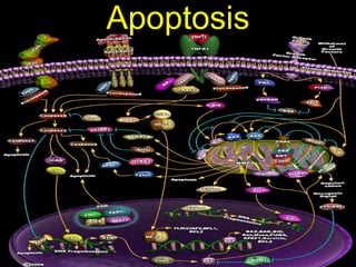
Cell Death by Apoptosis
Activation of caspase is the fundamental event of apoptosis
19. Caspase activation
a. Proteolytic cleavage
e.g. pro-caspase 3
b. Induced proximity
e.g. pro-caspase 8
c. Oligomerization,
e.g. cyt c, Apaf-1 &
caspase 9
20. Inflammatory Caspases: -1, -4, and -5
Initiator Caspases: -2, -8, -9, and -10
Long N-terminal domain
Interact with effector caspases
Effector Caspases: -3, -6, and -7
Little to no N-terminal domain
Initiate cell death
21.
22. But become avid targets to phagocytosis by flip
out of phosphotidyl serine on the inner leaflet of
plasma membrane
These are recognized by macrophages
Also secrete soluble factors that recruit
phagocytes
Express adhesive glycoproteins recognized by
phagocytes and macrophages
23.
24. CASPASE
.
Cleavage of nuclear LAMINS is involved in chromatin condensation
and nuclear shrinkage.
Cleavage of the inhibitor of the DNase CAD (caspase-activated
deoxyribonuclease), ICAD causes the release of the endonuclease,
which fragment DNA.
Cleavage of cytoskeletal proteins such as actin, plectin, Rho
kinase 1 (ROCK1) and gelsolin leads to cell fragmentation, blebbing
and the formation of apoptotic bodies.
25.
26. Constant number of cells in an organism.
Cell death = Cell proliferation
Cell
Death
Growth
Survival
Proliferation
27. What happens when things become imbalanced?
Cell
Death
Growth
Survival
Proliferation
28. Resistance of tumor cells to apoptosis is an
essential feature of cancer development
Loss of apoptosis can promote tumor
Initiation
Progression
Treatment resistance
29. p53 induce the expression of proteins involved in
the mitochondrial pathway — BAX, NOXA, PUMA and
P53aip1
death receptor pathway — CD95, TRAIL-R1 and TRAIL-
R2.
G1 S G2 M
p53
MalignancyMutation
30. overexpression of anti-apoptotic genes
follicular B-cell lymphoma t(14;18), couples BCL2
gene to immunoglobulin heavy chain locus, leading
to enhanced BCL2 expression.
EBV and HHV8 encode proteins that are
homologues of BCL2.
BHRF1 from EBV and KSbcl-2 (vBcl-2) from HHV8 —
have an anti-apoptotic function and enhance
survival of the infected cells.
31. Adenovirus induce cells to bypass apoptosis
and replicate by E1B oncoprotein inducing cells
to enter S phase
Cytokines like IL6 can inhibit apoptosis
32. soluble receptors that act as decoys for death
ligands.
soluble CD95 (sCD95) and decoy receptor 3 (DcR3)
shown to competitively inhibit CD95 signaling.
sCD95 is expressed in various malignancies, and
elevated levels can be found in the sera of cancer
patients.
33. associated with poor prognosis in melanoma
patients
It is genetically amplified in several lung and
colon carcinomas
overexpressed in several adenocarcinomas,
glioma cell lines and glioblastomas.
34. the pro-apoptotic BCL2 family member BAX is
mutated.
Frame shift mutations
loss of expression and function.
Reduced BAX expression is associated with a
poor response rate to chemotherapy and
shorter survival
35. Expression of anti-apoptotic proteins
High levels of FLIP which interferes with
apoptosis induction at the level of the death
receptors
Human melanomas
murine B-cell lymphoma
FLIP:caspase-8 ratio cause resistance to
CD95-mediated apoptosis
EBV-positive Burkitt's lymphoma
Viral analogues of FLIP, called viral FLIPs
(v-FLIPs)
36. Expression of anti-apoptotic proteins
Expression of the IAP-family protein
survivin is highly tumour specific.
Found in most human tumours but not
in normal adult tissues.
In neuroblastoma expression correlates
with a more aggressive and
unfavourable disease.
37. Bcl2 shows discrepancies in effect on tumor
management
Studies have shown a correlation between
high levels of BCL2 expression and the
severity of malignancy of human tumours.
a high level of BCL2 expression is associated
with a poor response to chemotherapy and
seems to be predictive of shorter, disease-
free survival.
While in some cases Bcl-2 has been shown to
cause chemo resistance in other settings it is
shown to improve prognosis.
38. Clinical studies examine single alterations
Cannot exclude extragenic mutations in the
same pathway
Thus almost impossible to determine negative
results
Ex: murine lymphomas harbouring INK4a/ARF
have defective p53 but harbour wild type p53
hence classified as p53 normal
Extracellular survival factors affect cell
death
Cell density
microenvironment
39. Application in Treatment
Chemotherapy, irradiation and other stimuli
can initiate apoptosis through the
mitochondrial (intrinsic) pathway.
Pro-apoptotic BCL2 family proteins are
activated by treatment
40. Disrupted apoptotic cycle can affect sensitivity
of cancer drugs
As multiple drugs affect same mechanism it
causes multidrug resistance
Sufficient doses of almost all anticancer drugs
induce apoptosis by alternate pathway
independent of p53 pathway
Contribution of p53 is dependent on
Agent
Dose
mutational background
41. The best-defined mechanism of therapy-induced
cellular stress induced cancer cell death.
Chemotherapeutic drugs (for example, the
nucleotide analogue 5-FU) induce CD95 by a
transcriptionally regulated, p53-dependent
mechanism.
Leads to upregulation of CD95L
Allows the cells to either commit suicide or kill
neighbouring cells.
42. Targeting in therapy
TNF alpha and Fas are toxic to
Normal cells
Cancer cells
But….
TRAIL ---- tumor necrosis factor related
ligand preferentially attacks tumor cells
43. Most mutations occur upstream
Machinery is retained
Tumor specific alterations in apoptotic
programmes
Ex: Adenoviral gene transfer in ovarian cancer
Ad-DF3-Bax eradicated >99% of these tumors in nude
mice
Ex2: Taxanes known to phosphorylate and inactivate
bcl2
inactivating NF-kb enhances chemo induced cell death
Restoring of pro-apoptotic p53
Activating death ligands hence p53 independent
death
44. A Few Add Ons
Apoptosis limited role in solid tumors
Side effects of cancer drugs are because
along with cancer cells they also target
normal cells like intestine and bone marrow
45. Deregulated proliferation alone is not
sufficient for tumour formation
Overexpression of growth-promoting
oncogenes — such as c-MYC, E1A or E2F1 —
sensitizes cells to apoptosis.
48. BCL2 family members can be divided into anti-BCL2 family members can be divided into anti-
apoptotic (BCL2, BCL-Xapoptotic (BCL2, BCL-XLL, BCL-w, MCL1, A1/BFL1,, BCL-w, MCL1, A1/BFL1,
BOO/DIVA, NR-13) and pro-apoptotic proteinsBOO/DIVA, NR-13) and pro-apoptotic proteins
(BAX, BAK, BOK/MTD, BCL-X(BAX, BAK, BOK/MTD, BCL-XSS, BID, BAD,, BID, BAD,
BIK/NBK, BLK, HRK/DP5, BIM/BOD, NIP3, NIX,BIK/NBK, BLK, HRK/DP5, BIM/BOD, NIP3, NIX,
NOXA, PUMA, BMF). Most anti-apoptotic membersNOXA, PUMA, BMF). Most anti-apoptotic members
contain the BCL2 homology (BH) domains 1, 2contain the BCL2 homology (BH) domains 1, 2
and 4, whereas the BH3 domain seems to beand 4, whereas the BH3 domain seems to be
crucial for apoptosis induction. The pro-apoptoticcrucial for apoptosis induction. The pro-apoptotic
members can be subdivided into the BAXmembers can be subdivided into the BAX
subfamily (BAX, BAK, BOK) and the BH3-onlysubfamily (BAX, BAK, BOK) and the BH3-only
proteins (for example, BID, BAD and BIM).proteins (for example, BID, BAD and BIM).
Notes de l'éditeur
Entire mechanism triggered inside the cell. There is no inflammation, no residual or any sign that the cell existed. Infact its close to impossible to pick up under a microscope.
Lymphocytes not stimulated by antigens and cytokines Involution of hormone dependent tissues Neurons deprived of nerve growth factors
Mito membrane permeability important
More than 20 proteins
Prototype is Bcl-2
Binding of death ligands (CD95L is used here as an example) to their receptor leads to the formation of the death-inducing signalling complex (DISC). In the DISC, the initiator procaspase-8 is recruited by FADD (FAS-associated death domain protein) and is activated by autocatalytic cleavage. Death-receptor-mediated apoptosis can be inhibited at several levels by anti-apoptotic proteins: CD95L can be prevented from binding to CD95 by soluble 'decoy' receptors, such as soluble CD95 (sCD95) or DcR3 (decoy receptor 3). FLICE-inhibitory proteins (FLIPs) bind to the DISC and prevent the activation of caspase-8; and inhibitors of apoptosis proteins (IAPs) bind to and inhibit caspases. FLIPL and FLIPS refer to long and short forms of FLIP, respectively.
Viruses produce homologus of FLIP hence keeping cells alive
This process mainly involved in eliminating self reactive lymphocytes and killing targets by cytoxic T cells.
Fundamental event is activation of caspases
When p53 is damaged it cannot induce apoptosis so cell survive Subsequent mutations develop Progresses to malignancy
Pro-apoptotic BCL2 family proteins are activated by treatment ---- caspase cascade ---- death of cell