SlideShare une entreprise Scribd logo
1  sur  13
Télécharger pour lire hors ligne
CASE OF THE WEEK
                   PROFESSOR YASSER METWALLY
CLINICAL PICTURE

CLINICAL PICTURE:

29 years old female patient presented clinically with transverse myelitis with a high cervical sensory level. The
medical history of the patient started three years earlier with a clinical picture of transverse myelitis with a dorsal
level that gradually improve leaving some residual deficit, to be followed one year later by a history of left sided
optic neuritis that also gradually improved. (To inspect the patient's full radiological study, click on the attachment
icon (The paper clip icon in the left pane) of the acrobat reader then double click on the attached file) (Click here to
download the attached file)

RADIOLOGICAL FINDINGS

RADIOLOGICAL FINDINGS:


                                                                                Figure 1. MRI T2 images showing a
                                                                                longitudinally       extensive      T2
                                                                                hyperintensities involving the whole
                                                                                cervical spinal cord and the the upper
                                                                                four segments of the dorsal spine.


                                                                                Figure 2. Precontrast MRI T1 images
                                                                                showing a longitudinally extensive T1
                                                                                hypointensities involving the whole
                                                                                cervical spinal cord and the the upper
                                                                                four segments of the dorsal spine.
Figure 3. Precontrast MRI T1 image (A), and MRI T2 images (B,C) showing central hypointensity (A) and central
hyperintensity involving more than 2/3 of the spinal cord in cross section (B,C). Notice the central dot sign. The
MRI signal changes represent central cord vasogenic edema and the central dot sign represents compressed gray
matter.


                                                   Figure 4. Precontrast MRI 1 images showing optic nerve
                                                   enlargement on the left side.


In general encephalitis/myelitis is an acute inflammatory process that affects brain or spinal cord tissue and is
almost always accompanied by inflammation of the adjacent meninges. The disease is most commonly caused by
viral infection. Encephalitis resulting from viral infection manifests as either acute viral encephalitis or
postinfectious encephalomyelitis. Acute viral encephalitis is caused by direct viral infection of neural cells with
associated perivascular inflammation and destruction of gray matter. Postinfectious encephalomyelitis follows
infection with various viral or bacterial agents; the primary pathologic finding is demyelination of white matter.

Postinfectious encephalitis/myelitis is an immunological disorders in which peripheral blood lymphocytes cross-
react against myelin basic protein resulting in myelinolysis and inflammatory demyelination of the white matter.
Breakdown of the blood brain barrier results in the formation of vasogenic edema that migrate along white matter
tracts and is probably responsible for the MRI T2 hyperintensity observed in these disorders.

Histologically, the acute lesions in Postinfectious encephalitis/myelitis are characterized by an extensive loss of
myelin (perivenous cuffing with inflammatory cells, especially lymphocytes and macrophages, and loss of myelin).
This may be in the form of a well-demarcated area of demyelination, although in the acute situation, the edges of
the demyelinated lesions often are less well defined, and the demyelination and attendant cellular processes extend
into the surrounding rim. Demyelinated fibers may be recognized by an axon devoid of a sheath, as seen
histochemically, or immunohistochemically, or on electron microscopy by the presence of naked axons. In addition,
thinly myelinated fibers may be seen within the lesion, suggesting partially demyelinated or remyelinated fibers.
The presence of oligodendrocytes showing the re-expression of myelination proteins suggests the latter event is
occurring in a least a significant number of these fibers. Vasogenic edema (due to breakdown of blood brain
barrier) may be severe, and is seen as an expansion of the extracellular space, spreading apart both fibers and cells.

Accompanying the myelin loss is a large infiltrate of foamy or debris- filled macrophages lying in sheets that appear
to have replaced the normal neuropil. They also may be around the blood vessels, or infiltrating the more preserved
areas of tissue as single cells. Depending on the age of the lesion, the macrophages may contain some or none of the
myelin proteins described above, or may be LFB positive. The macrophages will stain for general markers such as
KPI but depending on the patient's age, early (MRP14) or late (27ElO) markers also may be present to help date
lesions. The inflammatory infiltrate varies, but in most acute cases will be of some significance. Lymphocytes
staining with the leukocyte common antigen comprise most cells, although polymorphonuclear leukocytes,
eosinophils, plasma cells, and even mast cells have been found, together with less well-characterized monocytes.
Although they may be present throughout the tissue, they are particularly prominent around the blood vessels, and
at times may be so severe as to mimic a vasculitis. Both CD4 helper cells and CD8 suppressor cells may be found in
the lesions. In the past, there have been suggestions that CD4 cells predominate in early lesions, with CD8 cells
taking over at later stages, but this is variable, and a fixed pattern has not been defined. Many workers also have
described the occurrence of gamma-delta lymphocytes in these lesions, and their association with acute phase
reactant or stress proteins such as heat shock protein on oligodendrocytes has been well recognized. (MS) [24]

Demyelination of the white matter is associated with breakdown of the blood brain barrier and the development of
vasogenic edema. Vasogenic edema is the most common type of edema results from local disruption of the blood
brain barrier. This leads to extravasation of protein-rich filtrate of plasma into the interstitial space, with
subsequent accumulation of vascular fluid. This disruption results from loosening of the tight junctions between
endothelial cells, and the neoformation of pinocytic vesicles. Once the barrier is breached, hydrostatic and osmotic
forces work together to extravasate intravascular fluid. Once extravasated, fluid is retained outside the vasculature,
mostly in the white matter of the brain, and within the bundles of myelinated axons of long tracts and commissural
fibers. This is because axons run in parallel bundles of fibres with loose extracellular space (that offer low resistance
and facilitates the extension of vasogenic edema along myelinated axons which are spreaded apart by the edema) as
opposed to gray matter, which has high cell density and is enmeshed in an interwoven network of connecting fibres
that offer high resistance to the formation and spread of edema. By definition, this type of edema is confined to the
extracellular space. Vasogenic edema is responsible for the MRI T2 hyperintensity and MRI T1 hypointensity and
The MRI T1 contrast enhancement frequently observed in these disorders. [24]

Vasogenic edema fluid is retained outside the vasculature, mostly in the white matter of the brain, and within the
bundles of myelinated axons of long tracts and commissural fibers. This is because axons run in parallel bundles of
fibres with loose extracellular space (that offer low resistance and facilitates the extension of vasogenic edema along
myelinated axons which are spreaded apart by the edema) as opposed to gray matter, which has high cell density
and is enmeshed in an interwoven network of connecting fibres that offer high resistance to the formation and
spread of edema.

Vasogenic edema is probably responsible for the multisegmental MRI T2 hyperintensity that are commonly seen in
postinfectious transverse myelitis that apparently spare gray matter (gray matter is commonly seen as the central
dot sign which represents the gray matter squeezed by edema). In postinfectious transverse myelitis vasogenic
edema travel up and down along white matter tracts resulting in the multisegmental involvement of the spinal cord
that is characteristic of postinfectious transverse myelitis. [24]

DIAGNOSIS:

DIAGNOSIS: NEUROMYELITIS OPTICA (DEVIC'S DISEASE)

DISCUSSION

DISCUSSION:

Neuromyelitis optica (NMO; also known as Devic’s syndrome or Devic’s disease) is an inflammatory disorder with
a striking predilection for the optic nerves and spinal cord. Acute transverse myelitis is often its initial
manifestation. The cardinal features of NMO (optic neuritis and myelitis) and tendency to recurrence led to its
classification as a subtype of multiple sclerosis (MS), but it has several unique features. Herein, [I] describe the
clinical, radiological, and pathological features of NMO, its pathogenesis, and its relationship to other forms of
central nervous system demyelinating disease.
   Clinical Features and Diagnostic Criteria

Devic’s syndrome consists of one or more clinical episodes of optic neuritis in combination with myelitis. These
clinical events also occur commonly in typical MS, however, in NMO they are usually more acute (sometimes
fulminant) and severe; these characteristics may raise initial diagnostic suspicion of NMO.

Paraclinical measures, such as magnetic resonance imaging (MRI) of the brain and spinal cord and cerebrospinal
fluid (CSF) examination, also frequently reveal findings that differ from those in prototypic MS. In retrospective
and small prospective series, most patients with NMO have no or few nonspecific white matter lesions on brain MR
imaging. Spinal cord MR imaging also shows distinctive findings: a majority of patients have longitudinally
extensive lesions extending over three or more vertebral segments. Furthermore,

NMO patients frequently have a CSF pleocytosis of more than 50 leukocytes, with or without the presence of
neutrophils. Three groups have proposed diagnostic criteria that employ some or all of these features (Table 1). [1-
3] With the development of these criteria the following key findings have become accepted:

1.The interval between the initial events of ON and myelitis is quite variable (several years, in some instances);

2.Some patients experience unilateral rather than bilateral optic neuritis; and

3.The course may be monophasic or relapsing.

Neuromyelitis optica may follow either a monophasic or relapsing course. [3] In monophasic NMO, patients
experience either unilateral or bilateral ON and a single episode of myelitis, typically but not always, within a very
short time of one another, but do not have further attacks. In contrast, patients with a relapsing course continue to
have discrete exacerbations of ON and/or myelitis after they meet NMO diagnostic criteria.

There are several important differences between the two disease courses that will be detailed further.

      Epidemiology

Neuromyelitis optica affects young adults, much like MS, but has been reported in infancy through the ninth
decade. The reported mean age of onset, especially for the relapsing type, may be greater than for typical MS. The
mean onset ages were 35 and 47years in two series of relapsing NMO. [1, 2] Wingerchuk et al reported a mean age
of onset of 29 years (range 1-54 y) for monophasic patients and 39 years (range 6-72 y) for relapsing patients. [3]

The ratio of women to men may also differ according to disease course. Most reports suggest a ratio of
approximately 1.4 to 1.8; the rate increases to 83-100% women in case series that consist predominantly of patients
with a relapsing course.

The incidence and prevalence of NMO are unknown. In Western nations, it has generally been considered a rare
disorder but is almost certainly under-recognized, in part due to the lack of clear diagnostic criteria and confusion
with MS. NMO appears to be more common in non-Caucasians such as African- Americans, Japanese, and other
Pacific Islanders. Demyelinating disease in Asia and India is often restricted to the optic nerves and spinal cord;
7.6% of Japanese MS patients had NMO, [4] however, this rate may be declining with a concomitant increase in the
frequency of “Western MS”. [5] Up to 6% of demyelinating disease cases in India are NMO. [6] Reports exist of
identical twins or siblings with NMO.

Despite these clues, the role of genetic factors in NMO is not known. Certain human leukocyte antigen (HLA) alleles
are also associated with opticospinal forms of MS. The HLA-DPB1*0501 allele was present in a higher frequency of
patients with opticospinal MS than prototypic MS in Japanese patients, whereas the DPB1*0301 allele may be
underrepresented. [7] These HLA associations differ from those described in patients with “Western” MS, which is
most consistently associated with HLA-DRB1*1501.

      Clinical picture

A viral prodrome precedes the onset of the disease in 30-50% of cases. The prodrome most often consists of
headache, pyrexia, fatigue, myalgias, and respiratory or gastrointestinal complaints. This suggests that infectious
agents may cause or trigger NMO. Many diseases have been associated with NMO, including most viral infections,
tuberculosis, hypothyroidism, lupus, Sjögren’s syndrome and other connective tissue disorders.

Textbook definitions of NMO generally require bilateral ON occurring in close conjunction with transverse
myelitis. It is now well established, however, that patients with unilateral ON pursue a course indistinguishable
from those with bilateral ON. [3] When bilateral ON and myelitis occur simultaneously or in rapid succession, it
usually predicts a monophasic course. The index events (those that herald the onset of NMO) also include unilateral
ON, myelitis, bilateral ON, or a combination of unilateral ON and myelitis. In one series, the initial presentation
was an isolated event of either ON or myelitis in 90% of patients destined for a relapsing course compared with
only 48% of those who had a monophasic illness. [3]

Acute transverse myelitis, defined as severe, bilateral inflammatory spinal cord injury with neurological
dysfunction worsening over several hours to days and involving motor, sensory, and sphincter function, is a typical
presentation of NMO. Deep or radicular pain, lower extremity paresthesias, or weakness may herald its onset.
Weakness rapidly evolves to paraplegia or quadriplegia, often causing complete sensory loss caudal to the lesion
and a flaccid bladder. The acute lesion usually traverses at least three contiguous vertebral segments of the spinal
cord and may result in “spinal shock” with flaccid weakness, absent deep tendon reflexes, and mute plantar
responses. A minority of patients experience less complete lesions that may present as Brown-Sequard or central
cord syndromes. Lhermitte’s symptom, paroxysmal tonic spasms, and radicular pain may occur, usually in those
patients with relapsing disease.

Partial recovery is common following the initial myelitis event. Seventy-eight to 88% of patients improved by one or
more levels on a seven-point ordinal scale of motor function regardless of eventual disease course. [3] Recurrent
episodes of myelitis increase the risk for permanent and severe morbidity from thromboembolic disease, urinary
tract infections, decubitus ulcerations, and pneumonia.. Acute cervical myelitis is associated with respiratory failure
and death, especially in relapsing NMO. [3]

Optic neuritis in NMO may be unilateral or bilateral. It is almost always acute, usually severe, and may or may not
be associated with retro-orbital pain. Field defects are variable and include central and paracentral scotomata as
well as altitudinal and chiasmatic defects. During the first episode of ON in NMO, nearly 40% of affected eyes
become completely blind (no light perception) at the nadir of the event; however, some cases involve only minor
visual deficits. Most patients experience some improvement in vision, especially if their disease course is
monophasic; accumulation of visual impairment occurs with successive recurrences of ON in relapsing cases.

Symptoms outside of the optic nerves and spinal cord are very uncommon. They are usually minor or subjective,
tend to occur later in the disease course, and are plausibly due to other causes than NMO. This includes symptoms
such as vertigo, facial numbness, nystagmus, headache, and postural tremor. These features may be related to the
brain MRI evidence that some patients have lesions that occur in the parenchyma or brain stem late in the disease
course.

      Etiology and Pathology

The cause of NMO is not known. Its clinical and pathological features have led most to consider it an autoimmune
variant of MS. The fact that multiple infectious and systemic autoimmune diseases have been associated with NMO
suggests that a single cause of the disorder is unlikely. A high prevalence of serum autoantibodies suggests that
NMO may be driven primarily by B cell dysfunction. Although there are some similarities between the distribution
of lesions in NMO and in myelin oligodendrocyte glycoprotein (MOG)- induced experimental allergic
encephalomyelitis (EAE), the target (auto)antigen in NMO is not known.

The pathology and immunology of NMO will be discussed in detail by Dr. Lucchinetti. Briefly, optic nerve
specimens typically reveal near-complete demyelination with modest inflammatory infiltrates. [12, 13]

Brain parenchyma is usually normal or reveals only scattered small perivascular infiltrates. Spinal cord lesions are
more distinctive but their characteristics depend on the stage of the disease. Macroscopic cord expansion with
softening and cavitation is often noted acutely; in chronic cases atrophy is present. Changes ranging from modest
perivascular inflammation and demyelination to complete necrotic destruction of both gray and white matter have
been described.[13-16] In acute lesions, necrosis, hemorrhage, and an intense inflammatory infiltrate with a
predominance of polymorphonuclear cells are often present; this may include large numbers of eosinophils. [17]
Some groups have described a hyalinized appearance of medium-sized spinal cord arteries as a hallmark of NMO.
[1, 17-19] The cause and significance of this vessel pathology is not known. Lucchinetti et al demonstrated
prominent deposition of IgG and C9 neoantigen (a marker of complement activation) at regions of active myelin
destruction and vessel walls, where there was vascular proliferation and fibrosis, suggesting that humoral
mechanisms may be of some importance.

      Natural History of NMO: Monophasic and Relapsing Disease Courses

Most patients, perhaps 70% or more, with NMO develop a relapsing course with recurrent events of ON and
myelitis. There are several differences in the demographics and outcomes of patients with the monophasic and
relapsing forms of NMO (Table 2). By definition, those who follow a monophasic course experience either unilateral
or bilateral ON and a single episode of myelitis without any further exacerbations. Several factors are associated
with a monophasic disease course. The index events of ON and myelitis are typically more severe, with over half of
patients experiencing complete loss of light perception with index ON compared with about 28% of relapsing
patients. Similarly, paraplegia occurs at the nadir of the index myelitis in 70% of monophasic patients compared
with 31% of those who eventually relapse. [3]
The tempo of NMO onset also has prognostic value. Patients who present with a combination of ON and myelitis
simultaneously or in rapid succession (over a few days) are much more likely have a monophasic course. In the
largest contemporary series, the median interval between the first clinical event and the development of bilateral
ON and myelitis (traditional definition of NMO) was 5 days (range 0 to 151 days) in the monophasic group versus
166 days (range 2 to 730 days) for the relapsing group. [3] To illustrate, a patient who presents with ON having had
severe myelitis six months earlier is much more likely to follow a relapsing disease course than the patient
presenting with simultaneous ON and myelitis. This prognostic information may be useful when considering
preventative immunotherapies.

Most patients with monophasic NMO experience some recovery from the index events of ON and myelitis and,
because no further relapses occur, subsequently remain stable. Although the index events that define the illness are
more severe in this group than for relapsing patients, recovery and long-term function are better, in part because
permanent remission spares the patient cumulative deficits. About 80% of ON events improve by a clinically
important degree (e.g., from no light perception to 20/200 vision) over the first six months. Wingerchuk et al found
that most patients with monophasic NMO recovered to 20/30 vision or better, however, 22% remained functionally
blind (20/200 vision or worse) in at least one eye.3 The brunt of disability in those with a monophasic course occurs
as a result of spinal cord injury; most patients experience at least moderate weakness of one or more limbs and
moderate sphincter dysfunction with occasional urinary incontinence. Permanent monoplegia or paraplegia
occurred in 31%. Five-year survival of this group is approximately 90%.

While the prognosis for most monophasic patients is to maintain some degree of independence (despite moderate
visual and motor deficits), patients who have relapsing disease face the prospect of incremental accumulation of
much more disability. The first events of ON and myelitis in this group are less severe, and the recovery better, than
those of the monophasic group, but recurrent severe episodes of ON and myelitis abolish any apparent advantages
of a relapsing course.

Most relapsing patients declare their disease course early. After meeting NMO diagnostic criteria, 55% have their
first optic nerve or spinal cord relapse within one year. [3] The proportion increases to 78% at three years and 90%
at five years. As in typical MS, relapse frequency is as extremely variable in NMO.

Several attacks may strike over a few months or remissions lasting a more than a decade may occur. Over a median
follow-up of 16.9 years, the median number of relapses was five (range 1 to 18). As in monophasic NMO, a
progressive phase of neurological deterioration is uncommon, although there are many patients who seem to have
rapid, sometimes stepwise, deterioration when they attempt to taper and discontinue corticosteroid therapy.

Severe cervical myelitis causing respiratory failure is more common in relapsing NMO, possibly affecting as many
as one-third of patients. In the largest contemporary series, the five-year survival of relapsing patients evaluated
between 1950 to 1955 was 68%. Respiratory failure was the sole cause of death. [3]

This was a frequent cause of death of NMO patients, but improvements in supportive care over the last two decades
have likely reduced these figures. For example, a patient under our care made an excellent recovery from NMO
after being ventilator dependent for several days following plasma exchange treatment.

      Diagnostic Evaluation

Several diagnostic tests can serve to support a diagnosis of NMO. Brain and spinal cord MRI features and CSF
findings have been incorporated into diagnostic criteria for NMO due to their ability to discriminate from typical
MS.

             Serological Tests.

One or more autoantibodies, including anti-nuclear antibody, anti-double-stranded DNA antibody, extractable
nuclear antigen, and anti-thyroid antibodies are commonly present at the time of diagnosis. [2, 3] The true
incidence is not known, but may approach 50%.

             Neurophysiological Tests.

Visual evoked potentials may occasionally detect subclinical optic nerve lesions when the clinical history and
examination confirm only unilateral deficits. Current clinical diagnostic criteria do not allow subclinical optic
neuropathy manifest as abnormalities on visual evoked potential to substitute for a history of optic neuritis, but this
may have to be readdressed pending further study. Electrophysiological studies otherwise have little diagnostic role.

             Neuroimaging findings

The diagnosis of NMO is strongly supported by the absence of brain parenchymal lesions (i.e. excluding the optic
nerves), or the presence of nonspecific white matter lesions that do not meet radiological criteria for MS. [1, 2, 8]
Some patients with relapsing disease accumulate white matter lesions over time, but these lesions tend to be
nonspecific punctate foci that fail to meet radiological criteria for MS. [3]

During acute ON, brain MR imaging may demonstrate swelling and/or gadolinium enhancement of an affected
optic nerve or the chiasm. While occasionally more severe and extensive than encountered in MS (e.g. involve the
entire chiasm), these nonspecific findings in the optic nerve do not distinguish NMO from isolated ON or typical
MS.

Episodes of myelitis in NMO are accompanied by striking spinal cord MR imaging abnormalities. During acute
myelitis, the affected region of the cord is usually expanded and swollen [9] and may enhance with gadolinium.
Heterogeneous T2 signal within the lesion may suggest cavitation and necrosis. The most distinct aspect of NMO
cord lesions is that they usually extend over three or more vertebral segments of the cord. [2, 3] Over time the
swelling and enhancement give way to persistent intramedullary T2 signal abnormality and/or cord atrophy.
Typically, the lesions are in the central part of the cord, rather than in the periphery of the cord as generally occurs
in patients with prototypic MS.

      The MRI picture characteristic of idiopathic transverse myelitis

         1. A centrally located multisegmental (3 to 8 spinal segments) MRI T2 hyperintensity that occupies more
            than two thirds of the cross-sectional area of the cord is characteristic of transverse myelitis. The MRI
            T2 hyperintensity commonly shows a slow regression with clinical improvement. The central spinal
            cord MRI T2 hyperintensity represents evenly distributed central cord edema. MRI T1 Hypointensity
            might be present in the same spinal segments that show T2 hyperintensity although to a lesser extent.
            The MRI T2 hyperintensity is central, bilateral, more or less symmetrical and multisegmental.

         2. MRI T2 central isointensity, or dot (within and in the core of the MRI T2 hyperintensity) might be
            present and is believed to represent central gray matter squeezed by the uniform, evenly distributed
            edematous changes of the cord. (central dot sign). It might not be of any clinical significance.

         3. Contrast enhancement is commonly focal or peripheral and maximal at or near the segmental MRI T2
            hyperintensity. In idiopathic transverse myelitis enhancement is peripheral to the centrally located area
            of high T2 signal intensity rather than in the very same area. The prevalence of cord enhancement is
            significantly higher in patients with cord expansion.

         4. Spinal cord expansion might or might not be present and when present is usually multisegmental and
            better appreciated on the sagittal MRI T1 images. Spinal cord expansion tapers smoothly to the normal
            cord, and is of lesser extent than the high T2 signal abnormality.

         5. Multiple sclerosis plaques (and subsequent T2 hyperintensity) are located peripherally, are less than 2
            vertebral segments in length, and occupies less than half the cross-sectional area of the cord. In contrast
            to transverse myelitis, enhancement in MS occurs in the same location of high-signal-intensity lesions
            seen on T2-weighted images.

Table 1. Differences between idiopathic transverse myelitis and spinal multiple sclerosis

                                         Number
                               T2           of
       Disease entity                                       Contrast element                     Pathology
                          hyperintensity segments
                                         involved
Idiopathic transverse    Central,       4-8         In       transverse     myelitis   Nonspecific    necrosis    that
myelitis                 multisegmental             enhancement is peripheral to       affects gray and white matter
                                                    the centrally located area of      indiscriminately and destroys
                                                    high T2 signal intensity rather    axons and cell bodies as well as
                                                    than in the very same area.        myelin.
Spinal multiple          Peripheral       1-2       In contrast to transverse White matter demyelination
sclerosis                                           myelitis, enhancement in MS only.
                                                    occurs in the same location of
                                                    high-signal-intensity   lesions
                                                    seen on T2-weighted images.
Figure 5. MRI T2 image of a patient with Neuromyelitis optica. The
                                            patient was presented clinically with transverse myelitis. Notice the
                                            longitudinally extensive T2 hyperintense lesions extending from C1 to
                                            D5. The cord is also diffusely expanded.


             Cerebrospinal Fluid.

CSF analysis may support the diagnosis of NMO. Occasional patients have a pleocytosis of more than 50
WBC/mm3 around the time of an acute myelitis exacerbation3; this degree of CSF cellularity is very rare in typical
MS. [10] The CSF leukocyte differential may also reveal the presence of neutrophils, another finding rarely seen in
MS. These abnormalities may reflect the severity of myelitis, which often results in necrosis.

Approximately 85% of patients with MS have detectable oligoclonal bands on CSF electrophoresis. [11] Oligoclonal
bands are far less common in NMO, occurring in 15-35% of patients in contemporary series. [1-3] Other
immunoglobulin abnormalities, such as increased rate of IgG synthesis, are also much less common in NMO than in
MS.

      Management of Neuromyelitis optica

All therapeutic recommendations in the literature represent anecdotal experience from small uncontrolled case
series. In the monophasic form of NMO and for index events and relapses of relapsing NMO, the mainstay of
therapy is treatment of acute attacks, prevention of medical complications, and rehabilitation. Specific measures
aimed at preventing future attacks are considered for patients who have demonstrated a propensity to relapse.

Most patients who present with NMO exacerbations receive intravenous corticosteroid treatment, for example,
1000 mg methylprednisolone per day for five consecutive days. A double-blind crossover study of plasmapheresis
versus sham exchanges documented that plasmapheresis (seven exchanges of approximately 55 ml/kg administered
every other day) is beneficial treating exacerbations of demyelinating disease (including NMO) that are not
responsive to methylprednisolone; this is used as second-line therapy. [20]

In one series, 6/10 patients treated with plasma exchange for severe, steroid-refractory NMO attacks experienced
moderate or marked improvement in close temporal proximity to the initiation of treatment. [21]

Intravenous immune globulin has also been used anecdotally. Prevention of medical complications is critically
important. Acute cervical cord attacks may cause respiratory failure. Patients at risk for this complication by virtue
of the location and severity of their acute myelitis require close intensive care unit observation with frequent
evaluation of respiratory and bulbar status.

Ventilatory assistance often becomes necessary. Medical measures to prevent thromboembolic complications,
aspiration pneumonia, decubiti, and urinary tract infections are also required. Preventative therapy is required for
patients with relapsing disease. Many North American NMO patients receive parenteral beta-interferon treatment
but some clinicians have felt that this treatment may be ineffective based on their uncontrolled experience. A
Japanese study, however, found that interferon beta-1b seemed to benefit the both the opticospinal form and
Western-type MS. [22] Some clinicians have noted that long-term oral corticosteroid monotherapy may
significantly reduce relapse frequency or severity.

It is not uncommon for patients to become steroid-dependent such that they are unable to taper their dose below a
certain level because of perceived worsening of lower extremity function; documentation of a new exacerbation in
this setting may or may not be possible. In the only published prospective treatment study, Mandler et al found that
seven newly diagnosed NMO patients seemed to stabilize for at least 18 months on a regimen of azathioprine and
oral prednisone. [23] Following an intravenous course of methylprednisolone, oral prednisone (1 mg/kg/d) was
started. Three weeks later, patients received azathioprine (2 mg/kg/d). At two months, the prednisone dose was
gradually tapered (by 10 mg every three weeks to 20 mg/d, then an even slower reduction to a maintenance dose of
10 mg/d). Most patients were maintained on prednisone 10 mg/d and azathioprine 75 to 100 mg/d. The authors [23]
noted that no exacerbations occurred and Expanded Disability Status Scale scores improved somewhat during the
18-month treatment period. Various other immunosuppressive drugs have been used but in limited numbers and
outside the realm of a structured study. For patients who “fail” the azathioprine and prednisone combination,
intravenous mitoxantrone, which is approved for use in rapidly worsening secondary progressive or relapsing-
remitting MS, might be tried. There are no data concerning the use of mitoxantrone in NMO.

      Summary

There is increasing evidence that NMO is a distinct entity. It differs from MS with respect to lesional topography,
exacerbation severity, MR imaging findings, CSF abnormalities, and immunopathology. Early diagnosis and
referral to tertiary care centers provides the best chance for complete case ascertainment and may make
multicenter efforts at studying this disease possible.


SUMMARY


SUMMARY

      Clinical picture or neuromyelitis optica

The cardinal clinical features of the disorder are transverse myelitis, which is often longitudinally extensive, and
optic neuritis. These two index events can occur simultaneously, in rapid succession, or they can be separated by
many years. The optic neuritis can be unilateral or bilateral. Some patients have repeated episodes of optic neuritis
before myelitis occurs and vice versa (the nomenclature of the disease at this stage is relapsing myelitis or relapsing
optic neuritis). Most of those affected (>80%) go on to have repeated relapses (relapsing NMO) though a minority
may have only the index events (monophasic disease). Relapses are generally more disabling than those in patients
with typical MS. Whereas in MS disability develops largely in the progressive phase of the disease, in neuromyelitis
optica disability is acquired as a consequence of relapses; progressive disability without relapses is rare in our
experience.

In white populations most patients presenting with optic neuritis and myelitis are likely to have MS rather than
neuromyelitis optica. Features which may help distinguish the disorder from MS clinically are the more severe
myelitis, optic neuritis with poor recovery, and no involvement of other parts of the neuraxis. However the disease
spectrum may be wider than currently accepted.

A variety of diagnostic criteria for the disorder have been formulated and are summarised elsewhere. [68] None is
perfect and it is likely that they will be revised in the light of emerging clinical and laboratory data. In general we
would consider the diagnosis in the presence of:

      Longitudinally extensive myelitis (usually more than three vertebral segments)

      Optic neuritis

      Normal brain MRI, or if abnormal, atypical for MS-see below.

Clinical, radiological, and immunopathological studies suggest neuromyelitis optica is distinct from MS

The prognosis also differs from MS. Early reports suggested a five year survival of 68%, death often resulting from
severe spinal cord disease and respiratory compromise. [67] More recent series suggest a better outcome, possibly
reflecting better case ascertainment or treatment. However there is no doubt that disability is acquired earlier in
neuromyelitis optica than MS; this and a mean relapse rate of around two per year make early diagnosis and
therapy imperative.
   Neuroimaging or neuromyelitis optica

The most characteristic radiological feature is a longitudinally extensive cord lesion, extending often over three or
more spinal segments and expanding the cord (fig 1). This is usually hypointense on T1 and hyperintense on T2
MRI. Lesions are centrally situated within the cord and patchy contrast enhancement is often seen. Occasionally
lesions can be identified in the optic nerves. Though classically NMO has been defined by lack of brain lesions or
symptoms, it has increasingly been noted that up 60% of patients with otherwise typical relapsing NMO (many with
positive NMO-IgG) can have lesions on brain MRI. These range from extension of high cervical cord lesions into
the brain stem, diencephalic, brainstem or cerebral lesions "atypical" for MS and in minority MS- like lesions.
However all these patients have long cord lesions which seems to be a specific feature, distinguishing these cases
from MS. [71]

      Laboratory investigations of neuromyelitis optica

Cerebrospinal fluid acutely may reveal a prominent pleocytosis of either lymphocytes or neutrophils and raised
protein. In contrast to MS, there are usually no oligoclonal bands (in over 80%). Lennon and co-workers at the
Mayo clinic recently reported the discovery of NMO- IgG, which may be the first "disease specific" antibody in
CNS demyelinating disease. [65]The antibody, identified initially from Western blots in patients screened for
possible paraneoplastic antibodies, is reported to have a sensitivity of 73% and a specificity 91% for neuromyelitis
optica and was also positive in a significant proportion of patients deemed to be at high risk of neuromyelitis optica
(that is, patients with recurrent optic neuritis or myelitis). Despite the apparent association of NMO- IgG with
neuromyelitis optica, independent confirmation of this finding and indeed evidence of pathogenesis is still awaited.
The Mayo group has also recently reported that the target antigen for NMO-IgG appears to be the aquaporin-4
water channel, located in astrocytic foot processes at the blood-brain barrier. [64] To date, testing for NMO-IgG is
only available through the Mayo clinic (Rochester, USA; at a cost of $500/sample). If the role of the antibody is
confirmed and the assay becomes readily available, the hope is clearly that the relation between neuromyelitis
optica and "neuromyelitis optica spectrum" disorders (see below) will be clarified, and predictive testing for
neuromyelitis optica in patients with isolated myelitis or severe optic neuritis may be possible.

A range of positive auto-antibodies (including ds-DNA) have been reported in up to 40% of patients. [66] This can
give rise to diagnostic difficulties. We would consider these to be epiphenomena occurring in the context of
disordered humoral immunity. However there are reports of patients with unambiguous systemic lupus
erythematosus, Sjögren's syndrome, and mixed connective tissue disease who have developed neuromyelitis optica.
Whether they have two separate autoimmune diseases, or neuromyelitis optica is a consequence of the primary
disorder, is impossible to determine clinically. Testing for NMO-IgG and pathological examination in such cases
may be able to clarify this.

      Pathology of of neuromyelitis optica

Extensive necrosis, demyelination, and often cavitation across multiple spinal cord segments, involving grey and
white matter with perivascular infiltrates, prominent macrophages, eosinophils, and vascular hyalinisation is
typical. Deposition of complement in a ring pattern on the outer surface of blood vessels and in a rosette
perivascular pattern has been elegantly demonstrated. Prominent perivascular IgG reactivity and IgM deposition
in a rosette pattern implicate these as sites of immune mediated damage. [70]

      Management of of neuromyelitis optica

The rarity of the condition inevitably limits the evidence for therapeutic interventions. As in most immune mediated
disorders management consists of treatment of relapses, therapy for the underlying disease, symptom control, and
rehabilitation. For the general neurologist managing a patient with neuromyelitis optica the approach to relapses
and indeed underlying disease therapy is perhaps most comparable to that of the more common antibody mediated
disorder myasthenia gravis.

             Management of relapses

High dose corticosteroids and supportive care remain the mainstays of management of relapse. In view of the
severity of relapses and likely need for maintenance treatment our policy is to follow a course of intravenous
methylprednisolone (1 gram daily for 3-5 days) with a gradual taper of oral prednisolone over several months, from
an initial dose of 1 mg/kg/ day. An alternate day maintenance dose of the order of 10-20 mg prednisolone is
generally our target in patients with relapsing disease.

A minority of patients fail to respond to adequate steroid therapy or relapse rapidly and in such cases there is a role
for therapeutic plasma exchange. We consider it early-within weeks of symptom onset. In the North American
randomised trial of plasma exchange in severe demyelinating events, patients with neuromyelitis optica were
overrepresented among the responders, with a 60% response rate (versus 6% overall for sham exchange). In this
study plasma exchange was undertaken within three months of onset of relapse.
   Prevention of relapses

Most patients follow a relapsing course, often acquiring substantial disability within two or three relapses.
Immunosuppression appears to reduce the relapse rate. The first report of successful treatment was a series of
seven patients treated with prednisolone (tailing dose as above) and azathioprine (at 2.5-3 mg/kg). In the author
experience, relapse rates were reduced by over 80% in patients established on immunosuppressive therapy (most
frequently azathioprine). This has therefore remained the author's initial therapy option in patients with relapsing,
but reasonably stable, disease. For patients intolerant of azathioprine, mycophenylate mofetil is a reasonable
alternative (but without any evidence base), with the possible advantage of more rapid onset of action.

      Practical points

         1. Although neuromyelitis optica is uncommon it is a rapidly disabling yet treatable disorder.

         2. Early recognition and diagnosis followed by prompt, carefully supervised immunosuppressive
            treatment in relapsing patients is paramount. A blind, quadriparetic, ventilator dependent person, from
            a treatable disorder, is a tragedy that one should strive to avoid at all costs.

         3. Management appears distinct from that of MS and should probably be in the hands of clinicians with an
            interest in demyelinating disease.

In patients who have "breakthrough" disease on azathioprine, or who present with frequent severe relapses, more
aggressive immunosuppression may be necessary. Recent small case series have reported on the successful use of
rituximab (a B cell depleting monoclonal antibody) and mitoxantrone. The author have used both of these agents in
small numbers of patients without complications. Interferon beta, the mainstay of treatment in relapsing MS, does
not appear to be effective.

      Symptoms control and rehabilitation

Pain, stiffness, bladder, and bowel symptoms need to be tackled. Tonic spasms seem to be much commoner than in
MS and in our experience usually respond to carbamazepine. Rehabilitation, physiotherapy, mobility, and visual
aids are often needed. Some patients with high cervical cord lesions will need long term home ventilatory support.
There is a UK based self-help group (telephone +44 (0)151529 6100) and a patient friendly website
(http://www.thewaltoncentre.co.uk/patients/ Neuromyelitis_Optica.html)

THE WIDENING SPECTRUM OF NEUROMYELITIS OPTICA

There may be a number of related disorders, a full discussion of which is beyond the scope of this review. Patients
with idiopathic relapsing myelitis, Asian "optico-spinal" MS, and chronic relapsing inflammatory optic neuropathy
may form part of the "neuromyelitis optica spectrum". We have seen several patients presenting with severe
myelitis without visual symptoms but delayed visual evoked responses, some of whom have subsequently developed
optic neuritis. It seems likely that such patients have neuromyelitis optica although they fall outside current criteria
for the disorder. Traditionally patients with clinical or radiological findings outside the optic nerves and spinal cord
have also been excluded from the diagnosis of neuromyelitis optica. There are however recent reports of patients
with clinically typical neuromyelitis optica who have developed brain lesions on MRI. Interestingly these patients
have been shown to have positive NMO-IgG, and histopathology (of the brain lesions) similar to classical
neuromyelitis optica, widening the spectrum still further. More studies with a validated disease marker in the near
future should clarify these relationships. Genetic links between neuromyelitis optica and MS have been
hypothesised and will perhaps open a window to the better understanding of both conditions. [24]


      Addendum

             A new version of this PDF file (with a new case) is uploaded in my web site every week (every Saturday and
              remains available till Friday.)

             To download the current version follow the link "http://pdf.yassermetwally.com/case.pdf".
             You can also download the current version from my web site at "http://yassermetwally.com".
             To download the software version of the publication (crow.exe) follow the link:
              http://neurology.yassermetwally.com/crow.zip
             The case is also presented as a short case in PDF format, to download the short case follow the link:
              http://pdf.yassermetwally.com/short.pdf
             At the end of each year, all the publications are compiled on a single CD-ROM, please contact the author to
              know more details.
             Screen resolution is better set at 1024*768 pixel screen area for optimum display.
   Also to view a list of the previously published case records follow the following link
             (http://wordpress.com/tag/case-record/) or click on it if it appears as a link in your PDF reader
            To inspect the patient's full radiological study, click on the attachment icon (The paper clip icon in the left
             pane) of the acrobat reader then double click on the attached file.
            Click here to download the short case version of this case record in PDF format


REFERENCES

References

1. Mandler RN, Davis LE, Jeffery DR, Kornfeld M. Devic’s neuromyelitis optica: a clinicopathological study of 8
patients. Ann Neurol 1993; 34:162-8.

2. O’Riordan JI, Gallagher HL, Thompson AJ, et al. Clinical, CSF, and MRI findings in Devic’s neuromyelitis
optica. J Neurol Neurosurg Psychiatr 1996; 60:382-7.

3. Wingerchuk DM, Hogancamp WF, O’Brien PC, Weinshenker BG. The clinical course of neuromyelitis optica
(Devic’s syndrome). Neurology 1999; 53:1107-14.

4. Kuroiwa Y, Igata A, Itahara K, Koshijima S, Tsubaki T. Nationwide survey of multiple sclerosis in Japan.
Clinical analysis of 1,084 cases. Neurology 1975; 25:845-51.

5. Kira J, Yamasaki K, Horiuchi I, Ohyagi Y, Taniwaki T, Kawano Y. Changes in the phenotypes of multiple
sclerosis over the last 50 years in Japan. J Neurol Sci 1999; 166:53-57.

6. Singhal BS. Multiple sclerosis–Indian experience. Annals of the Academy of Medicine, Singapore 1985; 14:32-6.

7. Fukazawa T, Yamasaki K, Ito H, et al. Both the HLA-CPB1 and -DRB1 alleles correlate with risk for multiple
sclerosis in Japanese: clinical phenotypes and gender as important factors. Tissue Antigens 2000; 55:199-205.

8. Fazekas F, Offenbacher H, Schmidt R, Strasser-Fuchs S. MRI of neuromyelitis optica: evidence for a distinct
entity [letter]. J Neurol Neurosurg Psychiatr 1994; 57:1140-2.

9. Tashiro K, Ito K, Maruo Y, et al. MR imaging of spinal cord in Devic disease. J Comp Assist Tomogr 1987;
11:516-7.

10. Tourtellotte WW, Staugaitis SM, Walsh MJ, et al. The basis of intra-blood-brain-barrier IgG synthesis. Ann
Neurol 1985; 17:21-27.

11. Paty DW, Oger JF, Kastrukoff LF, al e. MRI in the diagnosis of multiple sclerosis: prospective comparison of
clinical evaluation, EPs, oligoclonal banding, and CT. Neurology 1988; 38:180-185.

12. Achard C, Guinon L. Sur un cas de myelite aigue diffuse avec double nevrite optique. Arch de Med Exper er
d’Anat Path 1889; 1:696-710.

13. Stansbury FC. Neuromyelitis optica (Devic’s disease). Presentation of five cases with pathological study and
review of the literature. Arch Ophthalmol 1949; 42:292-335;465-501.

14. Goulden C. Optic neuritis and myelitis. Trans Ophthal Soc UK 1914; 34:229-252.

15. Beck GM. A case of diffuse myelitis associated with optic neuritis. Brain 1927; 50:687-703.

16. Kuroiwa Y. Neuromyelitis optica. In: Koetsler JC, ed. Handbook of Clinical Neurology. Vol. 47. New York:
Elsevier Science, 1985:397-408.

17. Lucchinetti C, Mandler R, Weinshenker B, Wingerchuk D, Parisi J, Lassmann H. Humoral mechanisms in the
pathogenesis of Devic’s neuromyelitis optica. Neurology 2000; 54(Suppl 3):A259-260.

18. Ortiz de Zarate JC, Tamaroff L, Sica RE, Rodriguez JA. Neuromyelitis optica versus subacute necrotic myelitis.
II. Anatomical study of two cases. J Neurol Neurosurg Psychiatr 1968; 31:641-5.

19. Lefkowitz D, Angelo JN. Neuromyelitis optica with unusual vascular changes. Arch Neurol 1984; 41:1103-5.
20. Weinshenker BG, O’Brien PC, Petterson TM, al e. A randomized trial of plasma exchange in acute central
nervous system inflammatory demyelinating disease. Ann Neurol 1999; 46:878-886.

21. Keegan BM, Pineda A, Rodriguez M, Weinshenker B. Predictive factors for response to plasma exchange in the
treatment of acute, severe attacks of central nervous system demyelinating disease. Ann Neurol 2000; 48:451.

22. Itoyama Y, Saida T, Tashiro K, Sato T, Ohashi Y, Japan TI-bMSCRGi. Japanese multicenter, randomized,
double-blind trial of interferon beta-1b in relapsing-remitting multiple sclerosis: two year results. Ann Neurol 2000;
48:487.

23. Mandler RN, Ahmed W, Dencoff JE. Devic’s neuromyelitis optica: a prospective study of seven patients treated
with prednisone and azathioprine. Neurology 1998; 51:1219-20.

24. Metwally, MYM: Textbook of neuroimaging, A CD-ROM publication, (Metwally, MYM editor) WEB-CD
agency for electronic publication, version 11.2a. April 2010

25. Case of the week...Acute postinfectious transverse myelitis (Transverse myelitis spectrum of disorders) (Click to
download in PDF format]

26. Online case record...Acute postinfectious transverse myelitis [Full text]

27. Case of the week...Acute postinfectious transverse myelitis [Click to download in PDF format]

28. Thesis section...Postinfectious monophasic demyelinating disorders of the CNS [Click to download in PDF
format]

29. Topic of the month...Transverse myelitis. [Click to download in PDF format]

30. Topic of the month...Neuromyelitis optica. [Click to download in PDF format]

31. Case of the week...Acute postinfectious transverse myelitis [Click to download in PDF format]

32. Case of the week...Acute postinfectious transverse myelitis [Click to download in PDF format]

33. Case of the week...Neuromyelitis optica [Click to download in PDF format]

34. A quick guide to management&ldots;Neuromyelitis optica [Click to download in PDF format]

35. Devic’s Disease (Neuromyelitis Optica [Full text]

Contenu connexe

Tendances

Presentation1.pptx, radiological imaging of bilaharziasis.
Presentation1.pptx, radiological imaging of bilaharziasis.Presentation1.pptx, radiological imaging of bilaharziasis.
Presentation1.pptx, radiological imaging of bilaharziasis.
Abdellah Nazeer
 
A large malignant peripheral nerve sheath tumour (mpsnt) of radial nerve
A large malignant peripheral nerve sheath tumour (mpsnt) of radial nerveA large malignant peripheral nerve sheath tumour (mpsnt) of radial nerve
A large malignant peripheral nerve sheath tumour (mpsnt) of radial nerve
dhanabir thangjam
 
Vertebral Osteomyelitis complicated with Epidural Absceses
Vertebral Osteomyelitis complicated with Epidural Absceses Vertebral Osteomyelitis complicated with Epidural Absceses
Vertebral Osteomyelitis complicated with Epidural Absceses
George Sapkas
 

Tendances (17)

Spinal neoplasm
Spinal neoplasmSpinal neoplasm
Spinal neoplasm
 
HemangioEndotelioma epiteloide de la silla turca
HemangioEndotelioma epiteloide de la silla turcaHemangioEndotelioma epiteloide de la silla turca
HemangioEndotelioma epiteloide de la silla turca
 
Short case...Multiple sclerosis
Short case...Multiple sclerosisShort case...Multiple sclerosis
Short case...Multiple sclerosis
 
Intracranial lesions mimicking neoplasms
Intracranial lesions mimicking neoplasmsIntracranial lesions mimicking neoplasms
Intracranial lesions mimicking neoplasms
 
Spinal Epidural Abscess
Spinal Epidural Abscess Spinal Epidural Abscess
Spinal Epidural Abscess
 
Presentation1.pptx, radiological imaging of bilaharziasis.
Presentation1.pptx, radiological imaging of bilaharziasis.Presentation1.pptx, radiological imaging of bilaharziasis.
Presentation1.pptx, radiological imaging of bilaharziasis.
 
A large malignant peripheral nerve sheath tumour (mpsnt) of radial nerve
A large malignant peripheral nerve sheath tumour (mpsnt) of radial nerveA large malignant peripheral nerve sheath tumour (mpsnt) of radial nerve
A large malignant peripheral nerve sheath tumour (mpsnt) of radial nerve
 
Epidural abscess
Epidural abscessEpidural abscess
Epidural abscess
 
tuberculosis of spine
tuberculosis of spinetuberculosis of spine
tuberculosis of spine
 
Short case...Friedreich ataxia
Short case...Friedreich ataxiaShort case...Friedreich ataxia
Short case...Friedreich ataxia
 
Pancoast tumour
Pancoast  tumourPancoast  tumour
Pancoast tumour
 
B017520718
B017520718B017520718
B017520718
 
SPINAL EPIDURAL, AND SUBDURAL - INTRAMEDULLAR ABSCESSES
SPINAL EPIDURAL,  AND SUBDURAL - INTRAMEDULLAR ABSCESSESSPINAL EPIDURAL,  AND SUBDURAL - INTRAMEDULLAR ABSCESSES
SPINAL EPIDURAL, AND SUBDURAL - INTRAMEDULLAR ABSCESSES
 
Discitis
DiscitisDiscitis
Discitis
 
TB & fungal infection of the spine adnan al bannna
TB &  fungal infection of the  spine adnan al bannnaTB &  fungal infection of the  spine adnan al bannna
TB & fungal infection of the spine adnan al bannna
 
Spinal tuberculosis
Spinal tuberculosisSpinal tuberculosis
Spinal tuberculosis
 
Vertebral Osteomyelitis complicated with Epidural Absceses
Vertebral Osteomyelitis complicated with Epidural Absceses Vertebral Osteomyelitis complicated with Epidural Absceses
Vertebral Osteomyelitis complicated with Epidural Absceses
 

En vedette (8)

SERVICIOS EN RED
SERVICIOS EN REDSERVICIOS EN RED
SERVICIOS EN RED
 
Cableado estructurado
Cableado estructuradoCableado estructurado
Cableado estructurado
 
Clase 03 Protocolos Y Servicios De Red
Clase 03   Protocolos Y Servicios De RedClase 03   Protocolos Y Servicios De Red
Clase 03 Protocolos Y Servicios De Red
 
Cálculo de un enlace de fibra óptica
Cálculo de un enlace de fibra ópticaCálculo de un enlace de fibra óptica
Cálculo de un enlace de fibra óptica
 
8.2 Transmision de datos por fibra óptica
8.2 Transmision de datos por fibra óptica8.2 Transmision de datos por fibra óptica
8.2 Transmision de datos por fibra óptica
 
Cableado Estructurado
Cableado EstructuradoCableado Estructurado
Cableado Estructurado
 
Cableado estructurado
Cableado estructuradoCableado estructurado
Cableado estructurado
 
Exposición instalaciones
Exposición instalacionesExposición instalaciones
Exposición instalaciones
 

Similaire à Case record...Neuromyelitis optica

Presentation2, radiological imaging of brachial plexus pathology.
Presentation2, radiological imaging of brachial plexus pathology.Presentation2, radiological imaging of brachial plexus pathology.
Presentation2, radiological imaging of brachial plexus pathology.
Abdellah Nazeer
 
Presentation1.pptx,radiological imaging of cord myelopathy.
Presentation1.pptx,radiological imaging of cord myelopathy.Presentation1.pptx,radiological imaging of cord myelopathy.
Presentation1.pptx,radiological imaging of cord myelopathy.
Abdellah Nazeer
 
Presentation1, radiological imaging of cavernous sinus lesions.
Presentation1, radiological imaging of cavernous sinus lesions.Presentation1, radiological imaging of cavernous sinus lesions.
Presentation1, radiological imaging of cavernous sinus lesions.
Abdellah Nazeer
 

Similaire à Case record...Neuromyelitis optica (20)

Case record...Neuromyelitis optica
Case record...Neuromyelitis opticaCase record...Neuromyelitis optica
Case record...Neuromyelitis optica
 
Case record...Idiopathic postinfectious transverse myelitis
Case record...Idiopathic postinfectious transverse myelitisCase record...Idiopathic postinfectious transverse myelitis
Case record...Idiopathic postinfectious transverse myelitis
 
Case record...Idiopathic postinfectious transverse myelitis'
Case record...Idiopathic postinfectious transverse myelitis'Case record...Idiopathic postinfectious transverse myelitis'
Case record...Idiopathic postinfectious transverse myelitis'
 
Case record...Cerebellopontine angle meningioma
Case record...Cerebellopontine angle meningiomaCase record...Cerebellopontine angle meningioma
Case record...Cerebellopontine angle meningioma
 
Case record...Multiple meningiomas
Case record...Multiple meningiomasCase record...Multiple meningiomas
Case record...Multiple meningiomas
 
Presentation2, radiological imaging of brachial plexus pathology.
Presentation2, radiological imaging of brachial plexus pathology.Presentation2, radiological imaging of brachial plexus pathology.
Presentation2, radiological imaging of brachial plexus pathology.
 
Case record...Spinal meningioma
Case record...Spinal meningiomaCase record...Spinal meningioma
Case record...Spinal meningioma
 
lecture for physiothrapy.pdf
lecture for physiothrapy.pdflecture for physiothrapy.pdf
lecture for physiothrapy.pdf
 
Tuberculosis of the Central Nervous system
Tuberculosis of the Central Nervous systemTuberculosis of the Central Nervous system
Tuberculosis of the Central Nervous system
 
Case record...Tectal plate glioma
Case record...Tectal plate gliomaCase record...Tectal plate glioma
Case record...Tectal plate glioma
 
Radiology of demyelinating diseases
Radiology of demyelinating diseases Radiology of demyelinating diseases
Radiology of demyelinating diseases
 
Presentation1.pptx,radiological imaging of cord myelopathy.
Presentation1.pptx,radiological imaging of cord myelopathy.Presentation1.pptx,radiological imaging of cord myelopathy.
Presentation1.pptx,radiological imaging of cord myelopathy.
 
Case record... Behcet disease
Case record... Behcet diseaseCase record... Behcet disease
Case record... Behcet disease
 
Acute transverse myelitis
Acute transverse myelitisAcute transverse myelitis
Acute transverse myelitis
 
Acute transverse myelitis.....
Acute transverse myelitis.....Acute transverse myelitis.....
Acute transverse myelitis.....
 
Presentation1, radiological imaging of cavernous sinus lesions.
Presentation1, radiological imaging of cavernous sinus lesions.Presentation1, radiological imaging of cavernous sinus lesions.
Presentation1, radiological imaging of cavernous sinus lesions.
 
Case record...Acute postinfectious transverse myelitis (Transverse myelitis s...
Case record...Acute postinfectious transverse myelitis (Transverse myelitis s...Case record...Acute postinfectious transverse myelitis (Transverse myelitis s...
Case record...Acute postinfectious transverse myelitis (Transverse myelitis s...
 
Demyelinating and inflammatory diseases
Demyelinating and inflammatory diseasesDemyelinating and inflammatory diseases
Demyelinating and inflammatory diseases
 
Case record...brain stem glioma
Case record...brain stem gliomaCase record...brain stem glioma
Case record...brain stem glioma
 
Cns TB
Cns TBCns TB
Cns TB
 

Plus de Professor Yasser Metwally

Plus de Professor Yasser Metwally (20)

The Egyptian Zoo in Cairo 2015
The Egyptian Zoo in Cairo 2015The Egyptian Zoo in Cairo 2015
The Egyptian Zoo in Cairo 2015
 
End of the great nile river in Ras Elbar
End of the great nile river in Ras ElbarEnd of the great nile river in Ras Elbar
End of the great nile river in Ras Elbar
 
The Lion and The tiger in Egypt
The Lion and The tiger in EgyptThe Lion and The tiger in Egypt
The Lion and The tiger in Egypt
 
The monkeys in Egypt
The monkeys in EgyptThe monkeys in Egypt
The monkeys in Egypt
 
The Snake, the Scorpion, the turtle in Egypt
The Snake, the Scorpion, the turtle in EgyptThe Snake, the Scorpion, the turtle in Egypt
The Snake, the Scorpion, the turtle in Egypt
 
The Egyptian Parrot
The Egyptian ParrotThe Egyptian Parrot
The Egyptian Parrot
 
The Egyptian Deer
The Egyptian DeerThe Egyptian Deer
The Egyptian Deer
 
The Egyptian Pelican
The Egyptian PelicanThe Egyptian Pelican
The Egyptian Pelican
 
The Flamingo bird in Egypt
The Flamingo bird in EgyptThe Flamingo bird in Egypt
The Flamingo bird in Egypt
 
Egyptian Cats
Egyptian CatsEgyptian Cats
Egyptian Cats
 
Radiological pathology of epileptic disorders
Radiological pathology of epileptic disordersRadiological pathology of epileptic disorders
Radiological pathology of epileptic disorders
 
Radiological pathology of cerebrovascular disorders
Radiological pathology of cerebrovascular disordersRadiological pathology of cerebrovascular disorders
Radiological pathology of cerebrovascular disorders
 
Radiological pathology of spontaneous cerebral hemorrhage
Radiological pathology of spontaneous cerebral hemorrhageRadiological pathology of spontaneous cerebral hemorrhage
Radiological pathology of spontaneous cerebral hemorrhage
 
Radiological pathology of cerebral amyloid angiography
Radiological pathology of cerebral amyloid angiographyRadiological pathology of cerebral amyloid angiography
Radiological pathology of cerebral amyloid angiography
 
Radiological pathology of cerebral microbleeds
Radiological pathology of cerebral microbleedsRadiological pathology of cerebral microbleeds
Radiological pathology of cerebral microbleeds
 
The Egyptian Zoo in Cairo
The Egyptian Zoo in CairoThe Egyptian Zoo in Cairo
The Egyptian Zoo in Cairo
 
Progressive multifocal leukoencephalopathy
Progressive multifocal leukoencephalopathyProgressive multifocal leukoencephalopathy
Progressive multifocal leukoencephalopathy
 
Progressive multifocal leukoencephalopathy
Progressive multifocal leukoencephalopathyProgressive multifocal leukoencephalopathy
Progressive multifocal leukoencephalopathy
 
Issues in radiological pathology: Radiological pathology of watershed infarct...
Issues in radiological pathology: Radiological pathology of watershed infarct...Issues in radiological pathology: Radiological pathology of watershed infarct...
Issues in radiological pathology: Radiological pathology of watershed infarct...
 
Radiological pathology of cortical laminar necrosis
Radiological pathology of cortical laminar necrosisRadiological pathology of cortical laminar necrosis
Radiological pathology of cortical laminar necrosis
 

Case record...Neuromyelitis optica

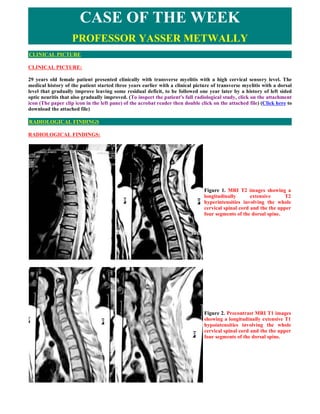
  • 1. CASE OF THE WEEK PROFESSOR YASSER METWALLY CLINICAL PICTURE CLINICAL PICTURE: 29 years old female patient presented clinically with transverse myelitis with a high cervical sensory level. The medical history of the patient started three years earlier with a clinical picture of transverse myelitis with a dorsal level that gradually improve leaving some residual deficit, to be followed one year later by a history of left sided optic neuritis that also gradually improved. (To inspect the patient's full radiological study, click on the attachment icon (The paper clip icon in the left pane) of the acrobat reader then double click on the attached file) (Click here to download the attached file) RADIOLOGICAL FINDINGS RADIOLOGICAL FINDINGS: Figure 1. MRI T2 images showing a longitudinally extensive T2 hyperintensities involving the whole cervical spinal cord and the the upper four segments of the dorsal spine. Figure 2. Precontrast MRI T1 images showing a longitudinally extensive T1 hypointensities involving the whole cervical spinal cord and the the upper four segments of the dorsal spine.
  • 2. Figure 3. Precontrast MRI T1 image (A), and MRI T2 images (B,C) showing central hypointensity (A) and central hyperintensity involving more than 2/3 of the spinal cord in cross section (B,C). Notice the central dot sign. The MRI signal changes represent central cord vasogenic edema and the central dot sign represents compressed gray matter. Figure 4. Precontrast MRI 1 images showing optic nerve enlargement on the left side. In general encephalitis/myelitis is an acute inflammatory process that affects brain or spinal cord tissue and is almost always accompanied by inflammation of the adjacent meninges. The disease is most commonly caused by viral infection. Encephalitis resulting from viral infection manifests as either acute viral encephalitis or postinfectious encephalomyelitis. Acute viral encephalitis is caused by direct viral infection of neural cells with associated perivascular inflammation and destruction of gray matter. Postinfectious encephalomyelitis follows infection with various viral or bacterial agents; the primary pathologic finding is demyelination of white matter. Postinfectious encephalitis/myelitis is an immunological disorders in which peripheral blood lymphocytes cross- react against myelin basic protein resulting in myelinolysis and inflammatory demyelination of the white matter. Breakdown of the blood brain barrier results in the formation of vasogenic edema that migrate along white matter tracts and is probably responsible for the MRI T2 hyperintensity observed in these disorders. Histologically, the acute lesions in Postinfectious encephalitis/myelitis are characterized by an extensive loss of
  • 3. myelin (perivenous cuffing with inflammatory cells, especially lymphocytes and macrophages, and loss of myelin). This may be in the form of a well-demarcated area of demyelination, although in the acute situation, the edges of the demyelinated lesions often are less well defined, and the demyelination and attendant cellular processes extend into the surrounding rim. Demyelinated fibers may be recognized by an axon devoid of a sheath, as seen histochemically, or immunohistochemically, or on electron microscopy by the presence of naked axons. In addition, thinly myelinated fibers may be seen within the lesion, suggesting partially demyelinated or remyelinated fibers. The presence of oligodendrocytes showing the re-expression of myelination proteins suggests the latter event is occurring in a least a significant number of these fibers. Vasogenic edema (due to breakdown of blood brain barrier) may be severe, and is seen as an expansion of the extracellular space, spreading apart both fibers and cells. Accompanying the myelin loss is a large infiltrate of foamy or debris- filled macrophages lying in sheets that appear to have replaced the normal neuropil. They also may be around the blood vessels, or infiltrating the more preserved areas of tissue as single cells. Depending on the age of the lesion, the macrophages may contain some or none of the myelin proteins described above, or may be LFB positive. The macrophages will stain for general markers such as KPI but depending on the patient's age, early (MRP14) or late (27ElO) markers also may be present to help date lesions. The inflammatory infiltrate varies, but in most acute cases will be of some significance. Lymphocytes staining with the leukocyte common antigen comprise most cells, although polymorphonuclear leukocytes, eosinophils, plasma cells, and even mast cells have been found, together with less well-characterized monocytes. Although they may be present throughout the tissue, they are particularly prominent around the blood vessels, and at times may be so severe as to mimic a vasculitis. Both CD4 helper cells and CD8 suppressor cells may be found in the lesions. In the past, there have been suggestions that CD4 cells predominate in early lesions, with CD8 cells taking over at later stages, but this is variable, and a fixed pattern has not been defined. Many workers also have described the occurrence of gamma-delta lymphocytes in these lesions, and their association with acute phase reactant or stress proteins such as heat shock protein on oligodendrocytes has been well recognized. (MS) [24] Demyelination of the white matter is associated with breakdown of the blood brain barrier and the development of vasogenic edema. Vasogenic edema is the most common type of edema results from local disruption of the blood brain barrier. This leads to extravasation of protein-rich filtrate of plasma into the interstitial space, with subsequent accumulation of vascular fluid. This disruption results from loosening of the tight junctions between endothelial cells, and the neoformation of pinocytic vesicles. Once the barrier is breached, hydrostatic and osmotic forces work together to extravasate intravascular fluid. Once extravasated, fluid is retained outside the vasculature, mostly in the white matter of the brain, and within the bundles of myelinated axons of long tracts and commissural fibers. This is because axons run in parallel bundles of fibres with loose extracellular space (that offer low resistance and facilitates the extension of vasogenic edema along myelinated axons which are spreaded apart by the edema) as opposed to gray matter, which has high cell density and is enmeshed in an interwoven network of connecting fibres that offer high resistance to the formation and spread of edema. By definition, this type of edema is confined to the extracellular space. Vasogenic edema is responsible for the MRI T2 hyperintensity and MRI T1 hypointensity and The MRI T1 contrast enhancement frequently observed in these disorders. [24] Vasogenic edema fluid is retained outside the vasculature, mostly in the white matter of the brain, and within the bundles of myelinated axons of long tracts and commissural fibers. This is because axons run in parallel bundles of fibres with loose extracellular space (that offer low resistance and facilitates the extension of vasogenic edema along myelinated axons which are spreaded apart by the edema) as opposed to gray matter, which has high cell density and is enmeshed in an interwoven network of connecting fibres that offer high resistance to the formation and spread of edema. Vasogenic edema is probably responsible for the multisegmental MRI T2 hyperintensity that are commonly seen in postinfectious transverse myelitis that apparently spare gray matter (gray matter is commonly seen as the central dot sign which represents the gray matter squeezed by edema). In postinfectious transverse myelitis vasogenic edema travel up and down along white matter tracts resulting in the multisegmental involvement of the spinal cord that is characteristic of postinfectious transverse myelitis. [24] DIAGNOSIS: DIAGNOSIS: NEUROMYELITIS OPTICA (DEVIC'S DISEASE) DISCUSSION DISCUSSION: Neuromyelitis optica (NMO; also known as Devic’s syndrome or Devic’s disease) is an inflammatory disorder with a striking predilection for the optic nerves and spinal cord. Acute transverse myelitis is often its initial manifestation. The cardinal features of NMO (optic neuritis and myelitis) and tendency to recurrence led to its classification as a subtype of multiple sclerosis (MS), but it has several unique features. Herein, [I] describe the clinical, radiological, and pathological features of NMO, its pathogenesis, and its relationship to other forms of central nervous system demyelinating disease.
  • 4. Clinical Features and Diagnostic Criteria Devic’s syndrome consists of one or more clinical episodes of optic neuritis in combination with myelitis. These clinical events also occur commonly in typical MS, however, in NMO they are usually more acute (sometimes fulminant) and severe; these characteristics may raise initial diagnostic suspicion of NMO. Paraclinical measures, such as magnetic resonance imaging (MRI) of the brain and spinal cord and cerebrospinal fluid (CSF) examination, also frequently reveal findings that differ from those in prototypic MS. In retrospective and small prospective series, most patients with NMO have no or few nonspecific white matter lesions on brain MR imaging. Spinal cord MR imaging also shows distinctive findings: a majority of patients have longitudinally extensive lesions extending over three or more vertebral segments. Furthermore, NMO patients frequently have a CSF pleocytosis of more than 50 leukocytes, with or without the presence of neutrophils. Three groups have proposed diagnostic criteria that employ some or all of these features (Table 1). [1- 3] With the development of these criteria the following key findings have become accepted: 1.The interval between the initial events of ON and myelitis is quite variable (several years, in some instances); 2.Some patients experience unilateral rather than bilateral optic neuritis; and 3.The course may be monophasic or relapsing. Neuromyelitis optica may follow either a monophasic or relapsing course. [3] In monophasic NMO, patients experience either unilateral or bilateral ON and a single episode of myelitis, typically but not always, within a very short time of one another, but do not have further attacks. In contrast, patients with a relapsing course continue to have discrete exacerbations of ON and/or myelitis after they meet NMO diagnostic criteria. There are several important differences between the two disease courses that will be detailed further.  Epidemiology Neuromyelitis optica affects young adults, much like MS, but has been reported in infancy through the ninth decade. The reported mean age of onset, especially for the relapsing type, may be greater than for typical MS. The mean onset ages were 35 and 47years in two series of relapsing NMO. [1, 2] Wingerchuk et al reported a mean age of onset of 29 years (range 1-54 y) for monophasic patients and 39 years (range 6-72 y) for relapsing patients. [3] The ratio of women to men may also differ according to disease course. Most reports suggest a ratio of approximately 1.4 to 1.8; the rate increases to 83-100% women in case series that consist predominantly of patients with a relapsing course. The incidence and prevalence of NMO are unknown. In Western nations, it has generally been considered a rare disorder but is almost certainly under-recognized, in part due to the lack of clear diagnostic criteria and confusion with MS. NMO appears to be more common in non-Caucasians such as African- Americans, Japanese, and other Pacific Islanders. Demyelinating disease in Asia and India is often restricted to the optic nerves and spinal cord; 7.6% of Japanese MS patients had NMO, [4] however, this rate may be declining with a concomitant increase in the frequency of “Western MS”. [5] Up to 6% of demyelinating disease cases in India are NMO. [6] Reports exist of identical twins or siblings with NMO. Despite these clues, the role of genetic factors in NMO is not known. Certain human leukocyte antigen (HLA) alleles are also associated with opticospinal forms of MS. The HLA-DPB1*0501 allele was present in a higher frequency of patients with opticospinal MS than prototypic MS in Japanese patients, whereas the DPB1*0301 allele may be underrepresented. [7] These HLA associations differ from those described in patients with “Western” MS, which is most consistently associated with HLA-DRB1*1501.  Clinical picture A viral prodrome precedes the onset of the disease in 30-50% of cases. The prodrome most often consists of headache, pyrexia, fatigue, myalgias, and respiratory or gastrointestinal complaints. This suggests that infectious agents may cause or trigger NMO. Many diseases have been associated with NMO, including most viral infections, tuberculosis, hypothyroidism, lupus, Sjögren’s syndrome and other connective tissue disorders. Textbook definitions of NMO generally require bilateral ON occurring in close conjunction with transverse myelitis. It is now well established, however, that patients with unilateral ON pursue a course indistinguishable from those with bilateral ON. [3] When bilateral ON and myelitis occur simultaneously or in rapid succession, it usually predicts a monophasic course. The index events (those that herald the onset of NMO) also include unilateral ON, myelitis, bilateral ON, or a combination of unilateral ON and myelitis. In one series, the initial presentation
  • 5. was an isolated event of either ON or myelitis in 90% of patients destined for a relapsing course compared with only 48% of those who had a monophasic illness. [3] Acute transverse myelitis, defined as severe, bilateral inflammatory spinal cord injury with neurological dysfunction worsening over several hours to days and involving motor, sensory, and sphincter function, is a typical presentation of NMO. Deep or radicular pain, lower extremity paresthesias, or weakness may herald its onset. Weakness rapidly evolves to paraplegia or quadriplegia, often causing complete sensory loss caudal to the lesion and a flaccid bladder. The acute lesion usually traverses at least three contiguous vertebral segments of the spinal cord and may result in “spinal shock” with flaccid weakness, absent deep tendon reflexes, and mute plantar responses. A minority of patients experience less complete lesions that may present as Brown-Sequard or central cord syndromes. Lhermitte’s symptom, paroxysmal tonic spasms, and radicular pain may occur, usually in those patients with relapsing disease. Partial recovery is common following the initial myelitis event. Seventy-eight to 88% of patients improved by one or more levels on a seven-point ordinal scale of motor function regardless of eventual disease course. [3] Recurrent episodes of myelitis increase the risk for permanent and severe morbidity from thromboembolic disease, urinary tract infections, decubitus ulcerations, and pneumonia.. Acute cervical myelitis is associated with respiratory failure and death, especially in relapsing NMO. [3] Optic neuritis in NMO may be unilateral or bilateral. It is almost always acute, usually severe, and may or may not be associated with retro-orbital pain. Field defects are variable and include central and paracentral scotomata as well as altitudinal and chiasmatic defects. During the first episode of ON in NMO, nearly 40% of affected eyes become completely blind (no light perception) at the nadir of the event; however, some cases involve only minor visual deficits. Most patients experience some improvement in vision, especially if their disease course is monophasic; accumulation of visual impairment occurs with successive recurrences of ON in relapsing cases. Symptoms outside of the optic nerves and spinal cord are very uncommon. They are usually minor or subjective, tend to occur later in the disease course, and are plausibly due to other causes than NMO. This includes symptoms such as vertigo, facial numbness, nystagmus, headache, and postural tremor. These features may be related to the brain MRI evidence that some patients have lesions that occur in the parenchyma or brain stem late in the disease course.  Etiology and Pathology The cause of NMO is not known. Its clinical and pathological features have led most to consider it an autoimmune variant of MS. The fact that multiple infectious and systemic autoimmune diseases have been associated with NMO suggests that a single cause of the disorder is unlikely. A high prevalence of serum autoantibodies suggests that NMO may be driven primarily by B cell dysfunction. Although there are some similarities between the distribution of lesions in NMO and in myelin oligodendrocyte glycoprotein (MOG)- induced experimental allergic encephalomyelitis (EAE), the target (auto)antigen in NMO is not known. The pathology and immunology of NMO will be discussed in detail by Dr. Lucchinetti. Briefly, optic nerve specimens typically reveal near-complete demyelination with modest inflammatory infiltrates. [12, 13] Brain parenchyma is usually normal or reveals only scattered small perivascular infiltrates. Spinal cord lesions are more distinctive but their characteristics depend on the stage of the disease. Macroscopic cord expansion with softening and cavitation is often noted acutely; in chronic cases atrophy is present. Changes ranging from modest perivascular inflammation and demyelination to complete necrotic destruction of both gray and white matter have been described.[13-16] In acute lesions, necrosis, hemorrhage, and an intense inflammatory infiltrate with a predominance of polymorphonuclear cells are often present; this may include large numbers of eosinophils. [17] Some groups have described a hyalinized appearance of medium-sized spinal cord arteries as a hallmark of NMO. [1, 17-19] The cause and significance of this vessel pathology is not known. Lucchinetti et al demonstrated prominent deposition of IgG and C9 neoantigen (a marker of complement activation) at regions of active myelin destruction and vessel walls, where there was vascular proliferation and fibrosis, suggesting that humoral mechanisms may be of some importance.  Natural History of NMO: Monophasic and Relapsing Disease Courses Most patients, perhaps 70% or more, with NMO develop a relapsing course with recurrent events of ON and myelitis. There are several differences in the demographics and outcomes of patients with the monophasic and relapsing forms of NMO (Table 2). By definition, those who follow a monophasic course experience either unilateral or bilateral ON and a single episode of myelitis without any further exacerbations. Several factors are associated with a monophasic disease course. The index events of ON and myelitis are typically more severe, with over half of patients experiencing complete loss of light perception with index ON compared with about 28% of relapsing patients. Similarly, paraplegia occurs at the nadir of the index myelitis in 70% of monophasic patients compared with 31% of those who eventually relapse. [3]
  • 6. The tempo of NMO onset also has prognostic value. Patients who present with a combination of ON and myelitis simultaneously or in rapid succession (over a few days) are much more likely have a monophasic course. In the largest contemporary series, the median interval between the first clinical event and the development of bilateral ON and myelitis (traditional definition of NMO) was 5 days (range 0 to 151 days) in the monophasic group versus 166 days (range 2 to 730 days) for the relapsing group. [3] To illustrate, a patient who presents with ON having had severe myelitis six months earlier is much more likely to follow a relapsing disease course than the patient presenting with simultaneous ON and myelitis. This prognostic information may be useful when considering preventative immunotherapies. Most patients with monophasic NMO experience some recovery from the index events of ON and myelitis and, because no further relapses occur, subsequently remain stable. Although the index events that define the illness are more severe in this group than for relapsing patients, recovery and long-term function are better, in part because permanent remission spares the patient cumulative deficits. About 80% of ON events improve by a clinically important degree (e.g., from no light perception to 20/200 vision) over the first six months. Wingerchuk et al found that most patients with monophasic NMO recovered to 20/30 vision or better, however, 22% remained functionally blind (20/200 vision or worse) in at least one eye.3 The brunt of disability in those with a monophasic course occurs as a result of spinal cord injury; most patients experience at least moderate weakness of one or more limbs and moderate sphincter dysfunction with occasional urinary incontinence. Permanent monoplegia or paraplegia occurred in 31%. Five-year survival of this group is approximately 90%. While the prognosis for most monophasic patients is to maintain some degree of independence (despite moderate visual and motor deficits), patients who have relapsing disease face the prospect of incremental accumulation of much more disability. The first events of ON and myelitis in this group are less severe, and the recovery better, than those of the monophasic group, but recurrent severe episodes of ON and myelitis abolish any apparent advantages of a relapsing course. Most relapsing patients declare their disease course early. After meeting NMO diagnostic criteria, 55% have their first optic nerve or spinal cord relapse within one year. [3] The proportion increases to 78% at three years and 90% at five years. As in typical MS, relapse frequency is as extremely variable in NMO. Several attacks may strike over a few months or remissions lasting a more than a decade may occur. Over a median follow-up of 16.9 years, the median number of relapses was five (range 1 to 18). As in monophasic NMO, a progressive phase of neurological deterioration is uncommon, although there are many patients who seem to have rapid, sometimes stepwise, deterioration when they attempt to taper and discontinue corticosteroid therapy. Severe cervical myelitis causing respiratory failure is more common in relapsing NMO, possibly affecting as many as one-third of patients. In the largest contemporary series, the five-year survival of relapsing patients evaluated between 1950 to 1955 was 68%. Respiratory failure was the sole cause of death. [3] This was a frequent cause of death of NMO patients, but improvements in supportive care over the last two decades have likely reduced these figures. For example, a patient under our care made an excellent recovery from NMO after being ventilator dependent for several days following plasma exchange treatment.  Diagnostic Evaluation Several diagnostic tests can serve to support a diagnosis of NMO. Brain and spinal cord MRI features and CSF findings have been incorporated into diagnostic criteria for NMO due to their ability to discriminate from typical MS.  Serological Tests. One or more autoantibodies, including anti-nuclear antibody, anti-double-stranded DNA antibody, extractable nuclear antigen, and anti-thyroid antibodies are commonly present at the time of diagnosis. [2, 3] The true incidence is not known, but may approach 50%.  Neurophysiological Tests. Visual evoked potentials may occasionally detect subclinical optic nerve lesions when the clinical history and examination confirm only unilateral deficits. Current clinical diagnostic criteria do not allow subclinical optic neuropathy manifest as abnormalities on visual evoked potential to substitute for a history of optic neuritis, but this may have to be readdressed pending further study. Electrophysiological studies otherwise have little diagnostic role.  Neuroimaging findings The diagnosis of NMO is strongly supported by the absence of brain parenchymal lesions (i.e. excluding the optic nerves), or the presence of nonspecific white matter lesions that do not meet radiological criteria for MS. [1, 2, 8]
  • 7. Some patients with relapsing disease accumulate white matter lesions over time, but these lesions tend to be nonspecific punctate foci that fail to meet radiological criteria for MS. [3] During acute ON, brain MR imaging may demonstrate swelling and/or gadolinium enhancement of an affected optic nerve or the chiasm. While occasionally more severe and extensive than encountered in MS (e.g. involve the entire chiasm), these nonspecific findings in the optic nerve do not distinguish NMO from isolated ON or typical MS. Episodes of myelitis in NMO are accompanied by striking spinal cord MR imaging abnormalities. During acute myelitis, the affected region of the cord is usually expanded and swollen [9] and may enhance with gadolinium. Heterogeneous T2 signal within the lesion may suggest cavitation and necrosis. The most distinct aspect of NMO cord lesions is that they usually extend over three or more vertebral segments of the cord. [2, 3] Over time the swelling and enhancement give way to persistent intramedullary T2 signal abnormality and/or cord atrophy. Typically, the lesions are in the central part of the cord, rather than in the periphery of the cord as generally occurs in patients with prototypic MS.  The MRI picture characteristic of idiopathic transverse myelitis 1. A centrally located multisegmental (3 to 8 spinal segments) MRI T2 hyperintensity that occupies more than two thirds of the cross-sectional area of the cord is characteristic of transverse myelitis. The MRI T2 hyperintensity commonly shows a slow regression with clinical improvement. The central spinal cord MRI T2 hyperintensity represents evenly distributed central cord edema. MRI T1 Hypointensity might be present in the same spinal segments that show T2 hyperintensity although to a lesser extent. The MRI T2 hyperintensity is central, bilateral, more or less symmetrical and multisegmental. 2. MRI T2 central isointensity, or dot (within and in the core of the MRI T2 hyperintensity) might be present and is believed to represent central gray matter squeezed by the uniform, evenly distributed edematous changes of the cord. (central dot sign). It might not be of any clinical significance. 3. Contrast enhancement is commonly focal or peripheral and maximal at or near the segmental MRI T2 hyperintensity. In idiopathic transverse myelitis enhancement is peripheral to the centrally located area of high T2 signal intensity rather than in the very same area. The prevalence of cord enhancement is significantly higher in patients with cord expansion. 4. Spinal cord expansion might or might not be present and when present is usually multisegmental and better appreciated on the sagittal MRI T1 images. Spinal cord expansion tapers smoothly to the normal cord, and is of lesser extent than the high T2 signal abnormality. 5. Multiple sclerosis plaques (and subsequent T2 hyperintensity) are located peripherally, are less than 2 vertebral segments in length, and occupies less than half the cross-sectional area of the cord. In contrast to transverse myelitis, enhancement in MS occurs in the same location of high-signal-intensity lesions seen on T2-weighted images. Table 1. Differences between idiopathic transverse myelitis and spinal multiple sclerosis Number T2 of Disease entity Contrast element Pathology hyperintensity segments involved Idiopathic transverse Central, 4-8 In transverse myelitis Nonspecific necrosis that myelitis multisegmental enhancement is peripheral to affects gray and white matter the centrally located area of indiscriminately and destroys high T2 signal intensity rather axons and cell bodies as well as than in the very same area. myelin. Spinal multiple Peripheral 1-2 In contrast to transverse White matter demyelination sclerosis myelitis, enhancement in MS only. occurs in the same location of high-signal-intensity lesions seen on T2-weighted images.
  • 8. Figure 5. MRI T2 image of a patient with Neuromyelitis optica. The patient was presented clinically with transverse myelitis. Notice the longitudinally extensive T2 hyperintense lesions extending from C1 to D5. The cord is also diffusely expanded.  Cerebrospinal Fluid. CSF analysis may support the diagnosis of NMO. Occasional patients have a pleocytosis of more than 50 WBC/mm3 around the time of an acute myelitis exacerbation3; this degree of CSF cellularity is very rare in typical MS. [10] The CSF leukocyte differential may also reveal the presence of neutrophils, another finding rarely seen in MS. These abnormalities may reflect the severity of myelitis, which often results in necrosis. Approximately 85% of patients with MS have detectable oligoclonal bands on CSF electrophoresis. [11] Oligoclonal bands are far less common in NMO, occurring in 15-35% of patients in contemporary series. [1-3] Other immunoglobulin abnormalities, such as increased rate of IgG synthesis, are also much less common in NMO than in MS.  Management of Neuromyelitis optica All therapeutic recommendations in the literature represent anecdotal experience from small uncontrolled case series. In the monophasic form of NMO and for index events and relapses of relapsing NMO, the mainstay of therapy is treatment of acute attacks, prevention of medical complications, and rehabilitation. Specific measures aimed at preventing future attacks are considered for patients who have demonstrated a propensity to relapse. Most patients who present with NMO exacerbations receive intravenous corticosteroid treatment, for example, 1000 mg methylprednisolone per day for five consecutive days. A double-blind crossover study of plasmapheresis versus sham exchanges documented that plasmapheresis (seven exchanges of approximately 55 ml/kg administered every other day) is beneficial treating exacerbations of demyelinating disease (including NMO) that are not responsive to methylprednisolone; this is used as second-line therapy. [20] In one series, 6/10 patients treated with plasma exchange for severe, steroid-refractory NMO attacks experienced moderate or marked improvement in close temporal proximity to the initiation of treatment. [21] Intravenous immune globulin has also been used anecdotally. Prevention of medical complications is critically important. Acute cervical cord attacks may cause respiratory failure. Patients at risk for this complication by virtue of the location and severity of their acute myelitis require close intensive care unit observation with frequent evaluation of respiratory and bulbar status. Ventilatory assistance often becomes necessary. Medical measures to prevent thromboembolic complications, aspiration pneumonia, decubiti, and urinary tract infections are also required. Preventative therapy is required for patients with relapsing disease. Many North American NMO patients receive parenteral beta-interferon treatment but some clinicians have felt that this treatment may be ineffective based on their uncontrolled experience. A Japanese study, however, found that interferon beta-1b seemed to benefit the both the opticospinal form and Western-type MS. [22] Some clinicians have noted that long-term oral corticosteroid monotherapy may significantly reduce relapse frequency or severity. It is not uncommon for patients to become steroid-dependent such that they are unable to taper their dose below a certain level because of perceived worsening of lower extremity function; documentation of a new exacerbation in
  • 9. this setting may or may not be possible. In the only published prospective treatment study, Mandler et al found that seven newly diagnosed NMO patients seemed to stabilize for at least 18 months on a regimen of azathioprine and oral prednisone. [23] Following an intravenous course of methylprednisolone, oral prednisone (1 mg/kg/d) was started. Three weeks later, patients received azathioprine (2 mg/kg/d). At two months, the prednisone dose was gradually tapered (by 10 mg every three weeks to 20 mg/d, then an even slower reduction to a maintenance dose of 10 mg/d). Most patients were maintained on prednisone 10 mg/d and azathioprine 75 to 100 mg/d. The authors [23] noted that no exacerbations occurred and Expanded Disability Status Scale scores improved somewhat during the 18-month treatment period. Various other immunosuppressive drugs have been used but in limited numbers and outside the realm of a structured study. For patients who “fail” the azathioprine and prednisone combination, intravenous mitoxantrone, which is approved for use in rapidly worsening secondary progressive or relapsing- remitting MS, might be tried. There are no data concerning the use of mitoxantrone in NMO.  Summary There is increasing evidence that NMO is a distinct entity. It differs from MS with respect to lesional topography, exacerbation severity, MR imaging findings, CSF abnormalities, and immunopathology. Early diagnosis and referral to tertiary care centers provides the best chance for complete case ascertainment and may make multicenter efforts at studying this disease possible. SUMMARY SUMMARY  Clinical picture or neuromyelitis optica The cardinal clinical features of the disorder are transverse myelitis, which is often longitudinally extensive, and optic neuritis. These two index events can occur simultaneously, in rapid succession, or they can be separated by many years. The optic neuritis can be unilateral or bilateral. Some patients have repeated episodes of optic neuritis before myelitis occurs and vice versa (the nomenclature of the disease at this stage is relapsing myelitis or relapsing optic neuritis). Most of those affected (>80%) go on to have repeated relapses (relapsing NMO) though a minority may have only the index events (monophasic disease). Relapses are generally more disabling than those in patients with typical MS. Whereas in MS disability develops largely in the progressive phase of the disease, in neuromyelitis optica disability is acquired as a consequence of relapses; progressive disability without relapses is rare in our experience. In white populations most patients presenting with optic neuritis and myelitis are likely to have MS rather than neuromyelitis optica. Features which may help distinguish the disorder from MS clinically are the more severe myelitis, optic neuritis with poor recovery, and no involvement of other parts of the neuraxis. However the disease spectrum may be wider than currently accepted. A variety of diagnostic criteria for the disorder have been formulated and are summarised elsewhere. [68] None is perfect and it is likely that they will be revised in the light of emerging clinical and laboratory data. In general we would consider the diagnosis in the presence of:  Longitudinally extensive myelitis (usually more than three vertebral segments)  Optic neuritis  Normal brain MRI, or if abnormal, atypical for MS-see below. Clinical, radiological, and immunopathological studies suggest neuromyelitis optica is distinct from MS The prognosis also differs from MS. Early reports suggested a five year survival of 68%, death often resulting from severe spinal cord disease and respiratory compromise. [67] More recent series suggest a better outcome, possibly reflecting better case ascertainment or treatment. However there is no doubt that disability is acquired earlier in neuromyelitis optica than MS; this and a mean relapse rate of around two per year make early diagnosis and therapy imperative.
  • 10. Neuroimaging or neuromyelitis optica The most characteristic radiological feature is a longitudinally extensive cord lesion, extending often over three or more spinal segments and expanding the cord (fig 1). This is usually hypointense on T1 and hyperintense on T2 MRI. Lesions are centrally situated within the cord and patchy contrast enhancement is often seen. Occasionally lesions can be identified in the optic nerves. Though classically NMO has been defined by lack of brain lesions or symptoms, it has increasingly been noted that up 60% of patients with otherwise typical relapsing NMO (many with positive NMO-IgG) can have lesions on brain MRI. These range from extension of high cervical cord lesions into the brain stem, diencephalic, brainstem or cerebral lesions "atypical" for MS and in minority MS- like lesions. However all these patients have long cord lesions which seems to be a specific feature, distinguishing these cases from MS. [71]  Laboratory investigations of neuromyelitis optica Cerebrospinal fluid acutely may reveal a prominent pleocytosis of either lymphocytes or neutrophils and raised protein. In contrast to MS, there are usually no oligoclonal bands (in over 80%). Lennon and co-workers at the Mayo clinic recently reported the discovery of NMO- IgG, which may be the first "disease specific" antibody in CNS demyelinating disease. [65]The antibody, identified initially from Western blots in patients screened for possible paraneoplastic antibodies, is reported to have a sensitivity of 73% and a specificity 91% for neuromyelitis optica and was also positive in a significant proportion of patients deemed to be at high risk of neuromyelitis optica (that is, patients with recurrent optic neuritis or myelitis). Despite the apparent association of NMO- IgG with neuromyelitis optica, independent confirmation of this finding and indeed evidence of pathogenesis is still awaited. The Mayo group has also recently reported that the target antigen for NMO-IgG appears to be the aquaporin-4 water channel, located in astrocytic foot processes at the blood-brain barrier. [64] To date, testing for NMO-IgG is only available through the Mayo clinic (Rochester, USA; at a cost of $500/sample). If the role of the antibody is confirmed and the assay becomes readily available, the hope is clearly that the relation between neuromyelitis optica and "neuromyelitis optica spectrum" disorders (see below) will be clarified, and predictive testing for neuromyelitis optica in patients with isolated myelitis or severe optic neuritis may be possible. A range of positive auto-antibodies (including ds-DNA) have been reported in up to 40% of patients. [66] This can give rise to diagnostic difficulties. We would consider these to be epiphenomena occurring in the context of disordered humoral immunity. However there are reports of patients with unambiguous systemic lupus erythematosus, Sjögren's syndrome, and mixed connective tissue disease who have developed neuromyelitis optica. Whether they have two separate autoimmune diseases, or neuromyelitis optica is a consequence of the primary disorder, is impossible to determine clinically. Testing for NMO-IgG and pathological examination in such cases may be able to clarify this.  Pathology of of neuromyelitis optica Extensive necrosis, demyelination, and often cavitation across multiple spinal cord segments, involving grey and white matter with perivascular infiltrates, prominent macrophages, eosinophils, and vascular hyalinisation is typical. Deposition of complement in a ring pattern on the outer surface of blood vessels and in a rosette perivascular pattern has been elegantly demonstrated. Prominent perivascular IgG reactivity and IgM deposition in a rosette pattern implicate these as sites of immune mediated damage. [70]  Management of of neuromyelitis optica The rarity of the condition inevitably limits the evidence for therapeutic interventions. As in most immune mediated disorders management consists of treatment of relapses, therapy for the underlying disease, symptom control, and rehabilitation. For the general neurologist managing a patient with neuromyelitis optica the approach to relapses and indeed underlying disease therapy is perhaps most comparable to that of the more common antibody mediated disorder myasthenia gravis.  Management of relapses High dose corticosteroids and supportive care remain the mainstays of management of relapse. In view of the severity of relapses and likely need for maintenance treatment our policy is to follow a course of intravenous methylprednisolone (1 gram daily for 3-5 days) with a gradual taper of oral prednisolone over several months, from an initial dose of 1 mg/kg/ day. An alternate day maintenance dose of the order of 10-20 mg prednisolone is generally our target in patients with relapsing disease. A minority of patients fail to respond to adequate steroid therapy or relapse rapidly and in such cases there is a role for therapeutic plasma exchange. We consider it early-within weeks of symptom onset. In the North American randomised trial of plasma exchange in severe demyelinating events, patients with neuromyelitis optica were overrepresented among the responders, with a 60% response rate (versus 6% overall for sham exchange). In this study plasma exchange was undertaken within three months of onset of relapse.
  • 11. Prevention of relapses Most patients follow a relapsing course, often acquiring substantial disability within two or three relapses. Immunosuppression appears to reduce the relapse rate. The first report of successful treatment was a series of seven patients treated with prednisolone (tailing dose as above) and azathioprine (at 2.5-3 mg/kg). In the author experience, relapse rates were reduced by over 80% in patients established on immunosuppressive therapy (most frequently azathioprine). This has therefore remained the author's initial therapy option in patients with relapsing, but reasonably stable, disease. For patients intolerant of azathioprine, mycophenylate mofetil is a reasonable alternative (but without any evidence base), with the possible advantage of more rapid onset of action.  Practical points 1. Although neuromyelitis optica is uncommon it is a rapidly disabling yet treatable disorder. 2. Early recognition and diagnosis followed by prompt, carefully supervised immunosuppressive treatment in relapsing patients is paramount. A blind, quadriparetic, ventilator dependent person, from a treatable disorder, is a tragedy that one should strive to avoid at all costs. 3. Management appears distinct from that of MS and should probably be in the hands of clinicians with an interest in demyelinating disease. In patients who have "breakthrough" disease on azathioprine, or who present with frequent severe relapses, more aggressive immunosuppression may be necessary. Recent small case series have reported on the successful use of rituximab (a B cell depleting monoclonal antibody) and mitoxantrone. The author have used both of these agents in small numbers of patients without complications. Interferon beta, the mainstay of treatment in relapsing MS, does not appear to be effective.  Symptoms control and rehabilitation Pain, stiffness, bladder, and bowel symptoms need to be tackled. Tonic spasms seem to be much commoner than in MS and in our experience usually respond to carbamazepine. Rehabilitation, physiotherapy, mobility, and visual aids are often needed. Some patients with high cervical cord lesions will need long term home ventilatory support. There is a UK based self-help group (telephone +44 (0)151529 6100) and a patient friendly website (http://www.thewaltoncentre.co.uk/patients/ Neuromyelitis_Optica.html) THE WIDENING SPECTRUM OF NEUROMYELITIS OPTICA There may be a number of related disorders, a full discussion of which is beyond the scope of this review. Patients with idiopathic relapsing myelitis, Asian "optico-spinal" MS, and chronic relapsing inflammatory optic neuropathy may form part of the "neuromyelitis optica spectrum". We have seen several patients presenting with severe myelitis without visual symptoms but delayed visual evoked responses, some of whom have subsequently developed optic neuritis. It seems likely that such patients have neuromyelitis optica although they fall outside current criteria for the disorder. Traditionally patients with clinical or radiological findings outside the optic nerves and spinal cord have also been excluded from the diagnosis of neuromyelitis optica. There are however recent reports of patients with clinically typical neuromyelitis optica who have developed brain lesions on MRI. Interestingly these patients have been shown to have positive NMO-IgG, and histopathology (of the brain lesions) similar to classical neuromyelitis optica, widening the spectrum still further. More studies with a validated disease marker in the near future should clarify these relationships. Genetic links between neuromyelitis optica and MS have been hypothesised and will perhaps open a window to the better understanding of both conditions. [24]  Addendum  A new version of this PDF file (with a new case) is uploaded in my web site every week (every Saturday and remains available till Friday.)  To download the current version follow the link "http://pdf.yassermetwally.com/case.pdf".  You can also download the current version from my web site at "http://yassermetwally.com".  To download the software version of the publication (crow.exe) follow the link: http://neurology.yassermetwally.com/crow.zip  The case is also presented as a short case in PDF format, to download the short case follow the link: http://pdf.yassermetwally.com/short.pdf  At the end of each year, all the publications are compiled on a single CD-ROM, please contact the author to know more details.  Screen resolution is better set at 1024*768 pixel screen area for optimum display.
  • 12. Also to view a list of the previously published case records follow the following link (http://wordpress.com/tag/case-record/) or click on it if it appears as a link in your PDF reader  To inspect the patient's full radiological study, click on the attachment icon (The paper clip icon in the left pane) of the acrobat reader then double click on the attached file.  Click here to download the short case version of this case record in PDF format REFERENCES References 1. Mandler RN, Davis LE, Jeffery DR, Kornfeld M. Devic’s neuromyelitis optica: a clinicopathological study of 8 patients. Ann Neurol 1993; 34:162-8. 2. O’Riordan JI, Gallagher HL, Thompson AJ, et al. Clinical, CSF, and MRI findings in Devic’s neuromyelitis optica. J Neurol Neurosurg Psychiatr 1996; 60:382-7. 3. Wingerchuk DM, Hogancamp WF, O’Brien PC, Weinshenker BG. The clinical course of neuromyelitis optica (Devic’s syndrome). Neurology 1999; 53:1107-14. 4. Kuroiwa Y, Igata A, Itahara K, Koshijima S, Tsubaki T. Nationwide survey of multiple sclerosis in Japan. Clinical analysis of 1,084 cases. Neurology 1975; 25:845-51. 5. Kira J, Yamasaki K, Horiuchi I, Ohyagi Y, Taniwaki T, Kawano Y. Changes in the phenotypes of multiple sclerosis over the last 50 years in Japan. J Neurol Sci 1999; 166:53-57. 6. Singhal BS. Multiple sclerosis–Indian experience. Annals of the Academy of Medicine, Singapore 1985; 14:32-6. 7. Fukazawa T, Yamasaki K, Ito H, et al. Both the HLA-CPB1 and -DRB1 alleles correlate with risk for multiple sclerosis in Japanese: clinical phenotypes and gender as important factors. Tissue Antigens 2000; 55:199-205. 8. Fazekas F, Offenbacher H, Schmidt R, Strasser-Fuchs S. MRI of neuromyelitis optica: evidence for a distinct entity [letter]. J Neurol Neurosurg Psychiatr 1994; 57:1140-2. 9. Tashiro K, Ito K, Maruo Y, et al. MR imaging of spinal cord in Devic disease. J Comp Assist Tomogr 1987; 11:516-7. 10. Tourtellotte WW, Staugaitis SM, Walsh MJ, et al. The basis of intra-blood-brain-barrier IgG synthesis. Ann Neurol 1985; 17:21-27. 11. Paty DW, Oger JF, Kastrukoff LF, al e. MRI in the diagnosis of multiple sclerosis: prospective comparison of clinical evaluation, EPs, oligoclonal banding, and CT. Neurology 1988; 38:180-185. 12. Achard C, Guinon L. Sur un cas de myelite aigue diffuse avec double nevrite optique. Arch de Med Exper er d’Anat Path 1889; 1:696-710. 13. Stansbury FC. Neuromyelitis optica (Devic’s disease). Presentation of five cases with pathological study and review of the literature. Arch Ophthalmol 1949; 42:292-335;465-501. 14. Goulden C. Optic neuritis and myelitis. Trans Ophthal Soc UK 1914; 34:229-252. 15. Beck GM. A case of diffuse myelitis associated with optic neuritis. Brain 1927; 50:687-703. 16. Kuroiwa Y. Neuromyelitis optica. In: Koetsler JC, ed. Handbook of Clinical Neurology. Vol. 47. New York: Elsevier Science, 1985:397-408. 17. Lucchinetti C, Mandler R, Weinshenker B, Wingerchuk D, Parisi J, Lassmann H. Humoral mechanisms in the pathogenesis of Devic’s neuromyelitis optica. Neurology 2000; 54(Suppl 3):A259-260. 18. Ortiz de Zarate JC, Tamaroff L, Sica RE, Rodriguez JA. Neuromyelitis optica versus subacute necrotic myelitis. II. Anatomical study of two cases. J Neurol Neurosurg Psychiatr 1968; 31:641-5. 19. Lefkowitz D, Angelo JN. Neuromyelitis optica with unusual vascular changes. Arch Neurol 1984; 41:1103-5.
  • 13. 20. Weinshenker BG, O’Brien PC, Petterson TM, al e. A randomized trial of plasma exchange in acute central nervous system inflammatory demyelinating disease. Ann Neurol 1999; 46:878-886. 21. Keegan BM, Pineda A, Rodriguez M, Weinshenker B. Predictive factors for response to plasma exchange in the treatment of acute, severe attacks of central nervous system demyelinating disease. Ann Neurol 2000; 48:451. 22. Itoyama Y, Saida T, Tashiro K, Sato T, Ohashi Y, Japan TI-bMSCRGi. Japanese multicenter, randomized, double-blind trial of interferon beta-1b in relapsing-remitting multiple sclerosis: two year results. Ann Neurol 2000; 48:487. 23. Mandler RN, Ahmed W, Dencoff JE. Devic’s neuromyelitis optica: a prospective study of seven patients treated with prednisone and azathioprine. Neurology 1998; 51:1219-20. 24. Metwally, MYM: Textbook of neuroimaging, A CD-ROM publication, (Metwally, MYM editor) WEB-CD agency for electronic publication, version 11.2a. April 2010 25. Case of the week...Acute postinfectious transverse myelitis (Transverse myelitis spectrum of disorders) (Click to download in PDF format] 26. Online case record...Acute postinfectious transverse myelitis [Full text] 27. Case of the week...Acute postinfectious transverse myelitis [Click to download in PDF format] 28. Thesis section...Postinfectious monophasic demyelinating disorders of the CNS [Click to download in PDF format] 29. Topic of the month...Transverse myelitis. [Click to download in PDF format] 30. Topic of the month...Neuromyelitis optica. [Click to download in PDF format] 31. Case of the week...Acute postinfectious transverse myelitis [Click to download in PDF format] 32. Case of the week...Acute postinfectious transverse myelitis [Click to download in PDF format] 33. Case of the week...Neuromyelitis optica [Click to download in PDF format] 34. A quick guide to management&ldots;Neuromyelitis optica [Click to download in PDF format] 35. Devic’s Disease (Neuromyelitis Optica [Full text]