Case record...Tectal plate glioma
•
2 j'aime•1,836 vues

Case record...Tectal plate glioma http://yassermetwally.com http://yassermetwally.net

Recommandé
Recommandé
Contenu connexe
Tendances
Tendances (20)
En vedette
En vedette (20)
Similaire à Case record...Tectal plate glioma
Similaire à Case record...Tectal plate glioma (20)
Plus de Professor Yasser Metwally
Plus de Professor Yasser Metwally (20)
Dernier
Dernier (20)
Case record...Tectal plate glioma
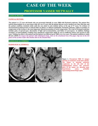
- 1. CASE OF THE WEEK PROFESSOR YASSER METWALLY CLINICAL PICTURE CLINICAL PICTURE: The patient is a 37 years old female who was presented clinically in years 2000 with Parinaud syndrome, The patient first noticed abnormalities in eye movement while she was 12 years old, the patient did not receive medical care since that time and the patient condition remained stable until she was asked to seek medical advice at the age of 37 years. No evidence of increased intracranial pressure is noticed either by history or clinical examination. Parinaud syndrome, which is secondary to compression of the tectum, is the most important clinical presentation of tectal compression. The triad of Parinaud syndrome includes palsy of upward gaze, dissociation of light and accommodation, and failure of convergence. In addition, findings secondary to hydrocephalus resulting from aqueductal compression might be seen in midbrain tumors (not present in this case). Nothing was done to the patient and the patient was followed up by MRI every two years. The patient condition is stable till now (2010) (To inspect the patient's full radiological study, click on the attachment icon (The paper clip icon in the left pane) of the acrobat reader then double click on the attached file) RADIOLOGICAL FINDINGS RADIOLOGICAL FINDINGS: Figure 1. Precontrast MRI T1 images showing an isointense tectal plate glioma. The tumor is showing posterior exophytosis to the left side of the quadrigeminal cistern. The posterior parts of the tumor are showing some hyperintense zones, probably representing hemorrhagic spots.
- 2. Figure 2. Postcontrast MRI T1 images showing dense ring enhancement within the tectal tumor mass. The mass is seen protruding to the left side of the quadrigeminal cistern. Figure 3. The tectal plate glioma is hyperintense in the MRI T2 images, with central hypointensities
- 3. Figure 4. The tectal plate glioma on postcontrast MRI T1 image (A) and MRI T2 image (B) DIAGNOSIS: DIAGNOSIS: TECTAL PLATE GLIOMA (FOCAL MIDBRAIN GLIOMA) DISCUSSION DISCUSSION: The diagnosis of brainstem glioma was long considered a single entity. However, since the advent of magnetic resonance imaging in the late 1980s, neoplasms within this anatomic region are now recognized to include several tumors of varying behavior and natural history. More recent reports of brainstem tumors include diverse sites such as the cervicomedullary junction, pons, midbrain, or the tectum. Today, these tumors are broadly categorized as either diffuse intrinsic gliomas, most often in the pons, or the nondiffuse brainstem tumors originating at the tectum, focally in the midbrain, dorsal and exophytic to the brainstem, or within the cervicomedullary junction. Although we briefly discuss the nondiffuse tumors, we focus specifically on those diffuse brainstem tumors that regrettably still carry a bleak prognosis. Epidemiology Brainstem gliomas account for approximately 20% of all CNS tumors among children younger than age 15.1 This increase from 10% in the late 1970s reflects an increase in detection of the nondiffuse brainstem tumors by magnetic resonance imaging (MRI) rather than a true increase in incidence. The median age at presentation for all brainstem gliomas in children is 6 to 7 years, with males and females equally affected. [2] Among adults, brainstem gliomas are considerably less common, but reported up to age 70 years, sometimes with a less aggressive course. There is an increased frequency of brainstem tumors among patients with neurofibromatosis type 1, and these tumors have a more indolent behavior. [3,4] Anatomy and function of the brain stem The human brainstem comprises three connected parts: the midbrain, pons, and medulla, each composed of numerous axonal tracts (white matter), cranial nerve nuclei, and noncranial nerve brain-stem nuclei (gray matter). Thus, the brainstem serves as the conduit through which axonal tracts pass to the spinal cord, cerebrum, or exit as cranial nerves. Within the brainstem, axonal tracts course in longitudinal, oblique, and transverse directions and may synapse within the brainstem nuclei. Newer imaging techniques such as diffusion tensor imaging allow these white matter tracts to be visualized. Neurologic deficits resulting from brainstem tumors result from mass effect or invasion of white matter tracts or nuclei. Midbrain gray matter includes the periaque-ductal gray circumferential around the aqueduct of Sylvius and reticular formation in the tegmentum. Tumors arising in this area tend to be lower grade and focal, and may cause aqueductal obstruction and subsequent hydrocephalus. The reticular formation extends the length of the dorsal brainstem, controlling vital functions such as blood pressure and respiration, while also regulating general level of alertness. Oculomotor (III) and trochlear nuclei (IV) reside in the midbrain. The third nerve exits ventrally from the medial aspects of the crus cerebri in the interpeduncular fossa. Lesions of the oculomotor nerve result in external strabismus, ptosis, and dilation of the pupil from loss of parasympathetic innervation of the radial muscles of the iris. The fourth cranial nerve exits the posterior brain-stem near the lower margin of the inferior colliculus. An isolated lesion of the trochlear nerve results in failure of downward gaze when the eye is turned to the nose and a patient may complain of vertical or skew (diagonal) diplopia. The tectum of the mid-brain is dorsal to the aqueduct of Sylvius and contains nuclei within the superior and inferior
- 4. colliculi, which participate in visual and auditory processing, respectively. Tectal tumors are typically low-grade lesions, often affecting the cerebral aqueduct. In addition to the reticular formation, gray matter structures in the pontine tegmentum include the trigeminal (V), abducens (VI), facial (VII), and vestibulocochlear (VIII) cranial nerve nuclei. Numerous unnamed pontine nuclei reside in the basilar pons. The nuclei of the trigeminal nerve are large, with a motor nucleus in the pons and sensory nuclei extending from the midbrain to the upper cervical spinal cord. The three divisions of the trigeminal nerve exit the lateral pons superior to the middle cerebellar peduncle. The motor nucleus controls muscles of mastication while larger sensory components distribute to the face, mouth, nasal cavity, orbit, anterior half of the scalp, and dura matter. Cranial nerves VI, VII, and VIII exit the brainstem anteriorly near the pontomedullary junction. The abducens nerve is susceptible to injury because it has the longest intracranial course passing between two layers of dura on the floor of the posterior fossa, to the cavernous sinus, and innervates the lateral rectus muscle after passing through the superior orbital fissure. Injury to the abducens nerve results in horizontal diplopia. Injury to the facial nerve results in ipsilateral facial weakness and loss of corneal reflex when the lesion is peripheral. In addition, when seventh nerve injury is proximal or in the cerebellopontine angle, ipsilateral hyperacusis, absent taste sensation in the anterior tongue, and disturbed secretion of tears and saliva are present. Tumors within the pons are often diffuse, infiltrative of white matter, and effect adjacent cranial nerves and white matter tracts passing through the brainstem. Gray matter structures in the medulla include the dorsal sensory nuclei and the nucleus ambiguous, in the anterolateral part of the reticular formation. Motor fibers from this nucleus leave the medulla as fibers of the glossopharyngeal nerve (IX), vagus (X), and bulbar accessory (XI) nerves. Lesions of these nuclei or nerves affect swallowing and vocalization and exit the medulla in the dorsolateral medullary groove. The hypoglossal nucleus (XII) is located medially in the inferior medulla and innervates ipsilateral striated muscle of the tongue after exiting from the ventrolateral medullary grove. The dorsal motor nucleus of the vagus nerve (X) is located lateral to the hypoglossal nucleus and is an important part of the parasympathetic nervous system. Inferior vestibular nuclei (VIII) are positioned dorsolateral in the medulla near the anterior fourth ventricle. Pathology of brain stem glioma Diffuse intrinsic tumors account for approximately 80% of all brain-stem gliomas. [5] These tumors are generally high-grade anaplastic astrocytoma (WHO grade 3), glioblastoma multiforme (WHO grade 4), or occasionally, well-differentiated diffuse astrocytoma (WHO grade 2). [2] At autopsy, most pontine tumors are high-grade gliomas and may disseminate within the neuraxis. [6,7] In contrast, the focal, the dorsal exophytic, and the cervicomedullary tumors are pilocytic astrocytomas (WHO grade 1) or, less often, ganglioglioma (WHO grade 1) or diffuse astrocytoma (WHO grade 2). [2,8] Because of their anatomic location, it is often difficult to obtain tissue for histopathologic confirmation of tumor type and grade. The differential diagnosis for a brainstem tumor at an atypical age, or with an unusual presentation or neuroimaging also includes the atypical teratoidrhabdoid tumor as well as the embryonal tumors, (ie, primitive neuroectodermal tumor).9,10 Hemangioblastoma is less common, and occurs in adolescents or adults, particularly in association with von Hippel-Lindau disease. Vascular malformation, demyelinating disease, and focal brainstem encephalitis seldom masquerade as a brainstem tumor. Clinical presentations of brain stem glioma The most common presentation of a brainstem glioma is that of the diffuse brainstem glioma, rising in the pons and causing diffuse enlargement. Patients typically report a short duration of symptoms, median one month, and have a triad of signs including cranial neuropathy, long tract signs (hyperreflexia, a Babinski sign, and weakness), and cerebellar signs (ataxia, dysmetria, or dysarthria). The cranial nerve impairment is often with multiple, unilateral or bilateral, cranial nerve deficits, particularly cranial nerve VI and VII paresis. The tumor commonly engulfs the basilar artery, and many have axial or exophytic extension to the midbrain, cerebral peduncles, cerebellum, or medulla. Hydrocephalus and metastatic disease from diffuse tumors of the pons are rare. In contrast, patients with a focal brainstem tumor have a more insidious presentation, with a long history of localizing signs, such as isolated cranial nerve deficit and contralateral hemiparesis. Elevated intracranial pressure is uncommon. These tumors are usually discrete, well circumscribed, and often times dorsal and exophytic, adjacent to or arising from the floor of the fourth ventricle. They may also involve the medulla or the midbrain, and may present with emesis and failure to thrive. Tumors arising in the tectal region obstruct the aqueduct of Sylvius, causing increased intracranial pressure and hydrocephalus.
- 5. Figure 1. Sagittal T2 weighted magnetic resonance image demonstrating a well circumscribed, low-grade, dorsally-exophytic brainstem tumor. (*) Indicates the solid portion of the tumor enhanced homogeneously on T1 weighted images, while the cystic portions of the tumor above and below enhanced less. Figure 2. Sagittal T1 magnetization prepared rapid gradient echo (A) and axial T2 weighted; and (B) magnetic resonance images showing the infiltrative nature of a diffuse brainstem glioma as it involves the entire pons. The left pons is more expanded as the tumor infiltrates between transverse bands of darker, myelinated white matter tracts. Neuroimaging of brain stem glioma High-quality MRI of brainstem tumors is essential in clinical management to localize the lesion and to differentiate focal from diffuse tumor involvement. A focal tumor is typically well marginated, enhances with contrast, and occupies less than 50% of the axial diameter of the midbrain or medulla (Fig 1). A diffuse tumor is poorly marginated, rarely enhances, occupies more than 50% of the axial diameter of the pons and commonly engulfs the basilar artery (Fig 2). The diffuse>>>tumor, which infiltrates and expands the brainstem, is shown as increased signal on T2-weighted MRI sequences. Treatment-related changes may be difficult to differentiate from progressive disease. Early cystic or necrotic changes within 3 to 4 months of radiation therapy often represent treatment effects. As intralesional hemorrhage may occur spontaneously or following irradiation, imaging studies should include pulse sequences sensitive to detecting intratumoral bleeding (eg, gradient echo sequences). Computed tomography brain scans can also be useful in detecting and characterizing intralesional bleeding.
- 6. Prospective studies testing new imaging technology such as perfusion and diffusion MR sequences, MR spectroscopy (MRS), and positron emission tomography (PET) are accumulating data helpful to differentiate treatment change from disease progression. [11] These imaging methods provide quantitative physiologic and functional information to complement the anatomic visualization provided by conventional imaging. Imaging protocols addressing perfusion and metabolic characteristics of diffuse brainstem tumors are under study by the Pediatric Brain Tumor Consortium. MR perfusion measures regional blood volume and flow reflecting the vascular nature of neoplasms. [12] Serial changes in tumor vascularity may be a valuable method to monitor effectiveness of therapy, especially as new biologic agents with antiangiogenic properties are introduced. Diffusion tensor imaging provides visualization and quantitative characterization of the major white matter pathways in the brain (Fig 3). It has been used to study supratentorial tumors, [13-19] and recent studies demonstrate its value in brainstem lesions to guide operative intervention in focal low-grade gliomas, and to characterize white matter tracts in the diffuse gliomas. [20,21] Figure 3 shows diffusion tensor imaging with color hue demonstrating the principal direction of anisotropic water diffusion in white matter tracts: normal axonal tract orientation in the pons; corticospinal tracts, transverse pontine (TP) tracts, middle cerebellar peduncles, and tracts of the central tegmentum; tumor within the fourth ventricle changing tract orientation in the tracts of the central tegmentum and middle cerebellar peduncles; tumor expanding the TP tracts, but not affecting direction; and tumor expanding the pons with loss of direction in the TP tracts suggesting invasion and destruction of axon fibers. Proton MRS may provide insight into the biology of brainstem tumors based on the small set of metabolites detected in the human brain. [22] The most prominent peaks in the brain spectrum are N-acetyl aspartate, creatine, and choline. NAA is a neuronal marker and is generally decreased in tumors. The creatine peak includes creatine and phosphocreatine and is often equal to, normal or diminished in neoplastic tissue. Choline reflects the metabolism of membrane turnover, and often is increased in tumors. In addition, peaks from lactate and mobile lipids are often elevated in malignant tumors, especially with necrosis. MRS is being used to differentiate brainstem glioma from brainstem enlargement associated with neurofibromatosis type 1. [23] [18F]fluorodeoxyglucose (FDG) -PET scanning has not been widely applied to the study of brainstem tumors. The principle indications for FDG-PET are to distinguish viable tumor from posttherapeutic changes and to detect tumor transformation to a higher grade. High FDG accumulation compared with adjacent brain indicates residual or recurrent tumor, whereas low or absent FDG uptake is observed in areas of necrosis. However, FDG-PET findings are not consistent in differentiating tumor from treatment effects. Other tracers may prove more useful in the study of brainstem gliomas. Carbon-11 methionine PET images show higher tumor to nontumor ratios in CNS tumors than FDG images do. Other tracers for future study include 18F-fluoro thymidine, a marker of DNA synthesis and cellular replication. Figure 3. Diffusion tensor imaging (DTI) color-mapped images compared with corresponding T2-weighted axial images. (A) DTI from a normal adult volunteer; normal axonal tract orientation in the pons; corticospinal tracts (CST), transverse pontine (TP) tracts, middle cerebellar peduncles (MCP), and tracts of the central tegmentum (CTT); (B) DTI from a patient with cervicomedullary juvenile pilocytic astrocytoma; (C) DTI from a patient with a diffuse pontine glioma; and (D) DTI in a patient with a diffuse pontine glioma with pontine expansion. Blue depicts tracts oriented in craniocaudad direction, red shows right to left, and green shows anterior to posterior. Color intensity indicates the degree of diffusion anisotropy.
- 7. Management of brain stem glioma The choice of therapy for a brainstem glioma rests largely on whether the tumor is diffuse or nondiffuse. Prognosis for a patient with a diffuse brainstem tumor is poor, with median survival less than 1 year and less than 20% of children alive by 2 years. [5] Conventional radiotherapy is standard treatment; the role of chemotherapy, radiosensitizing, and other biologic agents remain unclear. Among the nondiffuse tumors, there is a relatively good outcome with more than half of the patients surviving beyond 5 years. This finding and the site of the tumor affect selection of therapy. [1] Surgery At present, surgery no longer has a role in the diagnosis or treatment of diffuse pontine glioma. The MRI findings at presentation are highly specific, and histologic findings do not influence treatment. [24] Meaningful resection is not possible, as the diffuse tumor is interwoven within white matter tracts traversing the brainstem and resection does not confer a survival advantage. [25,26] In contrast, some nondiffuse tumors, especially those that are focal or cystic, are surgically accessible with modern neurosurgical techniques. [27] Many patients may require no additional postoperative treatment, even after subtotal resection.25 Extensive resection of a dorsal exophytic tumor, which leaves a layer of tumor on the surface of the brainstem, may prolong progression-free survival without additional treatment. [8,25,28] Whether these surgical results are an improvement over those achieved with conventional local-field irradiation is unclear. A lesion confined to the tectum does not require resection or biopsy, simply shunt placement or third ventriculostomy on development of obstructive hydrocephalus, followed by close observation. [29] Such a patient may remain stable for years or decades, and seldom requires radiotherapy. Radiotherapy Radiotherapy had been the recommended treatment for all brainstem gliomas, but now is used more selectively. For a patient with a diffusely infiltrative brainstem glioma, standard treatment remains conventional external beam, local field radiotherapy to a dose of 54 to 60 Gy in 6 weeks. Without radiation, median survival is approximately 20 weeks.30 Radiotherapy results in a worthwhile, albeit temporary, improvement in neurologic function, though the overall prognosis for such a patient remains dismal. Increasing the radiation dosage beyond 60 Gy has not proved effective. With hyperfractionation, using total doses of 64 Gy or higher, delivered in twice daily, smaller dose fractions over 6 weeks, failed to yield additional survival benefit. For a patient whose focal brainstem glioma is of favorable low-grade pathology, radiotherapy may be delivered postoperatively to residual disease or deferred until there is disease progression. [8,25] No controlled or prospective trials have been performed in these patients. For a patient whose tumor recurs after surgical resection, or for a patient with a WHO grade 3 or 4 astrocytoma, radiotherapy should be considered. Radiotherapy should also be considered for a patient with a symptomatic or inoperable tumor. Chemotherapy Currently, there is little, if any, evidence to suggest that chemotherapy has affected the outcome of children with diffuse brainstem gliomas. Response rates to various single-agent and multiple-agent regimens have been disappointingly low, even when chemotherapy was given as initial treatment. Cytotoxic, biologic, or radiosensitizing agents have yet to improve outcome. For a focal brainstem glioma, weekly carboplatin with vincristine has shown activity in a limited number of young patients. [31] Clinical trials in diffuse brain stem gliomas For many years clinicians believed that brainstem tumors in children were so rare, and had such a poor prognosis, that it would be impossible to perform a national clinical trial. However in 1983, radiation oncologists from 13 institutions within the Pediatric Oncology Group (POG) participated in a questionnaire survey examining the results of radiation treatment over the 10-year period January 1972 through December 1981. While the outcome of these 62 patients was disappointingly low, with 3- year survival of only 23%, several conclusions were apparent. [32] The role of surgery was limited to confirmation of diagnosis, or volume reduction in selected patients, while radiotherapy was the mainstay of treatment. Failure of local control was the major obstacle to cure, with no evidence to support whole-brain or whole-craniospinal axis irradiation. Investigators soon realized that a cooperative group effort was needed to address serial questions regarding biology and treatment for children afflicted with these kinds of tumors. [32] Thereafter a series of phase I/II trials were conducted in search for ways to improve results of treatment, including altered radiotherapy fractionation schedules, and other approaches such as the use of systemic chemotherapy in combination with radiotherapy and radiation sensitizers. Table 1 summarizes some of these trials. A series of radiation dose escalation studies used hyperfractionated radiotherapy (HRT) with doses from 64.8 Gy to 78 Gy, in a twice daily schedule with an interfractional interval of no less than 6 hours. [33-45] At the highest radiation dose levels, corticosteroid dependency, vascular events, white matter changes, hearing loss, hormone deficiencies, and seizures were observed. These important studies demonstrated that lower-radiation doses would need to be used for future studies. Table 1. Clinical Trials in Brainstem Gliomas
- 8. A trial of concurrent carboplatin and etoposide with 70.2 Gy HRT did not improve outcome over radiation alone, with only 11% of patients with 2-year survival. 37 This experience plus an earlier study of adjuvant chemotherapy of lomustine, vincristine, and prednisone compared with conventional radiotherapy alone showed no improvement in survival with the addition of chemotherapy.46 An important phase III randomized trial, POG 9239, tested radiation at the standard dose of 54 Gy in 1.8 Gy daily fractions versus 70.2 Gy in 1.17 Gy twice daily fractions, with all children receiving infusional cisplatin as a radiation sensitizer. There was no improvement in event-free survival or survival using HRT, thus leaving conventional radiotherapy as the radiotherapeutic regimen of choice for children with newly-diagnosed brainstem glioma.38,47 A comparison of children treated with 70.2 Gy HRT with or without cisplatin, demonstrated that patients receiving cisplatin plus HRT actually fared worse than those receiving HRT alone. [39] The addition of escalating doses of ? interferon following HRT produced dose-limiting toxicity, but survival of less than 10%.48 Groups have also tried approaches such as blood-brain barrier disruption (eg, the bradykinin RMP-7) and inhibition of drug resistance with cyclosporine to overwhelm P- glycoprotein mediated xenotoxin efflux. [49,50] Novel approaches are worthy of continual clinical trials in children with diffusely infiltrative pontine tumors. Future perspectives Effective experimental models and strategies that will improve survival for patients with diffuse brainstem gliomas are needed. At least three reasons account for our present lack of success. First, diffuse brainstem gliomas share some similarities in biology and behavior as the adult high-grade gliomas, where therapeutic advances have likewise been minimal. [51] The median survival for an adult with cerebral glioblastoma is approximately 41 weeks. Secondly, the generation of biologic studies of these tumors is now hampered by the current practice of not obtaining tissue for diagnosis. We cannot assume that biologic properties of brainstem tumors in children are completely identical to cerebral high-grade gliomas of adults. Limited prior studies using archival tissue from diffuse brainstem gliomas have demonstrated overexpression of the erbB1 receptor and presence of P53 mutations. [52,53] However, our future potential to more fully characterize the genetic or molecular nature of diffuse brainstem gliomas via conventional molecular or genetic methods is now thwarted by a lack of specimens. Finally, the location of these tumors, where the vital architecture of white matter tracts and gray matter nuclei are compactly juxtaposed with brain matter, has made resection or the placement of therapy directly into the tumor essentially impossible. Currently clinical trials employ chemotherapeutic and other biologic agents investigated in trials in adult gliomas, which unfortunately, are not sufficiently similar to the diffuse brainstem glioma of children. Nevertheless, immediate and future attention has turned to epidermal growth factor recptor signal inhibitors, antiangiogenesis agents (thalidomide), farnesyl
- 9. transferase inhibitors, radiation sensitizers (gadolinium texaphyrin, arsenic trioxide), inhibitors of chaperone proteins(eg,17- AAG),andhistonedeacetylaseinhibitors(valproicacid). The Children’s Oncology Group is studying the oral alkylating agent temozo-lomide concurrent with and following conventional radiotherapy, in an attempt to build on the small success demonstrated in adults with glioblastoma. [54] Phase I/II studies are being conducted in the Pediatric Brain Tumor Consortium with the EGFR inhibitor erlotinib and the farnesyl transferase inhibitor R115777. <<<<< Future advances in diffuse brainstem gliomas will likely require entirely new technological approaches. Advanced MRI techniques may begin to elucidate the biology of these tumors, especially over their clinical course. Local delivery of drugs by an osmotic pump is one example of an early effort to invoke novel engineering and an entirely different paradigm.55 Whether bioengineering or nanotechnology can provide therapeutic success in this tumor remains to be determined. SUMMARY SUMMARY In comparison to children, brain stem gliomas in adults is a less understood disease and present a much lower incidence (<2% of gliomas) [56]. The survival rate however can be very much longer with the peak incidence in the third and fourth decades [57]. The duration of symptoms is generally shorter in children while in adults it tends to be longer with tectal tumors, fourth ventricle dorsally exophytic tumors and cervicomedullary gliomas having a more favorable prognosis when compared to difuse lesions. Tectal plate gliomas have been reported as a particulary indolent lesion often remaining stable in size for many years. The majority of these lesions were described as low grade gliomas, presented with late-onset aqueductal stenosis often without associated brain stem signs [63]. The average age at the time of diagnosis is about 10 years in children, with the majority of cases presenting with raised intracranial pressure secondary to obstruction of the Sylvian aqueduct [69], with some other cases presented with Parinaud syndrome [70] like the patients observed in our ilustrative cases, although this is also an uncommon feature. Even before the development of computed tomography, tectal tumors were an unrecognized because of late onset hydrocephalus6, once said that this can be the smallest lesion that can lead to death of the patient. The radiologic investigation of these tumors have been up graded in the CT scan era with better visualization of the tectal region; but the majority of lesions continue to appear as noncommunicating hydrocephalus alone, although calcification or a hypodense lesion on the tectal plate can be observed. MRI of these tumors reveals tectal distortion or thickning caused by a localized mass, leading to aqueductal compression and hydrocephalus [71]; characteristic T1 hypointensity and T2 hyperintensity [72-76]. MRI is an accurate and noninvasive method of diagnosis that can be indicated in all cases of late onset hydrocephalus and aqueductal obstruction, especially in adults. Even when present, contrast enhancement after gadolinium injection was an independent factor of tumor grading [72]. The characteristics of the lesion seen on the MRI is of course dependent on its pathologic basis. The prognosis of tectal plate lesion is a much debated issue in neurosurgical publications. It is reasonable to expect that a malignant lesion, brain stem metastasis and rapidally infiltrating lesions present a poor prognosis. But even in cases of tectal gliomas some controversies had arise. Initially the pathology of intrinsic tectal lesions were considered to be similar to other brainstem tumors. In fact this was later discovered as not true. Many papers have oriented the presence of this tumor as a special site of low grade gliomas [59- 61,64,65,73] . These tumors appear to present a better prognosis given to a slow growing rate and boundaries displacement instead of an infiltrative behaviour. However, the management of the patients remains controversial. The majority of authors oriented the management including a mandatory histopatological analysis to acertain the low grade mark, and just afterwords the adequate treatment is traced. Surgical treatment consists on tumor resection or open biopsy; generally the surgical approach is that described for pineal region lesions, like the suboccipital-transtentorial or supracerebellar-infratentorial approach, prefered by this paper´s senior author and routinelly performed in this institution adopting the semi-sitting position. Ultimatelly, with the advance of neuroendoscopy, a new perspective in dealing with these lesions had arisen. When the objective is to treat the hydrocephalus, a rigid endoscope can be utilized to perform a third ventriculostomy with no major technical complications; the visibility of the inner surface of the third ventricle, with all structures involved is very good [77]. Tectal tumors may extrude from the tectum into the lumen of the cerebral aqueduct and subsequently protrude to the third ventricle, pushing away the posterior commissure and enlarging the orifice. Some authors consider this lesion as intraventricular ones, treating each lesion individually [74].
- 10. With the advent of the flexible neuroendoscope other types of therapy could be performed. Potential treatment options include shunt placement [75] and aqueductal plasty [74,76]. The third ventriculostomy success and failure rate is similar to those cerebral-spinal fluid (CSF) shunts – about 30% [24]. In conclusion, tectal tumors represent a differentiated category in neuro-oncology. Despite a variety of lesion encountered in this region, most publications indicate that low grade glioma is most prevalent. However, the major difficulty is regarding the adequate treatment of these lesions. A number of articles orients an invasive approach consisting of surgical treatment, given the need for mass reduction or just to obtain histopatological sample [60,82,83]; other authors decline to conservative treatment, justified by the benign behavior and slow growth of this tumors [59,61,63,74,84,85]. Addendum A new version of this PDF file (with a new case) is uploaded in my web site every week (every Saturday and remains available till Friday.) To download the current version follow the link "http://pdf.yassermetwally.com/case.pdf". You can also download the current version from my web site at "http://yassermetwally.com". To download the software version of the publication (crow.exe) follow the link: http://neurology.yassermetwally.com/crow.zip The case is also presented as a short case in PDF format, to download the short case follow the link: http://pdf.yassermetwally.com/short.pdf At the end of each year, all the publications are compiled on a single CD-ROM, please contact the author to know more details. Screen resolution is better set at 1024*768 pixel screen area for optimum display. Also to view a list of the previously published case records follow the following link (http://wordpress.com/tag/case- record/) or click on it if it appears as a link in your PDF reader To inspect the patient's full radiological study, click on the attachment icon (The paper clip icon in the left pane) of the acrobat reader then double click on the attached file. Click here to download the short case version of this case record in PDF format REFERENCES References 1. Smith MA, Freidlin B, Ries LA, et al: Trends in reported incidence of primary malignant brain tu-mors in children in the United States. J Natl Cancer Inst 90:1269-1277, 1998 2. Fisher PG, Breiter SN, Carson BS, et al: A clinicopathologic reappraisal of brain stem tumor classification: Identification of pilocytic astrocytoma and fibrillary astrocytoma as distinct entities. Cancer 89:1569-1576, 2000 3. Pollack IF, Schultz B, Mulvihill JJ: The man-agement of brainstem gliomas in patients with neu-rofibromatosis 1. Neurology 46:1652-1660, 1996 4. Molloy PT, Bilaniuk LT, Vaughan SN, et al: Brainstem tumors in patients with neurofibromatosis type 1: A distinct clinical entity. Neurology 45:1897-1902, 1995 5. Freeman CR, Farmer JP: Pediatric brainstem gliomas: A review. Int J Radiat Oncol Biol Phys 40:265-271, 1998 6. Packer RJ, Allen J, Nielsen S, et al: Brainstem glioma: Clinical manifestations of meningeal glioma-tosis. Ann Neurol 14:177-182, 1983 7. Silbergeld D, Berger M, Griffin B, et al: Brain-stem glioma with multiple intraspinal metastases during life: Case report and review of the literature. Pediatr Neurosci 14:103-107, 1988 8. Khatib ZA, Heideman RL, Kovnar EH, et al: Predominance of pilocytic histology in dorsally exo-phytic brain stem tumors. Pediatr Neurosurg 20:2-10, 1994 9. Burger PC, Yu IT, Tihan T, et al: Atypical teratoid/rhabdoid tumor of the central nervous sys-tem: A highly malignant tumor of infancy and child-hood frequently mistaken for medulloblastoma: A Pediatric Oncology Group study. Am J Surg Pathol 22:1083-1092, 1998 10. Zagzag D, Miller DC, Knopp E, et al: Primitive neuroectodermal tumors of the brainstem: Investiga-tion of seven cases. Pediatrics 106:1045-1053, 2000
- 11. 11. Packer, RJ, Zimmerman, RA, Kaplan, et al: Early cystic/necrotic changes after hyperfraction-ated radiation therapy in children with brain stem gliomas. Data from the Children’s Cancer Group 4-15. 71:2666-2674, 1993 12. Tzika AA, Astrakas LG, Zarifi MK, et al: Spec-troscopic and perfusion magnetic resonance imag-ing predictors of progression in pediatric brain tumors. Cancer 100:1246-1256, 2004 13. Gauvain KM, McKinstry RC, Mukherjee P, et al: Evaluating pediatric brain tumor cellularity with diffusion-tensor imaging. Am J Roentgenol 177:449-454, 2001 14. Tien RD, Felsberg GJ, Friedman H, et al: MR imaging of high-grade cerebral gliomas: Value of diffusion-weighted echoplanar pulse sequences. Am J Roentgenol 162:671-677, 1994 15. Guo AC, Cummings TJ, Dash RC, et al: Lym-phomas and high-grade astrocytomas: Comparison of water diffusibility and histologic characteristics. Radiology 224:177-183, 2002 16. Sinha S, Bastin ME, Whittle IR, et al: Diffusion tensor MR imaging of high-grade cerebral gliomas. Am J Neuroradiol 23:520-527, 2002 17. Witwer BP, Moftakhar R, Hasan KM, et al: Diffusion-tensor imaging of white matter tracts in patients with cerebral neoplasm. J Neurosurg 97: 568-575, 2002 18. Clark CA, Barrick TR, Murphy MM, et al: White matter fiber tracking in patients with space-occupying lesions of the brain: A new technique for neurosurgical planning? Neuroimage 20:1601-1608, 2003 19. Field AS, Alexander AL, Wu YC, et al: Diffu-sion tensor eigenvector directional color imaging patterns in the evaluation of cerebral white matter tracts altered by tumor. J Magn Reson Imaging 20:555-562, 2004 20. Phillips NS, Sanford RA, Helton KJ, et al: Diffusion tensor imaging analysis of intraaxial tu-mors at the cervicomedullary and pontomedullary junctions: Report of two cases. J Neurosurg 103: 557-562, 2005 21. Helton KJ, Phillips NS, Sanford RA, et al: Diffusion tensor imaging analysis of tract involve-ment of diffuse brainstem gliomas in children. ANJR Am J Neuroradiol (in press) 22. Taylor JS, Ogg RJ, Langston JW: Proton MRS of pediatric brain tumors, in Castillo M (ed): Neuro-imaging Clinics of North America. Philadelphia, PA, WB Saunders 1998, pp 753-779 23. Broniscer A, Gajjar A, Bhargava R, et al: Brain-stem involvement in children with neurofibromato-sis type 1: Role of magnetic resonance imaging and spectroscopy in the distinction from diffuse pontine glioma. Neurosurgery 40:331-338, 1997 24. Albright AL, Packer RJ, Zimmerman R, et al: Magnetic resonance scans should replace biopsies for the diagnosis of diffuse brain stem gliomas: A report from the Children’s Cancer Group. Neurosurg 33:1026-1029, 1993 25. Pierre-Kahn A, Hirsch JF, Vinchon M, et al: Surgical management of brain-stem tumors in chil-dren: Results and statistical analysis of 75 cases. J Neurosurg 79:845-852, 1993 26. Epstein F, McCleary E: Intrinsic brain-stem tumors of childhood: Surgical indications. J Neuro-surg 64:309-317, 1988 27. Edwards MSB, Wara WM, Ciricillo SF, et al: Focal brain-stem astrocytomas causing symptoms of involvement of the facial nerve nucleus: Long-term survival in six pediatric cases. J Neurosurg 80:20-25, 1994 28. Pollack IF, Hoffman HJ, Humphreys RP, et al: The long-term outcome after surgical treatment of dorsally exophytic brain-stem gliomas. J Neurosurg 78:859-863, 1993 29. Pollack IF, Pang D, Albright AL: The long-term outcome in children with late-onset aqueductal ste-nosis resulting from benign intrinsic tectal tumors. J Neurosurg 80:681-688, 1994 30. Langmoen IA, Lundar T, Storm-Mathisen I, et al: Management of pediatric pontine gliomas. Childs Nerv Syst 7:13-15, 1991 31. Packer RJ, Lange B, Ater J, et al: Carboplatin and vincristine for recurrent and newly diagnosed low-grade gliomas of childhood. J Clin Oncol 11:850-856, 1993 32. Freeman CR, Suissa S: Brain stem tumours in children: Results of a survey of 62 patients treated with radiotherapy. Int J Radiat Oncol Biol Phys 12:1823-1828, 1986 33. Guiney MJ, Smith JG, Hughes P, et al: Con-temporary management of adult and pediatric brain stem gliomas. Int J Radiat Oncol Biol Phys 25:235-241, 1993
- 12. 34. Freeman CR, Krischer J, Sanford RA, et al: Hyperfractionated radiotherapy in brain stem tu-mors: Results of a Pediatric Oncology Group Study. Int J Radiat Oncol Biol Phys 15:311-318, 1988 35. Freeman CR, Krischer J, Sanford RA, et al: Hyperfractionated radiotherapy for brain stem tu-mors: Results at the 7020 cGy dose level of Pediatric Oncology Group Study #8495. Cancer 68:474-481, 1991 36. Freeman CR, Krischer JP, Sanford RA, et al: Final results of a study of escalating doses of hyperfractionated radiotherapy in brain stem tumors in children: A Pediatric Oncology Group Study. Int J Radiat Oncol Biol Phys 27:197-206, 1993 37. Walter AW, Gajjar A, Ochs JS, et al: Carbopla-tin and etoposide with hyperfractionated radiother-apy in children with newly diagnosed diffuse pontine gliomas: A phase I/II study. Med Pediatr Oncol 30:28-33, 1998 38. Mandell LR, Kadota R, Freeman C, et al: There is no role for hyperfractionated radiotherapy in the management of children with newly diagnosed dif-fuse intrinsic brainstem tumors: Results of a Pedi-atric Oncology Group Phase III Trial Comparing Conventional vs. Hyperfractionated Radiotherapy. Int J Radiat Oncol Biol Phys 43:959-964, 1999 39. Freeman CR, Kepner J, Kun LE, et al: A detrimental effect of a combined chemotherapy-radiotherapy approach in children with diffuse intrin-sic brain stem gliomas. Int J Radiat Oncol Biol Phys 47:561-564, 2000 40. Packer RJ, Littman PA, Sposto RM, et al: Results of a pilot study of hyperfractionated radia-tion therapy for children with brain stem gliomas. Int J Radiat Oncol Biol Phys 13:1647-1651, 1987 41. Packer RJ, Allen JC, Goldwein JL, et al: Hy-perfractionated radiotherapy for children with brain-stem gliomas: A pilot study using 7,200 cGy. Ann Neurol 27:167-173, 1990 42. Packer RJ, Boyett JM, Zimmerman RA, et al: Outcome of children with brain stem gliomas after treatment with 7800 cGy of hyperfractionated radio-therapy. Cancer 74:1827-1834, 1994 43. Packer RJ, Boyett JM, Zimmerman RA, et al: Hyperfractionated radiotherapy (72 Gy) for children with brain stem gliomas: A Children’s Cancer Group phase I/II trial. Cancer 72:1414-1421, 1993 44. Edwards MSB, Wara WM, Urtasun RC, et al: Hyperfractionated radiotherapy for brain-stem gli-oma: A phase I-II trial. J Neurosurg 70:691-700, 1989 45. Prados MD, Wara WM, Edwards MSB, et al: The treatment of brain stem and thalamic gliomas with 78 Gy of hyperfractionated radiation therapy. Int J Radiat Oncol Biol Phys 32:85-91, 1995 46. Jenkin RD, Boesel C, Ertel I, et al: Brain-stem tumors in childhood: A prospective randomized trial of irradiation with and without adjuvant CCNU, VCR and prednisone: A report from the Children’s Cancer Study Group. J Neurosurg 66:227- 233, 1987 47. Fisher PG, Donaldson SS: Hyperfractionated radiotherapy in the management of diffuse intrinsic brainstem tumors: When is enough enough? Int J Radiat Oncol Biol Phys 43:947-949, 1999 48. Packer RJ, Prados M, Phillips P, et al: Treatment of children with newly diagnosed brain stem gliomas with intravenous recombinant beta-interferon and hy-perfractionated radiation therapy: A Children’s Cancer Group phase I/II study. Cancer 45:644-648, 2005 49. Packer RJ, Krailo M, Mehta M, et al: Phase 1 study of concurrent RMP-7 and carboplatin with radiotherapy for children with newly diagnosed brainstem gliomas. Cancer 104:1281-1287, 2005 50. Greenberg ML, Fisher PG, Freeman C, et al: Etoposide, vincristine, and cyclosporin, a with standard-dose radiation radiation therapy in newly diagnosed diffuse intrinsic brainstem gliomas: A Pediatric Oncology Group phase I study. Pediatr Blood Cancer 77:2150-2156, 2005 51. Fisher PG, Buffler PA: Malignant gliomas in 2005: Where to GO from here? JAMA 293:615-617, 2005 52. Gilbertson RJ, Hill DA, Herman R, et al: ERBB1 is amplified and overexpresssed in high-grade diffusely infiltrative pediatric brain stem gli-oma. Clin Cancer Res 9:3620-3624, 2003 53. Louis D, Rubio MP, Correa KM, et al: Molec-ular genetics of pediatric brain stem gliomas: Appli-cation of PCR techniques to small and archival brain tumor specimens. J Neuropathol Exp Neurol 52: 507-515, 1993 54. Stupp R, Mason WP, Van Den Bent MJ, et al: Radiotherapy plus concomitant and adjuvant temo-zolomide for glioblastoma. N Engl Med 10:352987-996, 2005 55. Wu Q, Guarnieri M, Tyler B, et al: Local release of carboplatin via an Alzet mini-osmotic pump prolongs survival in a rat
- 13. brainstem tumor model. Clin Neurosurg 51:332-339, 2004 56. Guillamo JS, Monjour A, Taillandier L, et al. Brain stem gliomas in adults: prognostic factors and classification. Brain 2001;124:2528-2539. 57. Selvapandian S, Rajshekhar V, Chandy MJ. Brain stem glioma: comparative study of clinico-radiological presentation, pathology and outcome in children and adults. Acta Neurochir 1999;141:721-727. 58. Pollack IF, Hoffman HJ, Humphreys RP, Becker L. The long-term outcome after surgical treatment of dorsally exophytic brain stem gliomas. J Neurosurg 1993;78:859-863. 59. May PL, Blaser SI, Hoffman HL, et al. Benign intrinsic tectal "tumors" in children. J Neurosurg 1991;74:867-871. 60. Lapras CI,. Bognar L, Turjman E, et al. Tectal plate gliomas. Part I: microsurgery of the tectal plate gliomas. Acta Neurochir 1994;126:76-83. 61. Yeh DD, Warnick RE, Ernst RJ. Management strategy for adult patients with dorsal midbrain gliomas. Neurosurgery 2002;50:740. 62. Epstein FJ, Farmer JP. Brain stem glioma growth patterns. J Neurosurg 1993;78:408-412. 63. Pollack IF, Pang D, Albright AL. The long term outcome in children with late-onset aqueductal stenosis resulting from benign intrinsic tectal tumors. J Neurosurg 1994;80:681-688. 64. Vandertop WP, Hoffman HJ, Drake JM, et al. Focal midbrain tumors in children. Neurosurgery 1992;31:186-194. 65. Wang C, Zhang J, Liu A, et al. Surgical treatment of primary midbrain gliomas. Surg Neurol 2000;53:41-51. 66. Uchino A, Hasuo K, Matsumoto S, Masuda K. MRI of dorsal mesensephalic lipomas. Clin Imaging 1993;17:12-16. 67. Weiding SM, Press GA, HasselinkJR. Am J Neuroradiol 1988;9:214-215. 68. Kurtkaya-Yapicier O, Elmaci I, Boran B, et al. Dysembrioplastic neuroepithelial tumor of the midbrain tectum: a case report. Brain Tumor Pathol 2002;19:97-100. 69. Gomez-Gonsalvez FA, Menor F, Morant A, et al. Tectal tumors in paediatrics. A review of eight patients. Rev Neurol 2001;33:605-611. 70. Moffie D, Ongerboer de Visser BW, Stefanko SZ. Parinaud syndrome. J Neurol Sci 1983;58:175-183. 71. Antunes NL, Tavora L, Souweidane M. Globular glioma of the tectum. Pediatr Neurol 1999;21:492-495. 72. Bognar L, Turjman F, Villanyi E, et al. Tectal plate gliomas Part II: CT scans and MRI imaging of tectal gliomas. Acta Neurochir 1994;127: 48-54. 73. Ramina R, Neto MC, Fernandes YB et al. Intrinsic tectal low grade astrocytomas. Arq Neuropsiquiatr 2005;63:40-45. 74. Bowers DC, Georgiades C, Aronson LJ et al. Tectal gliomas: natural history of an indolent lesion in pediatric patients. Pediatr Neurosurg 2000; 32:265-271. 75. Hamilton MG, Lauryssen C, Hagen N. Focal midbrain glioma: long term survival in a cohort of 16 patients and the implications for management. Can J Neurol Sci 1996;23:204-207. 76. Smith RR, Zimmerman RA, PackerRJ et al. Pediatric brain stem glioma; post-radiation clinical and radiographic follow- up. Neuroradiology 1990;32:265-271. 77. Oka K, Go Y, Kin Y, Tomonaga M. An observation of the third ventricle under flexible fiberoptic ventriculoscope: normal structure. Surg Neurol 1993;40:273-277. 78. Oka K, Kin Y, Go Y, et al. Neuroendoscopic approach to tectal tumors: a consecutive series. J Neurosurg 1999;91:964-970. 79. Bulsara KR, Villavicencio AT, Shah AJ, et al. Successful aqueductal plasty and stenting for tectal plate tumor after failed third ventricuostomy: a case report. Surg Neurol 2003;59:58-62. 80. Oka K, Yamamoto M, Ikeda K, Tomonaga M. Flexible endoneurosurgical therapy for aqueductal stenosis. Neurosurgery 1993;33:236-242. 81. Schroeder HW, Gaab MR. Endoscopic aqueductoplasty. Neurosurgery 1999;45:508-517.
- 14. 82. Koziarsky A, Zielinski G, Podgorski JK, Warczynska A. One stage removal of periaqueductal glioma in adult via infratentorial supracerebellar and transaqueductal approaches. Acta Neurochir 2004;146:169-173. 83. Robertson PL, Muraszko KM, Brumberg JA, Axtell RA, Dauser RC, Turrisi AT. Pediatric midbrain tumors: a benign subgroup of brain stem gliomas. Pediatr Neurosurg 1995;22:65-73. 84. Squires LA, Allen JC, Abbot R, Epstein FJ. Focal tectac tumors: management and prognosis. Neurology 1994;44:953-956. 85. Daglioglu E, Cataltepe O, Akalan N. Tectal gliomas in children: the implications for natural history and management strategy. Pediatr Neurosurg 2003;38:223-231. 86. Metwally, MYM: Textbook of neuroimaging, A CD-ROM publication, (Metwally, MYM editor) WEB-CD agency for electronic publication, version 11.1a. January 2010 87. Metwally, MYM (2001): Brain stem glioma, A clinico-radiological study: A classification system with prognostic significance is suggested. Ain Shams medical journal, VOL. 51, NO. 10,11,12, pp 1085-1115 [Click to download in PDF format] 88. Case of the week...Brain stem glioma. [Click to download in PDF format] 89. Case of the week...Focal midbrain tumor. [Click to download in PDF format]