SlideShare une entreprise Scribd logo
1  sur  17
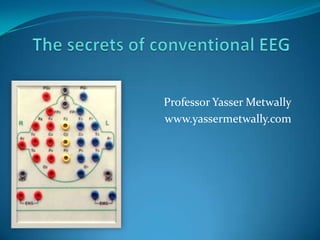
The secrets of conventional EEG Professor Yasser Metwally www.yassermetwally.com
POLYMORPHIC DELTA ACTIVITY Polymorphic delta activity…The EEG phenomenon characteristics of subcortical white matter destructive lesions © www.yassermetwally.com
Electrical criteria of the Polymorphic slow wave activity. Quite variable in wave shape morphology, frequency and amplitude. Commonly lateralized over a wide area of the scalp, persistent in eye closed, eye open state, during all sleep stages, with no visual reactivity. Polymorphic Delta activity that fails to persist into sleep or attenuates significantly with arousal or eye opening is less indicative of structural pathology. Persistent polymorphic delta activity may not precisely match the true location of the lesion, particularly since it presumably arises from physiological deranged neurons often lying on the margin of the destructive lesion. Persistent polymorphic delta activity is aetiologically nonspecific and is seen in a variety of subcortical (while matter) destructive lesions including neoplasms, infarctions, abscesses, trauma, and haemorrhage. It can also be seen in reversible processes such as focal ischemia in transient ischemic attacks or focal depression from a recent seizure. Commonly due to a subcortical white matter lesion inducing deafferentation of the cerebral cortex. A purely cortical lesion does not induce polymorphic slow wave activity. © www.yassermetwally.com
INTERMIT DELTA ACITIVY Intermittent rhythmic delta activity © www.yassermetwally.com
Electrical criteria of The intermittent rhythmic delta activity Consists of sinusoidal waveforms of approximately 2.5 Hz that occur intermittently in the EEG recording. It is most often symmetric but can be lateralized. In adults, the delta activity has a frontal predominance (frontal intermittent rhythmic delta activity [FIRDA]). In children, it is maximal posteriorly (occipital intermittent rhythmic delta activity [OIRDA]) The intermittent rhythmic delta activity shows visual reactivity and is commonly suppressed in the eye open state unless the patient is comatose. Intermittent rhythmic delta activity is associated with structural lesions, most commonly diencephalic, infratentorial or intraventricular tumors, or with diffuse encephalopathies. FIRDA occurring in patients with a normal EEG background suggests that the pattern is due to a structural lesion; when associated with EEG background abnormalities, it is likely to be due to encephalopathy. OIRDA is associated with absence epilepsy in children aged 6-10 years. © www.yassermetwally.com
THE EEG SHARP ACTIVITY © www.yassermetwally.com
Electroclinical criteria of spike/ sharp wave discharge A spike is a transient, clearly distinguished from the background activity, with pointed peak at conventional paper speeds and a duration from 20 to under 70 msec; the main component is generally negative. Amplitude is variable. Spikes represent the basic element of paroxysmal activity in the EEG A sharp wave is a transient, clearly distinguished from background activity, with pointed peak at conventional paper speeds and duration of 70 to 200 msec. The main component is generally negative relative to other areas. Both spikes and sharp waves have multiphasic characters, being composed of a sequence of a minor positive, a major negative, and a second minor positive component is typical in most instances. The long duration of a sharp wave permits better insight into the multiphasic character of this potential. The spike/sharp wave potentials are reliable indicators of a potential seizure focus because they result from the characteristic neurophysiological event "the paroxysmal depolarization shift" (PDS). This phenomenon consists of thousands of neurons simultaneously undergoing large depolarization with superimposed action potentials. Both synaptic events and intrinsic cellular currents have been implicated in this process. EEG spikes/sharp waves are due to the slow depolarization currents in the PDS. Neurons surrounding the focus are inhibited during the paroxysmal depolarization shift, and within the focus the the paroxysmal depolarization shift is followed by a hyperpolarization potential. Both an increase in depolarizing events and a loss of inhibitory mechanisms can lead to persistence and propagation of the discharge as a seizure. Spikes and sharp waves are neurophysiologically closely related phenomena; both of them are typical paroxysmal discharges and highly suggestive of an epileptic seizure disorder, although both phenomena may occur in patients without a history of seizure disorder. The largest and most pronounced spikes are not necessarily associated with more serious epileptic seizure disorders. On the contrary, Rolandic spikes in a child age 4 to 10 yr are very prominent; however, the seizure disorder is usually quite benign or there may be no clinical seizures at all. low voltage spiking in the frontal or anterior temporal regions is highly epileptogenic even though its amplitude can be so low to the point that these spikes might be completely drowned within the background waves and subsequently can not be easily detected. © www.yassermetwally.com
THE 3 C/S SPIKE WAVE DISCHARGE © www.yassermetwally.com
Electroclinical criteria of the 3 c/s spike/wave discharge It is bilateral fairly symmetrical and synchronous. It has a frontal midline maximum. It has a sudden onset and sudden offset. Readily activated by hyperventilation. It might be proceeded by intermittent, rhythmic, bisynchronous monomorphic slow waves in the occipital regions (occipital intermittent rhythmic delta activity OIRDA). The 3 c/s SWD is usually associated with an ictal absence episode when it lasts over 5 seconds. The 3 c/s SWD is an age specific electrophysiological phenomenon. It usually start at the age of 3.5 years and disappear at the age of 16 years. This discharge pattern is markedly enhanced during nonREM sleep, usually during stage II. However the morphological features of this discharge pattern are altered during sleep with the discharge occurring in a more fragmented and atypical fashion, occurring in bursts of spikes, polyspikes and atypical spike/wave complexes. This discharge pattern usually occurs in conjunction with sleep spindles and has an invariable frontal midline maximum. Background activity is within normal before and after termination of the paroxysmal discharge. © www.yassermetwally.com
THE FAST 4/6 SPIKE WAVE DISCHARGE The fast 4/6 C/S spike wave discharge of  Juvenile myoclonic epilepsy (JME) © www.yassermetwally.com
Electroclinical criteria of the fast 4-6 c/s spike/wave discharge This discharge occurs in patients older than 16 years. It is bilateral but less symmetrical and synchronous compared with the 3 c/s SWD and usually takes the morphological feature of polyspike wave discharge. It has a frontal midline maximum It has a sudden onset and sudden offset and lasts for a very short periods (usually less than 3 seconds) This discharge pattern is not activated hyperventilation, however phobic stimulation is a potent activator of this discharge pattern. The clinical correlate of this discharge pattern is myoclonus and grand mal fits (juvenile myoclonic epilepsy). Studies using video monitoring combined with EEG recording revealed that the spike components of this discharge coincide with the myoclonic jerks and the slow waves coincide with periods of relaxation between the myoclonic fits, accordingly the number of spikes in this polyspike/wave complexes were found to be proportional to the severity of the myoclonic fits. The fast spike/wave complexes of juvenile myoclonic epilepsy has a strong genetic background. The gene locus was mapped on the short arm of chromosome 6. © www.yassermetwally.com
SLOW 1-2.5 C/S SPIKE WAVE DISCHARGE  The slow spike/wave discharge of Lennox-Gastaut syndrome © www.yassermetwally.com
Electroclinical criteria of the slow 1-2.5c/s spike/wave discharge This EEG pattern is bilateral but asymmetrical and asynchronous with frequent lateralization and focalization. It has a frontal midline maximum. It is frequently continuous without any definite onset or offset and might extend through the whole record and is not associated with any clinical accompaniment. The discharge is not activated by hyperventilation The 1-2.5 c/s SWD is an age specific electrophysiological phenomenon. It usually start at the age of 6 months (earlier than the 3 c/s SWD) and disappear at the age of 16 years and is replaced by anterior temporal sharp activity and the clinical seizure manifestations merge into the main stream of temporal lobe epilepsies Background activity is often disorganized with frequent slow wave activity. The clinical correlate of this discharge is Lennox-Gastaut syndrome with multiseizure clinical presentation (grand mal fits, atonic fits, akinetic fits, atypical absence attacks, absence status). The occurrence of two or more than two types of seizures is almost the rule, mental retardation is very common. This discharge pattern could be idiopathic of genetic origin, cryptogenic with no overt cause , or symptomatic to a variety of brain diseases that include CNS infection, birth trauma, lipidosis, tuberous sclerosis, etc. © www.yassermetwally.com
THE HYPSARRHYTHMIA PATTERN Hypsarrhythmia pattern. © www.yassermetwally.com
Electroclinical criteria of Hypsarrhythmia discharge The word Hypsarrhythmia is originally derived from the Greek word hypsolos which means high and it refers to high voltage arrhythmia with a disorganized EEG pattern that consists of chaotic admixture of continuous, multifocal, high amplitude spikes, polyspikes, sharp waves and arrhythmic slow waves. This EEG pattern is dynamic and highly variable from one patient to anther and between one study and anther study for a single patient. Background activity is often disorganized with frequent slow wave activity Marked change in the Hypsarrhythmia pattern also occurs during sleep. In REM sleep there is marked reduction to total disappearance of this EEG pattern. There is also normalization of this discharge pattern immediately following awakening from sleep. This discharge pattern is seen in children between the age of 4 months to 4 years and after the age of 4 years this pattern of discharge usually merges into the slow spike/ slow wave complexes. Hypsarrhythmia pattern is frequently equated with infantile spasm (West syndrome), (characterized by massive flexion myoclonus of the head and neck called jack-knifing or Salaam attacks), however this pattern is not specific to any disease entity and is seen in response to any sever cerebral insult or sever multifocal disease process that occurs below the age of 1 year. Five different types of Hypsarrhythmia are present Hypsarrhythmia with increased interhemispheric synchronization. Asymmetrical Hypsarrhythmia. Hypsarrhythmia with a constant focus. Hypsarrhythmia with episodes of voltage attenuation. Hypsarrhythmia composed only of high voltage slow waves without spikes or sharp waves. © www.yassermetwally.com
HEPATIC TRIPHASIC WAVES The triphasic waves © www.yassermetwally.com
Electrical characteristics of triphasic waves Triphasic waves (TWs) are a distinctive but nonspecific electroencephalographic (EEG) pattern originally described in a stuporous patient in 1950 by Foley as "blunted spike and wave." In 1955, Bickford and Butt coined the term "triphasic wave." Since their findings were limited to patients with hepatic failure, triphasic wave encephalopathy (TWE) became synonymous with hepatic encephalopathy. More recently, TWE has been associated with a wide range of toxic, metabolic, and structural abnormalities. TWs are large-amplitude, generalized waves of 1.5-3.0 Hz. They are bilaterally synchronous and bifrontally predominant periodic waves with a characteristic morphology. Classic TWs have an initial small-amplitude, sharp-negative component followed by a large-amplitude, sharp-positive wave; they end with a slow negative wave. © www.yassermetwally.com

Contenu connexe

Tendances

Evoked potentials, clinical importance & physiological basis of consciousness...
Evoked potentials, clinical importance & physiological basis of consciousness...Evoked potentials, clinical importance & physiological basis of consciousness...
Evoked potentials, clinical importance & physiological basis of consciousness...
Rajesh Goit
 
Periodic lateralized epileptiform discharges
Periodic lateralized epileptiform dischargesPeriodic lateralized epileptiform discharges
Periodic lateralized epileptiform discharges
Manideep Malaka
 
neonatal cerebral function monitoring
neonatal cerebral function monitoringneonatal cerebral function monitoring
neonatal cerebral function monitoring
Ahmed Okasha
 

Tendances (20)

normal eeg
 normal eeg  normal eeg
normal eeg
 
Abnormal EEG patterns
Abnormal EEG patternsAbnormal EEG patterns
Abnormal EEG patterns
 
Eeg basics, part 1
Eeg basics, part 1Eeg basics, part 1
Eeg basics, part 1
 
Periodic Lateralizing Epileptiform Discharges
Periodic Lateralizing Epileptiform DischargesPeriodic Lateralizing Epileptiform Discharges
Periodic Lateralizing Epileptiform Discharges
 
Evoked potentials, clinical importance & physiological basis of consciousness...
Evoked potentials, clinical importance & physiological basis of consciousness...Evoked potentials, clinical importance & physiological basis of consciousness...
Evoked potentials, clinical importance & physiological basis of consciousness...
 
Generalised periodic epileptiform discharges
Generalised periodic epileptiform dischargesGeneralised periodic epileptiform discharges
Generalised periodic epileptiform discharges
 
EEG Variants with patterns by Murtaza Syed
EEG Variants with patterns by Murtaza SyedEEG Variants with patterns by Murtaza Syed
EEG Variants with patterns by Murtaza Syed
 
Brainstem auditory evoked potentials (baep)
Brainstem auditory evoked potentials (baep)Brainstem auditory evoked potentials (baep)
Brainstem auditory evoked potentials (baep)
 
Eeg basics.drjma
Eeg basics.drjmaEeg basics.drjma
Eeg basics.drjma
 
Neurophysiology of epilepsy
Neurophysiology of epilepsyNeurophysiology of epilepsy
Neurophysiology of epilepsy
 
Periodic lateralized epileptiform discharges
Periodic lateralized epileptiform dischargesPeriodic lateralized epileptiform discharges
Periodic lateralized epileptiform discharges
 
Artifacts & Normal variants in EEG
Artifacts & Normal variants in EEGArtifacts & Normal variants in EEG
Artifacts & Normal variants in EEG
 
neonatal cerebral function monitoring
neonatal cerebral function monitoringneonatal cerebral function monitoring
neonatal cerebral function monitoring
 
Approaching the eeg
Approaching the eegApproaching the eeg
Approaching the eeg
 
Continuous spike and wave during slow wave sleep(CSWS)
Continuous spike and wave during slow wave sleep(CSWS)Continuous spike and wave during slow wave sleep(CSWS)
Continuous spike and wave during slow wave sleep(CSWS)
 
EEG Epilepsy
EEG EpilepsyEEG Epilepsy
EEG Epilepsy
 
Eeg in pediatric (DNB PEDIATRIC)
Eeg in pediatric (DNB PEDIATRIC)Eeg in pediatric (DNB PEDIATRIC)
Eeg in pediatric (DNB PEDIATRIC)
 
PLEDS
PLEDSPLEDS
PLEDS
 
Eeg examples
Eeg examplesEeg examples
Eeg examples
 
Electroencephalography
ElectroencephalographyElectroencephalography
Electroencephalography
 

En vedette

Evoked potential - An overview
Evoked potential - An overviewEvoked potential - An overview
Evoked potential - An overview
Anbarasi rajkumar
 
Eeg normal inglish
Eeg normal inglishEeg normal inglish
Eeg normal inglish
guestf282ad
 
ID 352 –Sinus rhythm With premature atrial complex
ID 352 –Sinus rhythm With premature atrial complexID 352 –Sinus rhythm With premature atrial complex
ID 352 –Sinus rhythm With premature atrial complex
Anas Nader
 
Sinus Rhythms - BMH/Tele
Sinus Rhythms - BMH/TeleSinus Rhythms - BMH/Tele
Sinus Rhythms - BMH/Tele
TeleClinEd
 

En vedette (20)

E P I L E P S Y U P D A T E
E P I L E P S Y  U P D A T EE P I L E P S Y  U P D A T E
E P I L E P S Y U P D A T E
 
Encephalopathy with EEG based Grading
Encephalopathy with EEG based GradingEncephalopathy with EEG based Grading
Encephalopathy with EEG based Grading
 
EEG: Basics
EEG: BasicsEEG: Basics
EEG: Basics
 
Evoked potential - An overview
Evoked potential - An overviewEvoked potential - An overview
Evoked potential - An overview
 
Electroencephalogram(EEG)
Electroencephalogram(EEG)Electroencephalogram(EEG)
Electroencephalogram(EEG)
 
EEG
EEGEEG
EEG
 
Somatosensory evoked potential
Somatosensory evoked potentialSomatosensory evoked potential
Somatosensory evoked potential
 
Dr. kristen park kcnq2 Cure professional track - learn more at kcnq2cure.org
Dr. kristen park kcnq2 Cure professional track - learn more at kcnq2cure.orgDr. kristen park kcnq2 Cure professional track - learn more at kcnq2cure.org
Dr. kristen park kcnq2 Cure professional track - learn more at kcnq2cure.org
 
Eeg normal inglish
Eeg normal inglishEeg normal inglish
Eeg normal inglish
 
Chapter6
Chapter6Chapter6
Chapter6
 
Hidden context tree modeling of EEG data
Hidden context tree modeling of EEG dataHidden context tree modeling of EEG data
Hidden context tree modeling of EEG data
 
Epileptic encephalopathy -EEG
Epileptic encephalopathy -EEGEpileptic encephalopathy -EEG
Epileptic encephalopathy -EEG
 
General Principles Part 1 Historical And Classifications
General Principles Part 1 Historical And ClassificationsGeneral Principles Part 1 Historical And Classifications
General Principles Part 1 Historical And Classifications
 
West syndrome
West syndromeWest syndrome
West syndrome
 
Neurophysiological investigations
Neurophysiological investigationsNeurophysiological investigations
Neurophysiological investigations
 
EEG dr archana
EEG dr archanaEEG dr archana
EEG dr archana
 
BAEP, BERA, BEP, Brainstem auditory evoked potential By Murtaza Syed
BAEP, BERA, BEP, Brainstem auditory evoked potential By Murtaza SyedBAEP, BERA, BEP, Brainstem auditory evoked potential By Murtaza Syed
BAEP, BERA, BEP, Brainstem auditory evoked potential By Murtaza Syed
 
ID 352 –Sinus rhythm With premature atrial complex
ID 352 –Sinus rhythm With premature atrial complexID 352 –Sinus rhythm With premature atrial complex
ID 352 –Sinus rhythm With premature atrial complex
 
Sinus Rhythms - BMH/Tele
Sinus Rhythms - BMH/TeleSinus Rhythms - BMH/Tele
Sinus Rhythms - BMH/Tele
 
Antiepileptics (New) - drdhriti
Antiepileptics (New) - drdhritiAntiepileptics (New) - drdhriti
Antiepileptics (New) - drdhriti
 

Similaire à Issues in brainmapping...The secrets of conventional EEG

west syndrome ppt hypsarrhythmia infantile.pptx
west syndrome  ppt hypsarrhythmia infantile.pptxwest syndrome  ppt hypsarrhythmia infantile.pptx
west syndrome ppt hypsarrhythmia infantile.pptx
priya99148
 

Similaire à Issues in brainmapping...The secrets of conventional EEG (20)

Issues in brainmapping...The role of EEG in epileptic syndromes associated wi...
Issues in brainmapping...The role of EEG in epileptic syndromes associated wi...Issues in brainmapping...The role of EEG in epileptic syndromes associated wi...
Issues in brainmapping...The role of EEG in epileptic syndromes associated wi...
 
Issues in brainmapping...Generalized epilepsies
Issues in brainmapping...Generalized epilepsiesIssues in brainmapping...Generalized epilepsies
Issues in brainmapping...Generalized epilepsies
 
Issues in brainmapping...Age dependant epileptic syndromes
Issues in brainmapping...Age dependant epileptic syndromesIssues in brainmapping...Age dependant epileptic syndromes
Issues in brainmapping...Age dependant epileptic syndromes
 
Issues in brainmapping...EEG in brain tumors
Issues in brainmapping...EEG in brain tumorsIssues in brainmapping...EEG in brain tumors
Issues in brainmapping...EEG in brain tumors
 
Issues in brainmapping...EEG quantification ...basic methodology
Issues in brainmapping...EEG quantification ...basic methodologyIssues in brainmapping...EEG quantification ...basic methodology
Issues in brainmapping...EEG quantification ...basic methodology
 
Issues in brainmapping...Neonatal EEG
Issues in brainmapping...Neonatal EEGIssues in brainmapping...Neonatal EEG
Issues in brainmapping...Neonatal EEG
 
seizure semiology
seizure semiologyseizure semiology
seizure semiology
 
Ch23
Ch23Ch23
Ch23
 
EPILEPTIC ENCEPHALOPATHY
 EPILEPTIC ENCEPHALOPATHY  EPILEPTIC ENCEPHALOPATHY
EPILEPTIC ENCEPHALOPATHY
 
EPILEPTIC ENCEPHALOPATHY
 EPILEPTIC ENCEPHALOPATHY  EPILEPTIC ENCEPHALOPATHY
EPILEPTIC ENCEPHALOPATHY
 
Issues in brain mapping...EEG patterns of encephalopathies and altered state ...
Issues in brain mapping...EEG patterns of encephalopathies and altered state ...Issues in brain mapping...EEG patterns of encephalopathies and altered state ...
Issues in brain mapping...EEG patterns of encephalopathies and altered state ...
 
Pediatric posterior head region epilepsy
Pediatric posterior head region epilepsyPediatric posterior head region epilepsy
Pediatric posterior head region epilepsy
 
Issues in brainmapping...EEG in inflammatory brain disease
Issues in brainmapping...EEG in inflammatory brain diseaseIssues in brainmapping...EEG in inflammatory brain disease
Issues in brainmapping...EEG in inflammatory brain disease
 
Epileptic encephalopathies during infancy
Epileptic encephalopathies during infancyEpileptic encephalopathies during infancy
Epileptic encephalopathies during infancy
 
Epilepsy
EpilepsyEpilepsy
Epilepsy
 
west syndrome ppt hypsarrhythmia infantile.pptx
west syndrome  ppt hypsarrhythmia infantile.pptxwest syndrome  ppt hypsarrhythmia infantile.pptx
west syndrome ppt hypsarrhythmia infantile.pptx
 
EEG in idiopathic generalised epilepsies
EEG in idiopathic generalised epilepsiesEEG in idiopathic generalised epilepsies
EEG in idiopathic generalised epilepsies
 
Mesial temporal sclerosis cme
Mesial temporal sclerosis cmeMesial temporal sclerosis cme
Mesial temporal sclerosis cme
 
NEONATAL SEIZURES
NEONATAL SEIZURESNEONATAL SEIZURES
NEONATAL SEIZURES
 
Dravet syndrome
Dravet syndromeDravet syndrome
Dravet syndrome
 

Plus de Professor Yasser Metwally

Plus de Professor Yasser Metwally (20)

The Egyptian Zoo in Cairo 2015
The Egyptian Zoo in Cairo 2015The Egyptian Zoo in Cairo 2015
The Egyptian Zoo in Cairo 2015
 
End of the great nile river in Ras Elbar
End of the great nile river in Ras ElbarEnd of the great nile river in Ras Elbar
End of the great nile river in Ras Elbar
 
The Lion and The tiger in Egypt
The Lion and The tiger in EgyptThe Lion and The tiger in Egypt
The Lion and The tiger in Egypt
 
The monkeys in Egypt
The monkeys in EgyptThe monkeys in Egypt
The monkeys in Egypt
 
The Snake, the Scorpion, the turtle in Egypt
The Snake, the Scorpion, the turtle in EgyptThe Snake, the Scorpion, the turtle in Egypt
The Snake, the Scorpion, the turtle in Egypt
 
The Egyptian Parrot
The Egyptian ParrotThe Egyptian Parrot
The Egyptian Parrot
 
The Egyptian Deer
The Egyptian DeerThe Egyptian Deer
The Egyptian Deer
 
The Egyptian Pelican
The Egyptian PelicanThe Egyptian Pelican
The Egyptian Pelican
 
The Flamingo bird in Egypt
The Flamingo bird in EgyptThe Flamingo bird in Egypt
The Flamingo bird in Egypt
 
Egyptian Cats
Egyptian CatsEgyptian Cats
Egyptian Cats
 
Radiological pathology of epileptic disorders
Radiological pathology of epileptic disordersRadiological pathology of epileptic disorders
Radiological pathology of epileptic disorders
 
Radiological pathology of cerebrovascular disorders
Radiological pathology of cerebrovascular disordersRadiological pathology of cerebrovascular disorders
Radiological pathology of cerebrovascular disorders
 
Radiological pathology of spontaneous cerebral hemorrhage
Radiological pathology of spontaneous cerebral hemorrhageRadiological pathology of spontaneous cerebral hemorrhage
Radiological pathology of spontaneous cerebral hemorrhage
 
Radiological pathology of cerebral amyloid angiography
Radiological pathology of cerebral amyloid angiographyRadiological pathology of cerebral amyloid angiography
Radiological pathology of cerebral amyloid angiography
 
Radiological pathology of cerebral microbleeds
Radiological pathology of cerebral microbleedsRadiological pathology of cerebral microbleeds
Radiological pathology of cerebral microbleeds
 
The Egyptian Zoo in Cairo
The Egyptian Zoo in CairoThe Egyptian Zoo in Cairo
The Egyptian Zoo in Cairo
 
Progressive multifocal leukoencephalopathy
Progressive multifocal leukoencephalopathyProgressive multifocal leukoencephalopathy
Progressive multifocal leukoencephalopathy
 
Progressive multifocal leukoencephalopathy
Progressive multifocal leukoencephalopathyProgressive multifocal leukoencephalopathy
Progressive multifocal leukoencephalopathy
 
Issues in radiological pathology: Radiological pathology of watershed infarct...
Issues in radiological pathology: Radiological pathology of watershed infarct...Issues in radiological pathology: Radiological pathology of watershed infarct...
Issues in radiological pathology: Radiological pathology of watershed infarct...
 
Radiological pathology of cortical laminar necrosis
Radiological pathology of cortical laminar necrosisRadiological pathology of cortical laminar necrosis
Radiological pathology of cortical laminar necrosis
 

Dernier

🌹Attapur⬅️ Vip Call Girls Hyderabad 📱9352852248 Book Well Trand Call Girls In...
🌹Attapur⬅️ Vip Call Girls Hyderabad 📱9352852248 Book Well Trand Call Girls In...🌹Attapur⬅️ Vip Call Girls Hyderabad 📱9352852248 Book Well Trand Call Girls In...
🌹Attapur⬅️ Vip Call Girls Hyderabad 📱9352852248 Book Well Trand Call Girls In...
Call Girls In Delhi Whatsup 9873940964 Enjoy Unlimited Pleasure
 
Call Girls in Gagan Vihar (delhi) call me [🔝 9953056974 🔝] escort service 24X7
Call Girls in Gagan Vihar (delhi) call me [🔝  9953056974 🔝] escort service 24X7Call Girls in Gagan Vihar (delhi) call me [🔝  9953056974 🔝] escort service 24X7
Call Girls in Gagan Vihar (delhi) call me [🔝 9953056974 🔝] escort service 24X7
9953056974 Low Rate Call Girls In Saket, Delhi NCR
 

Dernier (20)

Premium Bangalore Call Girls Jigani Dail 6378878445 Escort Service For Hot Ma...
Premium Bangalore Call Girls Jigani Dail 6378878445 Escort Service For Hot Ma...Premium Bangalore Call Girls Jigani Dail 6378878445 Escort Service For Hot Ma...
Premium Bangalore Call Girls Jigani Dail 6378878445 Escort Service For Hot Ma...
 
Trichy Call Girls Book Now 9630942363 Top Class Trichy Escort Service Available
Trichy Call Girls Book Now 9630942363 Top Class Trichy Escort Service AvailableTrichy Call Girls Book Now 9630942363 Top Class Trichy Escort Service Available
Trichy Call Girls Book Now 9630942363 Top Class Trichy Escort Service Available
 
Manyata Tech Park ( Call Girls ) Bangalore ✔ 6297143586 ✔ Hot Model With Sexy...
Manyata Tech Park ( Call Girls ) Bangalore ✔ 6297143586 ✔ Hot Model With Sexy...Manyata Tech Park ( Call Girls ) Bangalore ✔ 6297143586 ✔ Hot Model With Sexy...
Manyata Tech Park ( Call Girls ) Bangalore ✔ 6297143586 ✔ Hot Model With Sexy...
 
🌹Attapur⬅️ Vip Call Girls Hyderabad 📱9352852248 Book Well Trand Call Girls In...
🌹Attapur⬅️ Vip Call Girls Hyderabad 📱9352852248 Book Well Trand Call Girls In...🌹Attapur⬅️ Vip Call Girls Hyderabad 📱9352852248 Book Well Trand Call Girls In...
🌹Attapur⬅️ Vip Call Girls Hyderabad 📱9352852248 Book Well Trand Call Girls In...
 
Independent Call Girls In Jaipur { 8445551418 } ✔ ANIKA MEHTA ✔ Get High Prof...
Independent Call Girls In Jaipur { 8445551418 } ✔ ANIKA MEHTA ✔ Get High Prof...Independent Call Girls In Jaipur { 8445551418 } ✔ ANIKA MEHTA ✔ Get High Prof...
Independent Call Girls In Jaipur { 8445551418 } ✔ ANIKA MEHTA ✔ Get High Prof...
 
Call Girls Service Jaipur {9521753030} ❤️VVIP RIDDHI Call Girl in Jaipur Raja...
Call Girls Service Jaipur {9521753030} ❤️VVIP RIDDHI Call Girl in Jaipur Raja...Call Girls Service Jaipur {9521753030} ❤️VVIP RIDDHI Call Girl in Jaipur Raja...
Call Girls Service Jaipur {9521753030} ❤️VVIP RIDDHI Call Girl in Jaipur Raja...
 
Best Rate (Patna ) Call Girls Patna ⟟ 8617370543 ⟟ High Class Call Girl In 5 ...
Best Rate (Patna ) Call Girls Patna ⟟ 8617370543 ⟟ High Class Call Girl In 5 ...Best Rate (Patna ) Call Girls Patna ⟟ 8617370543 ⟟ High Class Call Girl In 5 ...
Best Rate (Patna ) Call Girls Patna ⟟ 8617370543 ⟟ High Class Call Girl In 5 ...
 
Call Girls Ahmedabad Just Call 9630942363 Top Class Call Girl Service Available
Call Girls Ahmedabad Just Call 9630942363 Top Class Call Girl Service AvailableCall Girls Ahmedabad Just Call 9630942363 Top Class Call Girl Service Available
Call Girls Ahmedabad Just Call 9630942363 Top Class Call Girl Service Available
 
Model Call Girls In Chennai WhatsApp Booking 7427069034 call girl service 24 ...
Model Call Girls In Chennai WhatsApp Booking 7427069034 call girl service 24 ...Model Call Girls In Chennai WhatsApp Booking 7427069034 call girl service 24 ...
Model Call Girls In Chennai WhatsApp Booking 7427069034 call girl service 24 ...
 
Top Quality Call Girl Service Kalyanpur 6378878445 Available Call Girls Any Time
Top Quality Call Girl Service Kalyanpur 6378878445 Available Call Girls Any TimeTop Quality Call Girl Service Kalyanpur 6378878445 Available Call Girls Any Time
Top Quality Call Girl Service Kalyanpur 6378878445 Available Call Girls Any Time
 
The Most Attractive Hyderabad Call Girls Kothapet 𖠋 9332606886 𖠋 Will You Mis...
The Most Attractive Hyderabad Call Girls Kothapet 𖠋 9332606886 𖠋 Will You Mis...The Most Attractive Hyderabad Call Girls Kothapet 𖠋 9332606886 𖠋 Will You Mis...
The Most Attractive Hyderabad Call Girls Kothapet 𖠋 9332606886 𖠋 Will You Mis...
 
Top Rated Bangalore Call Girls Mg Road ⟟ 9332606886 ⟟ Call Me For Genuine S...
Top Rated Bangalore Call Girls Mg Road ⟟   9332606886 ⟟ Call Me For Genuine S...Top Rated Bangalore Call Girls Mg Road ⟟   9332606886 ⟟ Call Me For Genuine S...
Top Rated Bangalore Call Girls Mg Road ⟟ 9332606886 ⟟ Call Me For Genuine S...
 
Call Girls Vadodara Just Call 8617370543 Top Class Call Girl Service Available
Call Girls Vadodara Just Call 8617370543 Top Class Call Girl Service AvailableCall Girls Vadodara Just Call 8617370543 Top Class Call Girl Service Available
Call Girls Vadodara Just Call 8617370543 Top Class Call Girl Service Available
 
Call Girls Gwalior Just Call 8617370543 Top Class Call Girl Service Available
Call Girls Gwalior Just Call 8617370543 Top Class Call Girl Service AvailableCall Girls Gwalior Just Call 8617370543 Top Class Call Girl Service Available
Call Girls Gwalior Just Call 8617370543 Top Class Call Girl Service Available
 
Night 7k to 12k Navi Mumbai Call Girl Photo 👉 BOOK NOW 9833363713 👈 ♀️ night ...
Night 7k to 12k Navi Mumbai Call Girl Photo 👉 BOOK NOW 9833363713 👈 ♀️ night ...Night 7k to 12k Navi Mumbai Call Girl Photo 👉 BOOK NOW 9833363713 👈 ♀️ night ...
Night 7k to 12k Navi Mumbai Call Girl Photo 👉 BOOK NOW 9833363713 👈 ♀️ night ...
 
Call Girls Raipur Just Call 9630942363 Top Class Call Girl Service Available
Call Girls Raipur Just Call 9630942363 Top Class Call Girl Service AvailableCall Girls Raipur Just Call 9630942363 Top Class Call Girl Service Available
Call Girls Raipur Just Call 9630942363 Top Class Call Girl Service Available
 
Call Girls in Gagan Vihar (delhi) call me [🔝 9953056974 🔝] escort service 24X7
Call Girls in Gagan Vihar (delhi) call me [🔝  9953056974 🔝] escort service 24X7Call Girls in Gagan Vihar (delhi) call me [🔝  9953056974 🔝] escort service 24X7
Call Girls in Gagan Vihar (delhi) call me [🔝 9953056974 🔝] escort service 24X7
 
Russian Call Girls Service Jaipur {8445551418} ❤️PALLAVI VIP Jaipur Call Gir...
Russian Call Girls Service  Jaipur {8445551418} ❤️PALLAVI VIP Jaipur Call Gir...Russian Call Girls Service  Jaipur {8445551418} ❤️PALLAVI VIP Jaipur Call Gir...
Russian Call Girls Service Jaipur {8445551418} ❤️PALLAVI VIP Jaipur Call Gir...
 
Top Rated Bangalore Call Girls Richmond Circle ⟟ 9332606886 ⟟ Call Me For Ge...
Top Rated Bangalore Call Girls Richmond Circle ⟟  9332606886 ⟟ Call Me For Ge...Top Rated Bangalore Call Girls Richmond Circle ⟟  9332606886 ⟟ Call Me For Ge...
Top Rated Bangalore Call Girls Richmond Circle ⟟ 9332606886 ⟟ Call Me For Ge...
 
Call Girls Hosur Just Call 9630942363 Top Class Call Girl Service Available
Call Girls Hosur Just Call 9630942363 Top Class Call Girl Service AvailableCall Girls Hosur Just Call 9630942363 Top Class Call Girl Service Available
Call Girls Hosur Just Call 9630942363 Top Class Call Girl Service Available
 

Issues in brainmapping...The secrets of conventional EEG

  • 1. The secrets of conventional EEG Professor Yasser Metwally www.yassermetwally.com
  • 2. POLYMORPHIC DELTA ACTIVITY Polymorphic delta activity…The EEG phenomenon characteristics of subcortical white matter destructive lesions © www.yassermetwally.com
  • 3. Electrical criteria of the Polymorphic slow wave activity. Quite variable in wave shape morphology, frequency and amplitude. Commonly lateralized over a wide area of the scalp, persistent in eye closed, eye open state, during all sleep stages, with no visual reactivity. Polymorphic Delta activity that fails to persist into sleep or attenuates significantly with arousal or eye opening is less indicative of structural pathology. Persistent polymorphic delta activity may not precisely match the true location of the lesion, particularly since it presumably arises from physiological deranged neurons often lying on the margin of the destructive lesion. Persistent polymorphic delta activity is aetiologically nonspecific and is seen in a variety of subcortical (while matter) destructive lesions including neoplasms, infarctions, abscesses, trauma, and haemorrhage. It can also be seen in reversible processes such as focal ischemia in transient ischemic attacks or focal depression from a recent seizure. Commonly due to a subcortical white matter lesion inducing deafferentation of the cerebral cortex. A purely cortical lesion does not induce polymorphic slow wave activity. © www.yassermetwally.com
  • 4. INTERMIT DELTA ACITIVY Intermittent rhythmic delta activity © www.yassermetwally.com
  • 5. Electrical criteria of The intermittent rhythmic delta activity Consists of sinusoidal waveforms of approximately 2.5 Hz that occur intermittently in the EEG recording. It is most often symmetric but can be lateralized. In adults, the delta activity has a frontal predominance (frontal intermittent rhythmic delta activity [FIRDA]). In children, it is maximal posteriorly (occipital intermittent rhythmic delta activity [OIRDA]) The intermittent rhythmic delta activity shows visual reactivity and is commonly suppressed in the eye open state unless the patient is comatose. Intermittent rhythmic delta activity is associated with structural lesions, most commonly diencephalic, infratentorial or intraventricular tumors, or with diffuse encephalopathies. FIRDA occurring in patients with a normal EEG background suggests that the pattern is due to a structural lesion; when associated with EEG background abnormalities, it is likely to be due to encephalopathy. OIRDA is associated with absence epilepsy in children aged 6-10 years. © www.yassermetwally.com
  • 6. THE EEG SHARP ACTIVITY © www.yassermetwally.com
  • 7. Electroclinical criteria of spike/ sharp wave discharge A spike is a transient, clearly distinguished from the background activity, with pointed peak at conventional paper speeds and a duration from 20 to under 70 msec; the main component is generally negative. Amplitude is variable. Spikes represent the basic element of paroxysmal activity in the EEG A sharp wave is a transient, clearly distinguished from background activity, with pointed peak at conventional paper speeds and duration of 70 to 200 msec. The main component is generally negative relative to other areas. Both spikes and sharp waves have multiphasic characters, being composed of a sequence of a minor positive, a major negative, and a second minor positive component is typical in most instances. The long duration of a sharp wave permits better insight into the multiphasic character of this potential. The spike/sharp wave potentials are reliable indicators of a potential seizure focus because they result from the characteristic neurophysiological event "the paroxysmal depolarization shift" (PDS). This phenomenon consists of thousands of neurons simultaneously undergoing large depolarization with superimposed action potentials. Both synaptic events and intrinsic cellular currents have been implicated in this process. EEG spikes/sharp waves are due to the slow depolarization currents in the PDS. Neurons surrounding the focus are inhibited during the paroxysmal depolarization shift, and within the focus the the paroxysmal depolarization shift is followed by a hyperpolarization potential. Both an increase in depolarizing events and a loss of inhibitory mechanisms can lead to persistence and propagation of the discharge as a seizure. Spikes and sharp waves are neurophysiologically closely related phenomena; both of them are typical paroxysmal discharges and highly suggestive of an epileptic seizure disorder, although both phenomena may occur in patients without a history of seizure disorder. The largest and most pronounced spikes are not necessarily associated with more serious epileptic seizure disorders. On the contrary, Rolandic spikes in a child age 4 to 10 yr are very prominent; however, the seizure disorder is usually quite benign or there may be no clinical seizures at all. low voltage spiking in the frontal or anterior temporal regions is highly epileptogenic even though its amplitude can be so low to the point that these spikes might be completely drowned within the background waves and subsequently can not be easily detected. © www.yassermetwally.com
  • 8. THE 3 C/S SPIKE WAVE DISCHARGE © www.yassermetwally.com
  • 9. Electroclinical criteria of the 3 c/s spike/wave discharge It is bilateral fairly symmetrical and synchronous. It has a frontal midline maximum. It has a sudden onset and sudden offset. Readily activated by hyperventilation. It might be proceeded by intermittent, rhythmic, bisynchronous monomorphic slow waves in the occipital regions (occipital intermittent rhythmic delta activity OIRDA). The 3 c/s SWD is usually associated with an ictal absence episode when it lasts over 5 seconds. The 3 c/s SWD is an age specific electrophysiological phenomenon. It usually start at the age of 3.5 years and disappear at the age of 16 years. This discharge pattern is markedly enhanced during nonREM sleep, usually during stage II. However the morphological features of this discharge pattern are altered during sleep with the discharge occurring in a more fragmented and atypical fashion, occurring in bursts of spikes, polyspikes and atypical spike/wave complexes. This discharge pattern usually occurs in conjunction with sleep spindles and has an invariable frontal midline maximum. Background activity is within normal before and after termination of the paroxysmal discharge. © www.yassermetwally.com
  • 10. THE FAST 4/6 SPIKE WAVE DISCHARGE The fast 4/6 C/S spike wave discharge of Juvenile myoclonic epilepsy (JME) © www.yassermetwally.com
  • 11. Electroclinical criteria of the fast 4-6 c/s spike/wave discharge This discharge occurs in patients older than 16 years. It is bilateral but less symmetrical and synchronous compared with the 3 c/s SWD and usually takes the morphological feature of polyspike wave discharge. It has a frontal midline maximum It has a sudden onset and sudden offset and lasts for a very short periods (usually less than 3 seconds) This discharge pattern is not activated hyperventilation, however phobic stimulation is a potent activator of this discharge pattern. The clinical correlate of this discharge pattern is myoclonus and grand mal fits (juvenile myoclonic epilepsy). Studies using video monitoring combined with EEG recording revealed that the spike components of this discharge coincide with the myoclonic jerks and the slow waves coincide with periods of relaxation between the myoclonic fits, accordingly the number of spikes in this polyspike/wave complexes were found to be proportional to the severity of the myoclonic fits. The fast spike/wave complexes of juvenile myoclonic epilepsy has a strong genetic background. The gene locus was mapped on the short arm of chromosome 6. © www.yassermetwally.com
  • 12. SLOW 1-2.5 C/S SPIKE WAVE DISCHARGE The slow spike/wave discharge of Lennox-Gastaut syndrome © www.yassermetwally.com
  • 13. Electroclinical criteria of the slow 1-2.5c/s spike/wave discharge This EEG pattern is bilateral but asymmetrical and asynchronous with frequent lateralization and focalization. It has a frontal midline maximum. It is frequently continuous without any definite onset or offset and might extend through the whole record and is not associated with any clinical accompaniment. The discharge is not activated by hyperventilation The 1-2.5 c/s SWD is an age specific electrophysiological phenomenon. It usually start at the age of 6 months (earlier than the 3 c/s SWD) and disappear at the age of 16 years and is replaced by anterior temporal sharp activity and the clinical seizure manifestations merge into the main stream of temporal lobe epilepsies Background activity is often disorganized with frequent slow wave activity. The clinical correlate of this discharge is Lennox-Gastaut syndrome with multiseizure clinical presentation (grand mal fits, atonic fits, akinetic fits, atypical absence attacks, absence status). The occurrence of two or more than two types of seizures is almost the rule, mental retardation is very common. This discharge pattern could be idiopathic of genetic origin, cryptogenic with no overt cause , or symptomatic to a variety of brain diseases that include CNS infection, birth trauma, lipidosis, tuberous sclerosis, etc. © www.yassermetwally.com
  • 14. THE HYPSARRHYTHMIA PATTERN Hypsarrhythmia pattern. © www.yassermetwally.com
  • 15. Electroclinical criteria of Hypsarrhythmia discharge The word Hypsarrhythmia is originally derived from the Greek word hypsolos which means high and it refers to high voltage arrhythmia with a disorganized EEG pattern that consists of chaotic admixture of continuous, multifocal, high amplitude spikes, polyspikes, sharp waves and arrhythmic slow waves. This EEG pattern is dynamic and highly variable from one patient to anther and between one study and anther study for a single patient. Background activity is often disorganized with frequent slow wave activity Marked change in the Hypsarrhythmia pattern also occurs during sleep. In REM sleep there is marked reduction to total disappearance of this EEG pattern. There is also normalization of this discharge pattern immediately following awakening from sleep. This discharge pattern is seen in children between the age of 4 months to 4 years and after the age of 4 years this pattern of discharge usually merges into the slow spike/ slow wave complexes. Hypsarrhythmia pattern is frequently equated with infantile spasm (West syndrome), (characterized by massive flexion myoclonus of the head and neck called jack-knifing or Salaam attacks), however this pattern is not specific to any disease entity and is seen in response to any sever cerebral insult or sever multifocal disease process that occurs below the age of 1 year. Five different types of Hypsarrhythmia are present Hypsarrhythmia with increased interhemispheric synchronization. Asymmetrical Hypsarrhythmia. Hypsarrhythmia with a constant focus. Hypsarrhythmia with episodes of voltage attenuation. Hypsarrhythmia composed only of high voltage slow waves without spikes or sharp waves. © www.yassermetwally.com
  • 16. HEPATIC TRIPHASIC WAVES The triphasic waves © www.yassermetwally.com
  • 17. Electrical characteristics of triphasic waves Triphasic waves (TWs) are a distinctive but nonspecific electroencephalographic (EEG) pattern originally described in a stuporous patient in 1950 by Foley as "blunted spike and wave." In 1955, Bickford and Butt coined the term "triphasic wave." Since their findings were limited to patients with hepatic failure, triphasic wave encephalopathy (TWE) became synonymous with hepatic encephalopathy. More recently, TWE has been associated with a wide range of toxic, metabolic, and structural abnormalities. TWs are large-amplitude, generalized waves of 1.5-3.0 Hz. They are bilaterally synchronous and bifrontally predominant periodic waves with a characteristic morphology. Classic TWs have an initial small-amplitude, sharp-negative component followed by a large-amplitude, sharp-positive wave; they end with a slow negative wave. © www.yassermetwally.com

Notes de l'éditeur

  1. The secrets of conventional EEG
  2. The secrets of conventional EEG
  3. The secrets of conventional EEG
  4. The secrets of conventional EEG
  5. The secrets of conventional EEG
  6. The secrets of conventional EEG
  7. The secrets of conventional EEG
  8. The secrets of conventional EEG
  9. The secrets of conventional EEG
  10. The secrets of conventional EEG
  11. The secrets of conventional EEG
  12. The secrets of conventional EEG
  13. The secrets of conventional EEG
  14. The secrets of conventional EEG
  15. The secrets of conventional EEG
  16. The secrets of conventional EEG
  17. The secrets of conventional EEG