การวัดความดันในหลอดเลือดดำกลาง Yui
•Télécharger en tant que DOC, PDF•
53 j'aime•116,221 vues


Recommandé
Contenu connexe
Tendances
Tendances (20)
Similaire à การวัดความดันในหลอดเลือดดำกลาง Yui
Similaire à การวัดความดันในหลอดเลือดดำกลาง Yui (20)
การวัดความดันในหลอดเลือดดำกลาง Yui
- 1. การวัด ความดัน ในหลอดเลือ ดดำา กลาง Central Venous Pressure Monitoring วัต ถุป ระสงค์ เพื่อประเมินสารนำ้าในร่างกาย ความหมาย Central Venous Pressure (CVP) หมายถึง ความดันในหลอดเลือดดำา Superior Vena Cava (SVC) ซึ่งมีค่าเท่ากับความดันของ right atrium (RA) และเป็นการแสดงถึง preload ของ right ventricle (RV) หรือ right ventricular end-diastolic pressure (RVEDP) ค่า CVP จะบอกได้ถึงปริมาณนำ้าและเลือดที่ไหลเวียนในร่างกาย ประสิทธิภาพของ right ventricle และ venous capacitance ปริมาณนำ้า หรือเลือดในหัวใจซีกซ้าย (Left Atrial Pressure, LAP) อาจวัดโดยการใส่สาย polyvinyl catheter เข้าไปใน left atrium โดยตรงระหว่างการผ่าตัดหัวใจ แบบเปิด หรือโดยใส่ Swan-Ganz catheter ผ่านทาง เส้นเลือดดำาใหญ่เข้าสู่ pulmonary artery และวัด Pulmonary Capillary Wedge Pressure (PCWP) ซึ่งมีค่าใกล้เคียงกับความดันใน left atrium ข้อ บ่ง ชีใ นการ monitor CVP มีด ง นี้ ้ ั 1. ในผู้ป่วยที่สูญเสียเลือดจากอุบัติเหตุหรือจากการผ่าตัด ภาวะ sepsis และ กรณีอื่นที่ทำาให้ปริมาณเลือดและนำ้าในร่างกายลดลง 2. ในผู้ป่วยที่มีภาวะนำ้าเกิน 3. ในกรณีที่ต้องการประเมินการทำางานของหัวใจและหลอดเลือด ตำา แหน่ง เส้น เลือ ดที่ใ ช้ส ำา หรับ monitor CVP เช่น Basilic vein Brachial vein Cephalic vein Saphenous vein นอกจากนี้สามารถ monitor CVP ทาง catheter ซึ่งแทงผ่านผิวหนัง เข้าไปในเส้นเลือดดำาใหญ่ ได้แก่ External/Internal jugular vein หรือทาง Subclavian vein catheter สาย catheter ซึ่งมักใช้ feeding tube No. 8 ยาว 100 cm. โดยปลายสาย จะอยู่ที่ Superior Vena Cava ก่อนเข้า right atrium ในกรณีที่ผู้ป่วยใส่ Swan-Ganz catheter สามารถ monitor CVP ได้ทาง proximal line ซึ่งมีรู เปิดอยู่ใน right atrium
- 2. วิธ ีก ารวัด CVP 1. บอกให้ผู้ป่วยทราบและล้างมือให้สะอาด 2. จัดท่าผู้ป่วยให้นอนหงายราบ (ผู้ป่วยบางรายมีข้อจำากัดในการนอนราบหรือ อาจหอบ เหนื่อยขณะที่นอนราบ จัดท่าศีรษะสูงได้ไม่เกิน 45 องศา) และแขนขาขณะที่วัด ควรเหยียดตรง 3. หาตำาแหน่งของ zero จุดตัดของ midaxillary line กับ fourth intercostal space และอาจขีดระดับ ไม่ว่าจะเป็น external jugular ,subclavian vein,cutdown ให้วัดที่ ตำาแหน่ง zero หรือ phlebostatic axis 4. หมุน three-way ให้ IV fluid ไหลเข้าไปในสาย iv ด้านไม้บรรทัด โดยปิด ด้านผู้ป่วยไว้ก่อน ควรให้ IV fluid อยู่ในสาย ในระดับเกือบเต็มสาย หรือ มากกว่าค่าเดิม (ประมาณ 5 cm) จากนั้นหมุนปิด three-way ด้านไม้บรรทัด 5. นำาไม้บรรทัดวางทาบที่ผู้ป่วย โดยให้ตำาแหน่งของ zero หรือเลขศูนย์ ซึ่งจุดที่ วางต้องอยู่ระดับเดียวกับ right atrium นั่นคือที่ตำาแหน่งจุดตัดของ midaxillary line กับ fourth intercostal space 6. หมุน three-way เปิดเฉพาะด้านผู้ป่วยกับไม้บรรทัด ปิดด้าน IV (กรณีที่มี three-way หลายอัน ให้ปรับเฉพาะอันที่อยู่ติดกับสาย cut down หรืออันที่มี ไม้บรรทัด)
- 3. 7. การอ่านค่า CVP ที่ work ดี จะต้อง fluctuate หรือมีการเต้นขึ้นลงของระดับ นำ้าในสายที่ไม้บรรทัดตามจังหวะการหายใจ (หากพบว่าเต้นขึ้นลงตามชีพจร แสดงว่าปลายสาย CVP อยู่ลึกเกินไปลงเข้าไปถึงในหัวใจ) ให้อ่านค่าเมื่อเริ่มคงที่ โดยอ่านค่าช่วงหายใจออกสุด (end of expiration) เนื่องจากความดันในช่อง ทรวงอกจะใกล้เคียงกับความดันบรรยากาศ *กรณีที่ผู้ป่วยใส่เครื่องช่วยหายใจ และสามารถหายใจเองได้ ไม่มีหอบเหนื่อย ขณะอ่านค่าให้ปลดเครื่องช่วยหายใจ เนื่องจากเครื่องช่วยหายใจจะทำาให้ได้ค่า CVP สูงกว่าค่าจริง *กรณีที่มีการใส่ PEEP จะทำาให้ค่า CVP สูงกว่าค่าจริงมากขึ้น เนื่องจากความดัน ในช่องทรวงอกมาก แต่ในการวัด CVP ผู้ป่วยที่ on PEEP โดยเฉพาะที่ค่า PEEP > 5 cmH2O จะวัด CVP โดยไม่ปลดเครื่องช่วยหายใจ ทั้งนี้เนื่องจากผู้ป่วยเหล่า นี้มักมีภาวะของ hypoxia และการปลดเครื่องบ่อยจะมีผลให้ประสิทธิภาพในการ ถ่างถุงลมปอดลดลง ดังนั้นในการอ่านค่า CVP ทุกครั้ง ควรบันทึกไว้ด้วยว่าวัดขณะใส่เครื่องช่วย หายใจหรือปลดเครื่อง นอนศีรษะสูงกี่องศา 9. เมื่ออ่านค่า CVP เสร็จแล้ว ให้หมุน three-way อยู่ในลักษณะเดิม คือ ปิดด้าน ไม้บรรทัด 10. ตรวจสอบความเรียบร้อยอีกครั้ง โดยเฉพาะการหมุน three-way, rate IV fluid และข้อต่อต่างๆ ไม่ให้หลวมหรือหลุด 11. จัดท่าผู้ป่วยให้เหมือนเดิมหรือตามความเหมาะสม การแปลค่า CVP - ใช้ pressure transducer ซึ่งจะมีหน่วยเป็น millimeters of mercury (mmHg) - ใช้ water manometer หรือใช้ไม้บรรทัดที่มีสายยาง (extension tube) ซึ่ง ใช้บ่อยบนคลินิก จะมีหน่วยเป็น centimeters of water (cmH2O) หมายเหตุ: 1 cmH2O=1.36 mmHg ,1mmHg.= 0.76 cmH2O ค่า CVP ปกติ อาจอยู่ในช่วง 6-12 cmH2O ทั้งนี้มักใช้ค่า CVP ในการ เปรียบเทียบการเปลี่ยนแปลงจากการรักษาในผู้ป่วยรายนั้นๆ มากกว่า ค่า CVP ตำ่า หมายถึง ปริมาณนำ้าและเลือดในร่างกายลดลง ค่า CVP สูงขึ้นมัก หมายถึงปริมาณนำ้าและเลือดในร่างกายมากขึ้น ที่สำาคัญ ในการแปลค่า CVP จะต้องดูอาการและอาการแสดงอื่นร่วมด้วย เช่น blood pressure, heart rate, urine output, urine specific gravity, intake/output, conscious, ฟังปอดได้ยินเสียงผิดปกติ อาการหอบเหนื่อย ความตึงตัว ความอุ่น เย็น ชื้นของผิวหนัง เป็นต้น
- 4. ค่า CVP สูง และตำ่า พบได้ใ นหลายๆ สาเหตุ ดัง นี้ สาเหตุท ท ำา ให้ CVP สูง ี่ Elevated vascular volume Increased cardiac output (hyperdynamic cardiac function) Depressed cardiac function (RV infarct, RV failure) Cardiac tamponade Constrictive pericarditis Pulmonary hypertension Chronic left ventricular failure สาเหตุท ท ำา ให้ CVP ตำ่า ี่ Reduced vascular volume Decreased mean systemic pressure (e.g., as in late shock state) Venodilation (drug induced) Central Venous Pressure Monitoring อุป กรณ์ set IV ,0.9NSS 100ml ,three way,extention ,transducer ,แป้น transducer syring 3 ml วิธ ีก าร 1.ขั้น ต่อ อุป กรณ์ ต่อ set iv เข้ากับ ขวดนำ้าเกลือ 0.9 NSS 100 ml. แล้วต่อ สาย IV เข้ากับตัว transducer แล้วต่อสาย extension เข้ากับ transducer ต่อแป้นสำาหรับวาง transducerg เข้ากับเสานำ้าเกลือ โดยตัวแป้นต้องอยู่ใน ตำาแหน่ง phlebostatic axis คือ midaxillary line กับ fourth intercostal space ดังภาพ Set IV Extension
- 5. จากนั้นเปิดนำ้าเกลือเพื่อไล่ air ที่อยู่ใน set ทั้งหมด เสร็จแล้วต่อสาย extension เข้ากับสาย cutdown หรือ subclavian vein โดย subclavian ต่อเข้ากับสาย สีนำ้าตาล หรือ proximal lumen 2.ต่อ monitor ต่อสาย cable เข้าที่จอ monitor สาย 3.การต่อ monitor เมื่อต่อสาย cable หน้าจอจะปรากฏดังภาพ เลือกชนิด cable ทีต่อเข้าไปเป็น CVP โดย เลือกไปที่ label เลือก CVP ่
- 6. ถึ ง ขั้ น ตอน calibration เมื่ อ วางตำา แหน่ ง transcuder ที่ จุ ด phlebostatic axis แล้ ว หมุ น three way ของ transducer ด้ า นผู้ ป่ ว ย แล้ ว เปิ ด จุ ก ออก “close patient open to air”ดังภาพ กด zero cal รอเครื่อง calibrate ให้ cvp = 0 mmHg เสร็จแล้วก็หมุน three way มาด้านจุก three way ตามเดิมและปิดจุก ห ม า ย เ ห ตุ ค ว ร ทำา ก า ร flush ส า ย CVP ทุก เ ว ร เ พื่ อ ป้ อ ง กั น ก า ร อุ ด ตั น ของสาย และ cribrate เมื่อ wave CVP เมื่อ มีก าร Overdamping การอ่า นค่า CVP wave
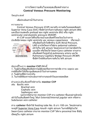
- 7. จากรูป เป็น ปกติข อง CVP A wave - occurs after the P wave of the ECG complex during the PR interval. It reflects the increased atrial pressure that occurs with atrial contraction. Note that the A wave will be absent in patients who do not have a distinct atrial contraction, such as those with atrial fibrillation. Since the CVP value should be a reflection of the Right Ventricular End-Diastolic Pressure, the CVP reading is taken at the last half of the A wave at the midpoint of the X descent. Calculate the CVP by averaging the pressure measured at the peak of the A wave and at the subsequent trough. due to atrial contraction. Absent in atrial fibrillation. Enlarged in tricuspid stenosis, pulmonary stenosis and pulmonary hypertension. The C wave - occurs at the end of the QRS complex at the beginning of the ST segment on the ECG tracing. It reflects closure of the tricuspid valve between the right atrium and right ventricle and the slight bulging of the tricuspid valve during ventricular contraction. The C wave is not always visualized. X descent - due to atrial relaxation.
- 8. The V wave occurs at the end of the T wave on the ECG tracing. It reflects the increased pressure during passive atrial filling. The Y descent occurs prior to the P wave on the ECG tracing. It reflects the opening of the tricuspid valve and the passive flow of blood from the right atrium into the right ventricle prior to atrial contraction. Canon waves - large waves not corresponding to a, v or c waves. Due to complete heart block or junctional arrhythmias. ตัว อย่า ง 1 Measure CVP here Inspiration Expiration ภาวะแทรกซ้อ นทัง จากขั้น ตอนการใส่ส าย CVP และการวัด มีด ัง นี้ ้ 1. Hemothorax 2. Pneumothorax 3. Nerve injury 4. Arterial puncture 5. Thoracic duct perforation 6. Arrhythmias 7. Systemic or local infection 8. Perforation or erosion of vascular structure 9. Thrombosis 10. Air embolism 11. Blood loss จากข้อต่อหลุด 12. Volume overload จากลืมปรับ rate IV หลังวัด CVP FIGURE 3 FIGURE เอกสารอ้า งอิง
- 9. AnaesthesiaUK.The central venous pressure trace.จาก http://www.frca.co.uk/article.aspx?articleid=100036 ค้น เมื่อ 10 ธันวาคม 2555 Elaine cole.Measuring central venous pressure.Senior lecturer ED/Trauma, City University Bartsand the London NHS Trust.จาก www.Cetl.org.uk .ค้นเมื่อ 10 ธันวาคม 2555 Olando health. Fundamentals of Hemodynamic Monitoring : ค้น เมื่อ 1 ธันวาคม 2555 ทนันชัย บุญบูรพงศ์.Central Venous Pressure Monitoring:ค้นเมื่อ 1 ธันวาคม 2555 นุชนารถ บุญจึงมงคล ,ตันหยง พิพานเมฆาภรณ์.Advanced Hemodynamic monitoring :จาก http://www.google.co.th/url? sa=t&rct=j&q=advanced%20hemodynamic%20monitoring %20&source= web&cd=1&cad=rja&ved=0CD0QFjAA&url=http %3A%2F%2Fthaists.org%2Fnews_files%2Fnews_file _385.pdf&ei=CkDHUI3GL8TorQfCgYHIDA&usg=AFQjCNFZOuIrMeF Ds5z0KlgpVQhx0eAbYA&bvm= bv.1354675689,d.bmk: ค้นเมื่อ 1 ธันวาคม 2555