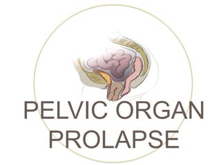
Pelvic organ prolapse
•Télécharger en tant que PPTX, PDF•
51 j'aime•20,393 vues

Seminar presentation

Recommandé
Contenu connexe
Tendances
Tendances (20)
Similaire à Pelvic organ prolapse
Similaire à Pelvic organ prolapse (20)
Plus de yuyuricci
Plus de yuyuricci (20)
Dernier
Dernier (20)
Pelvic organ prolapse
- 1. MUHAMMAD AFFAN SYAFIQI | NUR AMANINA NASIR | NIK NOR LIYANA SUHAIM PELVIC ORGAN PROLAPSE
- 2. WHAT IS PELVIC ORGAN PROLAPSE? Pelvic organ prolapse is the descent of the genital organs beyond their normal anatomical confines. It is caused by herniation through deficient pelvic fascia or due to weakness or deficiency of the ligaments or muscles or blood or nerve supply to the pelvic organs.
- 3. DEFINITION AND CLASSIFICATION A prolapse is protrusion of an organ or structure beyond its normal confines. Prolapses are classified according to their location and the organs contained within them Anterior vaginal wall prolapse POSTERIOR vaginal wall prolapse APICAL vaginal prolapse Urethrocele Urethral descent Cystocele Bladder descent Cystourethrocele Descent of bladder and urethra Rectocele Rectal descent Enterocele Small bowel descent Uterovaginal Uterine descent with inversion of vaginal apex Vault Post-hysterectomy inversion of vaginal apex
- 5. PREVALENCE 41-50 % of women over age of 40 years. Lifetime risk: - 7% operation for prolapse - 11% operation for incontinence/prolapse The annual incidence of surgery for pelvic organ prolapse within 15-49 per 10000 women.
- 6. GRADING 1ST Degree Descent within the vagina 2nd Degree Descent to the introitus 3rd Degree Descent outside the introitus
- 7. GRADING
- 8. The connective tissue, levator ani and intact nerve supply are vital for the maintenance of the position of the pelvic structures and are influenced by pregnancy, childbirth and ageing. Whether congenital or acquired, connective tissue defects appear to be important in the aetiology of prolapse and urinary stress incontinence. AETIOLOGY
- 9. AETIOLOGY 1. Congenital 2% of symptomatic prolapse occurs in nulliparous women Genital prolapse is rare in Afro-Caribbean women 2. Childbirth and Raised Intra-Abdominal Pressure Major factor – vaginal delivery Nerve and mechanical damage resulting from vaginal delivery Parity is associated with increasing prolapse Prolapse during pregnancy is rare, but may be mediated by the effects of progesterone and relaxin Increase in intra-abdominal pressure will put an added strain on the pelvic floor Conditions such as constipation or chronic cough can also raised intra- abdominal pressure
- 10. 3. Ageing Loss of collagen and weakening of fascia and connective tissue Particularly during the post-menopause as a consequence of oestrogen deficiency 4. Postoperative Poor attention to vaginal vault support can lead to vault prolapse Usage of mechanical displacement such as colposuspension may lead to development of rectocele or enterocele AETIOLOGY
- 11. PATHOPHYSIOLOGY There are three components that are responsible for supporting the position of the uterus and vagina LIGAMENTS AND FASCIA LEVATOR ANI MUSCLES POSTERIOR ANGULATION OF THE VAGINA By suspension from pelvic side walls By constricting thereby maintaining the position of the organ Which is enhanced by rises in intra-abdominal pressure causing closure of the ‘flap valve’
- 13. Levator ani
- 15. NORMAL CONDITION At rest, tonic contraction of levator ani muscles provides support to pelvic organs with their activity adjusting to variation in posture, increased vaginal distension, and intra-abdominal pressure. In presence of normal support by levator ani muscle, the supportive connective tissues of vagina pulled the vagina superiorly and back towards the sacrum placing the upper vagina at a nearly horizontal orientation over the levator ani muscle. With presence of intra-abdominal pressure, the upper vagina is compressed against the levator ani muscles and pelvic organ support is maintained.
- 16. PELVIC ORGAN PROLAPSE Damage to any component of vaginal connective tissue support changes the vaginal axis to a vertical position directly over the genital hiatus. Thus with increase in intra-abdominal pressure, the vagina is no longer compressed against the levator ani muscles but directed downward toward the genital hiatus thus can cause pelvic organ prolapse.
- 17. ANALOGY DEMONSTTRATING THE SUPPORT
- 19. Non-specific clinical features Pressure, pain or “fullness” in vagina or rectum or both Sensation of ‘your insides falling out’ – vaginal tissue bulge Urinary incontinence Urine retention Fecal incontinence Chronic constipation Back or pelvic pain Tampons pushing out Dyspareunia (painful / difficult sexual intercourse) Apareunia (inability to perform sexual intercourse) Coital incontinence (leakage of urine or stool during intimacy)
- 20. Specific clinical features CYSTOURET HROCELE RECTOCELE • Urinary frequency & urgency • Voiding difficulty • Urinary Tract Infection (UTI) • Stress incontinence • Incomplete bowel emptying • Digitation • Splinting • Passive anal incontinence
- 21. EXAMINATIONS ABDOMINAL EXAMINATION VAGINAL EXAMINATION COMBINED RECTAL AND VAGINAL EXAMINATION To exclude organomegaly / abdominopelvic mass Examine in dorsal position (if protrude beyond introitus) Assess with ptt straining in left lateral position & Sims speculum To differentiate rectocele from enterocele
- 22. DIFFERENTIAL DIAGNOSIS Anterior wall prolapse 1. Congenital / inclusion dermoid vaginal cyst 2. Uretheral diverticulum
- 23. DIFFERENTIAL DIAGNOSIS UTERINE PROLAPSE 1. Congenital elongation of cervix 2. Chronic inversion 3. Fibroid polyps
- 25. There is no single way to COMPLETELY prevent these problems. MANAGEMENT 1. Overweight women are at a significantly increased risk. 2. Avoid constipation and chronic straining – increase fiber and fluid intake. 3. Seek medical attention if chronic cough which increases abdominal and pelvic pressure. 4. Avoid heavy lifting and learn how to lift safely 5. Do not smoke. 6. Avoid repetitive strenuous activities.
- 26. MANAGEMENT To avoid injuries to supporting structures during time to vaginal delivery either spontaneously or instrumental. Encourage early ambulation and encourage pelvic floor exercise by squeezing the pelvic floor muscles during puerperium Avoid strenuous activity and avoid pregnancy too soon and too many by contraceptive practice Antenatal and Intranatal Care POSTNATAL Care GENERAL MEASURES
- 27. REMEMBER! 1. Antenatal physiotheraphy & relaxation exercises (attention to weight gain and anemia) 2. Proper supervision and management of second stage of labour 3. A generous episiotomy 4. Low forceps delivery if there is delay in second stage 5. Suture perineal tear 6. Postnatal exercises and physiotherapy 7. Early postnatal ambulation 8. Adequate spacing of births 9. Avoid multi-parity 10. Prophylatic HRT in postmenopausal women
- 28. REMEMBER! 1. Antenatal physiotheraphy & relaxation exercises (attention to weight gain and anemia) 2. Proper supervision and management of second stage of labour 3. A generous episiotomy 4. Low forceps delivery if there is delay in second stage 5. Suture perineal tear 6. Postnatal exercises and physiotherapy 7. Early postnatal ambulation 8. Adequate spacing of births 9. Avoid multi-parity 10. Prophylatic HRT in postmenopausal women
- 29. REMEMBER! If symptoms are mild, practice pelvic floor physiotherapy (Kegel Exercise). Once prolapse has developed, Kegel will not correct the problem but may prevent the prolapsed from worsen.
- 31. TYPES
- 32. Silicon rubber based ring pessaries most popular form Ring pessary is made of soft plastic polyvinyl chloride & available in different sizes.
- 33. INDICATIONS? A young woman planning a pregnancy During early pregnancy Puerperium Temporary use while clearing infection and decubitus ulcer A woman unfit for surgery In case a woman refuses for surgery
- 34. LIMITATIONS It is never curative and only be palliative It can cause vaginitis Pessary needs to be changed every 3 months The wearing of pessary is not comfortable to some women and may cause dyspareunia If the vaginal orifice is very patulous, the pessary is often not retained. A forgotten pessary can be the cause of ulcer, rarely carcinoma of vagina and vesicovaginal fistula A pessary does not cure urinary stress incontinence
- 35. SURGERY AIMING TO Relieve symptoms, Restore anatomy AND Restore sexual function
- 36. TYPES OF SURGERY OFFERED TO PATIENTS WITH PROLAPSE DEPENDS ON: 1. The age of patient 2. Desire to retain the uterus 3. Menstrual history 4. General condition 5. Degree of uterine prolapse 6. Uterine abnormality
- 38. 1. Anterior Colporrhaphy To correct cystocele & urethrocele or cystourethrocele
- 39. 2. Perineorrhaphy / Colpoperineorrhaphy To repair the prolapse of posterior vaginal wall
- 40. UTEROVAGINAL PROLAPSE UTERINE PRESERVING SURGERY 1. Hysterosacropexy Open or laparoscopic route Mesh is attached to the isthmus of cervix and uterus to other part of anterior longitudinal ligament on sacrum 2. Manchester repair Accessing uterus vaginally Amputate cervix Use uterosacral cardinal ligament complex to support uterus Rare method 3. Le Fort colpocleisis Partial closure of vagina while preserving the uterus
- 41. UTEROVAGINAL PROLAPSE Hysterosacropexy Manchester (cardinal ligament)
- 42. PROCEDURE INVOLVING HYSTERECTOMY To proceed as that of anterior colporraphy up to pushing up of bladder The UV fold of peritoneum incised The cervical incision is extended posteriorly along the cervicovaginal junction and the pouch of douglas is opened Uterus is delivered anteriorly First clamp on utero sacral and cardinal ligaments, tissues cut and ligated on both sides Second clamp involves uterine vessels which are cut and ligated 1. VAGINAL HYSTERECTOMY
- 43. PROCEDURE INVOLVING HYSTERECTOMY Third clamp on round ligament, fallopian tube and ovarian ligament which are cut and ligated Uterus removed Peritonium closed by purse string suture Enterocele correction done by McCall’s culdoplasty Anterior colporrhaphy is completed Posterior colpoperineorrhaphy performed if there is rectocele