SlideShare une entreprise Scribd logo
1  sur  71
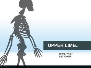
UPPER LIMB..
Dr MEHWISH
LECTURER
ANATOMY OF THE UPPER LIMB
1-Bones of the upper limb.
2-Joints of the upper limb.
3- Muscles of the upper limb.
4- Vessels of the upper limb.
5- Nerves of the upper limb.
.
ANATOMY OF THE UPPER LIMB
Surface anatomy of the upper limb.
The upper limb divided to
1- The Shoulder
2- The arm
3- The forearm
4- The hand
5- The axilla & the breast
6-Cubital fossa
Osteology of upper
limb.
Bones of the upper limb.
SHOULDER
 THE SHOULDER
 It contains THE
SCAPULA & THE
CLAVICLE which
articulate with the
sternum & humerus.
THE SCAPULA
THE SCAPULA
 It is a triangle flat bone with
lies on the posterlateral
aspect of the thorax.
 2 surfaces (ventral& dorsal).&
 3 angles (superior, lateral&
inferior).&
 3 borders (medial ,lateral &
superior).
 It has a spine , acromial
process & coracoid process.
THE SCAPULA
• The scapula (shoulder blade) is a triangular flat bone that lies
on the posterolateral aspest of the thorax, overlying the 2nd-7th
ribs.
• The spine of the scapula starts medially.
• Extend laterally where be wider to form acromial process
• Articulate with the lateral end of the clavicle.
• At the lateral end of the superior border is the coracoid process.
• The lateral angle forms the glenoid cavity.(SHOULDER JOINT)
Articulation of the scapula
 There are 2 synovial & 2 fibrous joints.
 The synovial joints
 The glenoid cavity with the head of the humerus to form the shoulder joint
 Acromio-clavicular joint
 The fibrous joints
 coraco- clavicular joint (strong joint covered with strong ligament)
 coraco- acromial joint (strong joint covered with strong ligament)
Anastamosis around the scapula
 It is an important anastamosis between branches of the first
part of the subclavian artery.& the third part of axillary artery.
 Branches of the first part of the subclavian artery.
 The supra scapular artery. which distributed to the supraspinous & the
infraspinous fossa.
 Deep branch of the transverse cervical artery. which go down along the
medial border of the scapula.
Anastamosis around the scapula
 The branches of the third part of axillary artery.
 The sub scapular artery. which go down along the lateral border of the scapula
 The circumflex scapular artery. which arise from the sub scapular artery
& go to the infraspinous fossa.
THE CLAVICLE
THE CLAVICLE
 It lies horizontally in the root of the neck.
 It covers the flat 1st rib
 its medial 2/3 are curved forward to give room for the passage of the
large vessels & nerves which leave the neck & pass behind & below
the clavicle on their way to upper limb.
 It has 2 important functions:
 To transmit forces from the upper limb to the bones of the axial
skeleton (sternum)
 To act as support, holding the arm free from the trunk.
THE CLAVICLE
 It is along bone with 2 ends:
 The sternal end articulate with the manuberium of the sternum forming the
Sterno- clavicular joint.
 The acromial end articulate with the acromial process of the scapula forming the
Acromio- clavicular joint.
 The body is convex in medial 2/3 concave in lateral 1/3.
THE CLAVICLE
Important relations of the clavicle
 The subclavian artery &vein &the trunk of the brachial pluxes pass behind the
middle /3 of the clavicle.
 The common carotid artery lies behind the left sternoclavicular joint
 The brachio-cephalic artery. divided & its branches behind the right sternoclavicular
joint.
 The internal jugular vein lies a little laterally on either side.
THE HUMERUS
THE HUMERUS
 It is a tubular long bone composed of upper end ,
body (shaft ) & lower end.
 The upper end formed.
 The head
 The neck ( anatomical &surgical )
 The tubercles (greater & lesser )
THE HUMERUS
 The head : which is less than ½ ball directed medially ,upward &
posteriorly .
 It is separated from the greater & lesser tuberosities by a shallow
groove called the anatomical neck .
 The greater tuberosity is a importance which projects posteriorly in
rest position.
 The lesser tuberosity is a small importance which project anteriorly.
 The greater & lesser tuberosities & head are separated from the
shaft by the surgical neck.
 There is small groove below them called the bicipital groove .
THE HUMERUS
 The anterior aspect :medial epicondyle , trochlea ,capitulum &
lateral epicondyle .
 2 fossae (coronoid & radial ) .
 The posterior aspect: medial epicondyle , trochlea & lateral epicondyle with
one fossa (olecranon) .The medial epicondyle is larger , more prominent &
extend downward more than the lateral .It carries a shallow groove in the
posterior surface for the ulner nerve .
THE HUMERUS
• Nerves related to the humerus :
• The circumflex (axillary) nerves may be injured in fracture of surgical
neck
• The radial nerves. (which lies in the spiral groove ) may be injured in
fracture of the middle of the shaft .
• The ulnar nerves may be injured in fracture of the lower end (the
medial epicondyle)
Bone of forarm
Bone of forarm.
• THE RADIUS
 The radius bone is the lateral and shorter bone.
 The lower end which is the styloid process laterally &
ulnar notch medially
• THE ULNA
 The ulna is the stabilizing bone of forearm and it is medial and longer bone
of forearm.
THE RADIUS
 The radius is the lateral bone of the forarm ,and
homologus with the tibiaof the lower limb. it has 2
end and shaft
Above:
 Upper end is having disc-shaped head while lower end
is styloid process.
 Shaft: 3borders,3 surface
BORDERS SURFACES
Anterior border anterior ,
Posterior border posterior,
Medial or interosseous border lateral
Below
 The lower end is the wides part of the bone.
it has 5 surfaces
 Anterior surfaces.
 Posterior surfaces.
 Medial surfaces.
 Lateral surface
 Inferior surfaces
THE ULNA
 The ulna is the bone of forearm,its
homologous with the fibula of the lower limb
its thas 2 end and shaft.
ABOVE:
 upper end is hook-like, upper end presented
the olecranon and coronoid processes, and
trochlear and radial notches.
 5 surfaces.(olecranon processes)
Anterior, Posterior, Medial, Lateral, Superior,
 4 surface.(coronoid processes)
Superior, Anterior, Lateral,medial
 Shaft 3 borders,3surfaces
Borders surfaces
Lateral Anterior
Anterior Medial
Posterior Posterior
THE ULNA
BELOW
lower end of ulna is made up of
head and styloid process..
 The head : small rounded articulating
with the medial side of the lower end of the
radius forming the inferior radio – ulnar
joint.
 The styloid process : projects
medially & below the head .
Ulna bone with border and surfaces
The interosseous membrane
 The radius & ulna are also joined by a fibrous membrane which
stretches between the interosseous borders of the 2 bones..
 It run obliquely downwards &medially from radius to ulna.
 It is pierce by the ant: Interosseous membrane .
Its function is
 Increase the area of origin of forearm.
 transmits forces received by the lower end of radius to ulna.
BONES OF HAND
The bones of the hand
 They consist of :carpus , metacarpus & phalanges
bones
 The carpus bones : they are 8 arrange in 2 rows
( proximal & distal ).
From lateral to medial:
 The proximal row : scaphoid ,lunate , triquetral &
pisiform
 The distal row : trapezium , trapezoid , capitate &
hamate .
The bones of the hand
• The Metacarpal bones : are 5 . One for each finger .
• Each metacarpal bone has : base ,shaft & head .
• The phalanges : all the fingers have 3 phalanges
• ( proximal , middle & distal )
• Each phalanx has base , shaft & head
• except the thumb has only 2 (proximal & distal ).
The articulation of the carpal bones
 The proximal row is convex toward the lower end of radius & ulna .
 The scaphoid & lunate articulate with the lower end of the radius
 The triquetral articulates with the lower end of the ulna .
 The bones of the proximal row articulate with the bones of the distal row in
mid carpal (transverese carpal) joint.
The bones of the distal row articulate with the base of the
metacarpals by
 The trapezium articulate with the1st metacarpal bone
 The trapazoid articulate with the 2nd metacarpal .
 The capitate articulate with the 3rd metacarpal .
 The hamate articulate with the 4th & 5th metacarpals..
MYOLOGY THE MUSCLES OF
THE
UPPER LIMB
THE MUSCLES OF THE UPPER LIMB
In general they divided to
Muscle attached the upper limb to axial
skeleton .
Muscles of the upper limb proper .
Muscle attached the upper limb to axial
skeleton .
 THE FRONT Mm.
Pectoralis major , Pectoralis minor & subclavius Muscles.
 AT SIDE Mm.
serratus ant Muscles.
 THE BACK Mm.
Latissmus dorsi , trapezius , levator scapulae , rhomboideus minor
& rhomboideus major muscles.
Only the pect: Major & latissmus dorsi are inserted in the humerus while all
the others are inserted in the shoulder girdle( scapula & clavicle) .
MOVEMENTS OF THE SHOULDER
GIRDLE
1- ELEVATION: by upper of trapezius & levator scapulae Mm
.2-DEPRETION: by pect.major , pect.minor &latissmus dorssi Mm.
3- RETRACTION: by middle of trapezius, rhomboideus major & minor Mm
.4- PROTRACTION: by serratus ant:,levator scapulae & pect. Minor.
5- ROTATION UP : by upper &lower of trapezius &serratus anterior Mm.
6-ROTATION DOWN :levator scapulae,rhomboideus major & rhom.minor Mm.
MUSCLES OF THE SHOUlDER REGION
• They are 6 Mm.: deltoid , teres major , teres minor ,supraspinatus ,
infraspinatus& subscabularis Mm.
• The Rotator cuff M. ,supraspinatus , infraspinatus& subscabularis.
teres minor
• Arise from the scapula ( all from the dorsal surface except subscapularis
M. from the anterior surface )
• Inserted in the tuberosities of humerus .They rotate the arm(medially or
laterally) & adduct the arm (except the deltoid &supraspinatus Mm.) all
suplied by C5, C6 Nn.
MUSCLES OF THE BACK OF THE ARM.:
Triceps Brachii: The triceps
brachii muscle “three-headed muscle
of the arm” is the large muscle on the
Posterior Compartment of the upper
limb. Triceps Brachii muscle is
principally responsible for extension of
the elbow joint.
• Attachments of the triceps brachii
muscle
• Origin:
• Long head – originates from the
infraglenoid tubercle
• Medial head – originates from
the humerus, inferior to the radial
groove
• Lateral head – originates from the
humerus, superior to the radial groove
• Insertion:
Distally, the heads converge onto
one tendon and insert into the
olecranon of the ulna.
• Actions of the
triceps brachii
muscle
• Extension of the arm
at the elbow
THE FOREARM MUSCLES THE FLEXOR –PRONATOR GROUP
 They divided to 2 groups:
1- The flexor- pronator group.
2- The extensor- supinator group.
*They divided to superficial & deep groups.
*The superficial group arise from the front of medial epicondyle of humerus,
pass in front of the forearm& the wrist to inserted in bones of the hand .
THE FLEXOR –PRONATOR GROUP
Anterior compartment of forearm
 They are :Pronator teres , Fl. Carpi-
radialis , Fl. Carpi-ulnaris ,Fl. Digitorum
superficialis &palmaris longus Mm .
 The deep group arise from the
front of interosseous membrane
of radius & ulna.
 They are : Fl.digitorum profundus ,
Fl.pollicis longus & pronator quadratus
Mm.
THE FLEXOR –PRONATOR GROUP :
• The pronators; are Pronator teres & Pronator quadratus Mm.
where inserted in the radius & pronate the forearm .
• The flexors of the wrist are ;Fl.carpi-radialis ,Fl. carpi-ulnaris &
Palmaris longus where inserted in the metacarpal bones .
• The flexor of the fingers; are Fl. digitorum superficialis & Fl. Digitorum
profundus where inserted in the phalanges.
• The flexor of the thumb; is Fl. Pollicis longus& inserted in phalanges of
the thumb .
THE EXTENSOR - SUPINATOR GROUP.
Posterior compartment of forearm
• *They extend the wrist & fingers & supinate the forearm .
• * They divided in to superficial & deep :
• The superficial group arise from the back of the lateral epicondyle of
humerus to pass on the back of the forearm
• inserted in the bones of the hand .
• There are 7 ; Brachio-radialis , extensor carpi – radialis longus ;ext.calpi-
radialis brevis ;ext.digitorum ; ext. digiti- minimi ;ext. carpi –ulnaris &
anconeus Mm.
THE EXTENSOR - SUPINATOR GROUP.
Posterior compartment of forearm
THE EXTENSOR - SUPINATOR GROUP.
THE EXTENSOR - SUPINATOR GROUP
Muscles of the thumb
 Muscles of the thumb are :
 Abductor pollicis longus ,
 Ext. pollicis brevis
 Ext. pollicis longus Mm.
 Inserted in the thumb bones
RETINACULUM
 Two types of
retinaculum
 Flexor retinaculum
 Extensor retinaculum
THE FLEXOR RETINACULUM
 They are the band of
deep fascia of the wrist.
 Attachments
Medially
 The pisiform bone &
 The hook of the hamate.
Laterally
 The tubercle of the scaphoid
 The crest of the trapezium
Function
 Hold the long flexor and extensor
 Tendons in positions at the wrist.
RELATIONS..
THE EXTENSOR RETINACULUM
 They are the band of deep fascia of
the wrist.
 Attachments
Medially
 Medially pisiform &triquterum
Laterally
 Distal end of radius
 Function
 Hold the long flexor and extensor
 Tendons in positions at the wrist.
RELATIONS..
CARPAL TUNNEL SYNDROME
• The Carpal Tunnel in your wrist.
• It is made up of the carpal bones
and the flexor retinaculum
(transverse carpal ligament).
Carpal Bones
•
Carpal tunnel syndrome
• Carpal tunnel syndrome is a common
condition that causes pain, numbness, and
tingling in the hand and arm.
• The condition occurs when one of the
major nerves compressed
• The median nerve; is squeezed or
compressed as it travels through the
wrist.
THENAR & HYPOTHENAR
HYPOTHENAR
• The hypothenar eminence is a group
of three muscles found at the base of
the little finger, on the ulnar side of the
palm.
• Similarly to the thenar muscles with
the thumb, these muscles control
movement in the little finger.
• This group consists of the following
three muscles:
• abductor digiti minimi,
• the flexor digiti minimi brevis
• opponens digiti minimi.
Thenar
 Thenar Eminence
Group of three short muscles located
at the base of the thumb.
This group of muscles forms a “fleshy
mass” on the lateral side of the palm
which is responsible for the thumb’s
fine movements.
• This group consists of the following
three muscles:
• Opponens Pollicis.
• Abductor Pollicis Brevis.
• Flexor Pollicis Brevis.
MOVEMENT OF THUMB
REFERENCES
B D CHURASIA’S HUMAN ANATOY.
SNELL
 THANK YOU

Contenu connexe

Tendances (20)

Pectoral region
Pectoral regionPectoral region
Pectoral region
 
Shoulder region
Shoulder regionShoulder region
Shoulder region
 
Clavicle bone Anatomy
Clavicle bone AnatomyClavicle bone Anatomy
Clavicle bone Anatomy
 
Anatomy of Back of fore arm and dorsum of hand
Anatomy of Back of fore arm and dorsum of handAnatomy of Back of fore arm and dorsum of hand
Anatomy of Back of fore arm and dorsum of hand
 
Muscles of arm
Muscles of armMuscles of arm
Muscles of arm
 
Introduction to upper limb
Introduction to upper limbIntroduction to upper limb
Introduction to upper limb
 
The anatomy of pectoral region
The  anatomy of pectoral regionThe  anatomy of pectoral region
The anatomy of pectoral region
 
Upper limb muscles
Upper limb musclesUpper limb muscles
Upper limb muscles
 
Anatomy of posterior compartment of the arm
Anatomy of  posterior compartment of the armAnatomy of  posterior compartment of the arm
Anatomy of posterior compartment of the arm
 
Ulna bone structure
Ulna bone structureUlna bone structure
Ulna bone structure
 
Axillary artey ppt
Axillary artey pptAxillary artey ppt
Axillary artey ppt
 
Trapezius Muscle
Trapezius MuscleTrapezius Muscle
Trapezius Muscle
 
Slideshow: Hip Bone
Slideshow: Hip BoneSlideshow: Hip Bone
Slideshow: Hip Bone
 
Slideshow: Ulna
Slideshow: UlnaSlideshow: Ulna
Slideshow: Ulna
 
STERNUM.pptx
STERNUM.pptxSTERNUM.pptx
STERNUM.pptx
 
Anatomy of Femoral nerve
Anatomy of Femoral nerve Anatomy of Femoral nerve
Anatomy of Femoral nerve
 
Cervical vertebrae
Cervical vertebraeCervical vertebrae
Cervical vertebrae
 
Muscles of upper
Muscles of upperMuscles of upper
Muscles of upper
 
Cubital fossa
Cubital fossaCubital fossa
Cubital fossa
 
Thorax
ThoraxThorax
Thorax
 

Similaire à Here are the key muscles of the back of the arm:- Triceps brachii: Three-headed muscle that extends the elbow. Originates from the scapula and humerus, inserts into the olecranon process. - Brachialis: Located underneath triceps. Flexes the elbow. Originates from the distal humerus, inserts into the ulna. - Anconeus: Small muscle on lateral aspect of triceps. Assists triceps in extending the elbow. Originates from lateral epicondyle, inserts into olecranon process.The triceps brachii is the main extensor of the elbow joint. The

UPPER LIMB 4 ppt of human body..........
UPPER LIMB 4 ppt of human body..........UPPER LIMB 4 ppt of human body..........
UPPER LIMB 4 ppt of human body..........aahanak787
 
Bones of upper limb, By Nida Manzoor.pptx
Bones of upper limb, By Nida Manzoor.pptxBones of upper limb, By Nida Manzoor.pptx
Bones of upper limb, By Nida Manzoor.pptxssuser1d41c52
 
UPPERLIMB bs1.pptx
UPPERLIMB bs1.pptxUPPERLIMB bs1.pptx
UPPERLIMB bs1.pptxTHIRUVAZAHAN
 
1-bones upper livvvvvvvvvvvvvvvvvvvvvvvvvvvvmb.pptx
1-bones upper livvvvvvvvvvvvvvvvvvvvvvvvvvvvmb.pptx1-bones upper livvvvvvvvvvvvvvvvvvvvvvvvvvvvmb.pptx
1-bones upper livvvvvvvvvvvvvvvvvvvvvvvvvvvvmb.pptxpaulfiredust
 
Pectoral Girdle and Shoulder
Pectoral Girdle and ShoulderPectoral Girdle and Shoulder
Pectoral Girdle and ShoulderSado Anatomist
 
Skeleton of the upper limb
Skeleton of the upper limbSkeleton of the upper limb
Skeleton of the upper limbKamal Deen
 
Skeletal System 2
Skeletal System 2Skeletal System 2
Skeletal System 2Marc Potter
 
Anatomy (anatomy of bone, joint & muscles of upper limb)
Anatomy (anatomy of bone, joint & muscles of upper limb)Anatomy (anatomy of bone, joint & muscles of upper limb)
Anatomy (anatomy of bone, joint & muscles of upper limb)Osama Al-Zahrani
 
Anatomy of brachial plexus
Anatomy of brachial plexusAnatomy of brachial plexus
Anatomy of brachial plexusPrince Lathiya
 
Anatomy of Upper extremity
Anatomy of Upper extremityAnatomy of Upper extremity
Anatomy of Upper extremitySunil Pahari
 
medicolegal aspects of anatomy of lower limb bones
medicolegal aspects of anatomy of lower limb bonesmedicolegal aspects of anatomy of lower limb bones
medicolegal aspects of anatomy of lower limb bonestsokos
 
Presentation1.pptx, radiological anatomy of the upper limb joint.
Presentation1.pptx, radiological anatomy of the upper limb joint.Presentation1.pptx, radiological anatomy of the upper limb joint.
Presentation1.pptx, radiological anatomy of the upper limb joint.Abdellah Nazeer
 
Purvi shah clavicle & scapula PPT {UL BONE}
Purvi shah clavicle & scapula PPT {UL BONE}Purvi shah clavicle & scapula PPT {UL BONE}
Purvi shah clavicle & scapula PPT {UL BONE}Purvi Shah
 
Anatomy upper limb scapulohumeral 24112010
Anatomy upper limb scapulohumeral  24112010Anatomy upper limb scapulohumeral  24112010
Anatomy upper limb scapulohumeral 24112010Lawrence James
 
muscels of the upper Limb edited(1).pptx
muscels of the upper Limb edited(1).pptxmuscels of the upper Limb edited(1).pptx
muscels of the upper Limb edited(1).pptxPhebeLois1
 
Vertebral Column And Contents Of The Vertebral Canal
Vertebral Column And Contents Of The Vertebral CanalVertebral Column And Contents Of The Vertebral Canal
Vertebral Column And Contents Of The Vertebral CanalMD Specialclass
 

Similaire à Here are the key muscles of the back of the arm:- Triceps brachii: Three-headed muscle that extends the elbow. Originates from the scapula and humerus, inserts into the olecranon process. - Brachialis: Located underneath triceps. Flexes the elbow. Originates from the distal humerus, inserts into the ulna. - Anconeus: Small muscle on lateral aspect of triceps. Assists triceps in extending the elbow. Originates from lateral epicondyle, inserts into olecranon process.The triceps brachii is the main extensor of the elbow joint. The (20)

UPPER LIMB 4 ppt of human body..........
UPPER LIMB 4 ppt of human body..........UPPER LIMB 4 ppt of human body..........
UPPER LIMB 4 ppt of human body..........
 
Appendicular Skeleton.pptx
Appendicular Skeleton.pptxAppendicular Skeleton.pptx
Appendicular Skeleton.pptx
 
Bones of upper limb, By Nida Manzoor.pptx
Bones of upper limb, By Nida Manzoor.pptxBones of upper limb, By Nida Manzoor.pptx
Bones of upper limb, By Nida Manzoor.pptx
 
UPPERLIMB bs1.pptx
UPPERLIMB bs1.pptxUPPERLIMB bs1.pptx
UPPERLIMB bs1.pptx
 
1-bones upper livvvvvvvvvvvvvvvvvvvvvvvvvvvvmb.pptx
1-bones upper livvvvvvvvvvvvvvvvvvvvvvvvvvvvmb.pptx1-bones upper livvvvvvvvvvvvvvvvvvvvvvvvvvvvmb.pptx
1-bones upper livvvvvvvvvvvvvvvvvvvvvvvvvvvvmb.pptx
 
Pectoral Girdle and Shoulder
Pectoral Girdle and ShoulderPectoral Girdle and Shoulder
Pectoral Girdle and Shoulder
 
Skeleton of the upper limb
Skeleton of the upper limbSkeleton of the upper limb
Skeleton of the upper limb
 
Skeletal System 2
Skeletal System 2Skeletal System 2
Skeletal System 2
 
Anatomy (anatomy of bone, joint & muscles of upper limb)
Anatomy (anatomy of bone, joint & muscles of upper limb)Anatomy (anatomy of bone, joint & muscles of upper limb)
Anatomy (anatomy of bone, joint & muscles of upper limb)
 
Anatomy of brachial plexus
Anatomy of brachial plexusAnatomy of brachial plexus
Anatomy of brachial plexus
 
Anatomy of Upper extremity
Anatomy of Upper extremityAnatomy of Upper extremity
Anatomy of Upper extremity
 
medicolegal aspects of anatomy of lower limb bones
medicolegal aspects of anatomy of lower limb bonesmedicolegal aspects of anatomy of lower limb bones
medicolegal aspects of anatomy of lower limb bones
 
THORACIC VERTEBARE.pdf
THORACIC VERTEBARE.pdfTHORACIC VERTEBARE.pdf
THORACIC VERTEBARE.pdf
 
Presentation1.pptx, radiological anatomy of the upper limb joint.
Presentation1.pptx, radiological anatomy of the upper limb joint.Presentation1.pptx, radiological anatomy of the upper limb joint.
Presentation1.pptx, radiological anatomy of the upper limb joint.
 
Anatomy of Humerus
Anatomy of HumerusAnatomy of Humerus
Anatomy of Humerus
 
Purvi shah clavicle & scapula PPT {UL BONE}
Purvi shah clavicle & scapula PPT {UL BONE}Purvi shah clavicle & scapula PPT {UL BONE}
Purvi shah clavicle & scapula PPT {UL BONE}
 
Anatomy upper limb scapulohumeral 24112010
Anatomy upper limb scapulohumeral  24112010Anatomy upper limb scapulohumeral  24112010
Anatomy upper limb scapulohumeral 24112010
 
muscels of the upper Limb edited(1).pptx
muscels of the upper Limb edited(1).pptxmuscels of the upper Limb edited(1).pptx
muscels of the upper Limb edited(1).pptx
 
Vertebral Column And Contents Of The Vertebral Canal
Vertebral Column And Contents Of The Vertebral CanalVertebral Column And Contents Of The Vertebral Canal
Vertebral Column And Contents Of The Vertebral Canal
 
axilla.pptx
axilla.pptxaxilla.pptx
axilla.pptx
 

Dernier

Presentation by Andreas Schleicher Tackling the School Absenteeism Crisis 30 ...
Presentation by Andreas Schleicher Tackling the School Absenteeism Crisis 30 ...Presentation by Andreas Schleicher Tackling the School Absenteeism Crisis 30 ...
Presentation by Andreas Schleicher Tackling the School Absenteeism Crisis 30 ...EduSkills OECD
 
Sanyam Choudhary Chemistry practical.pdf
Sanyam Choudhary Chemistry practical.pdfSanyam Choudhary Chemistry practical.pdf
Sanyam Choudhary Chemistry practical.pdfsanyamsingh5019
 
Grant Readiness 101 TechSoup and Remy Consulting
Grant Readiness 101 TechSoup and Remy ConsultingGrant Readiness 101 TechSoup and Remy Consulting
Grant Readiness 101 TechSoup and Remy ConsultingTechSoup
 
1029-Danh muc Sach Giao Khoa khoi 6.pdf
1029-Danh muc Sach Giao Khoa khoi  6.pdf1029-Danh muc Sach Giao Khoa khoi  6.pdf
1029-Danh muc Sach Giao Khoa khoi 6.pdfQucHHunhnh
 
The Most Excellent Way | 1 Corinthians 13
The Most Excellent Way | 1 Corinthians 13The Most Excellent Way | 1 Corinthians 13
The Most Excellent Way | 1 Corinthians 13Steve Thomason
 
Beyond the EU: DORA and NIS 2 Directive's Global Impact
Beyond the EU: DORA and NIS 2 Directive's Global ImpactBeyond the EU: DORA and NIS 2 Directive's Global Impact
Beyond the EU: DORA and NIS 2 Directive's Global ImpactPECB
 
Web & Social Media Analytics Previous Year Question Paper.pdf
Web & Social Media Analytics Previous Year Question Paper.pdfWeb & Social Media Analytics Previous Year Question Paper.pdf
Web & Social Media Analytics Previous Year Question Paper.pdfJayanti Pande
 
microwave assisted reaction. General introduction
microwave assisted reaction. General introductionmicrowave assisted reaction. General introduction
microwave assisted reaction. General introductionMaksud Ahmed
 
Disha NEET Physics Guide for classes 11 and 12.pdf
Disha NEET Physics Guide for classes 11 and 12.pdfDisha NEET Physics Guide for classes 11 and 12.pdf
Disha NEET Physics Guide for classes 11 and 12.pdfchloefrazer622
 
Software Engineering Methodologies (overview)
Software Engineering Methodologies (overview)Software Engineering Methodologies (overview)
Software Engineering Methodologies (overview)eniolaolutunde
 
Separation of Lanthanides/ Lanthanides and Actinides
Separation of Lanthanides/ Lanthanides and ActinidesSeparation of Lanthanides/ Lanthanides and Actinides
Separation of Lanthanides/ Lanthanides and ActinidesFatimaKhan178732
 
POINT- BIOCHEMISTRY SEM 2 ENZYMES UNIT 5.pptx
POINT- BIOCHEMISTRY SEM 2 ENZYMES UNIT 5.pptxPOINT- BIOCHEMISTRY SEM 2 ENZYMES UNIT 5.pptx
POINT- BIOCHEMISTRY SEM 2 ENZYMES UNIT 5.pptxSayali Powar
 
Measures of Central Tendency: Mean, Median and Mode
Measures of Central Tendency: Mean, Median and ModeMeasures of Central Tendency: Mean, Median and Mode
Measures of Central Tendency: Mean, Median and ModeThiyagu K
 
Kisan Call Centre - To harness potential of ICT in Agriculture by answer farm...
Kisan Call Centre - To harness potential of ICT in Agriculture by answer farm...Kisan Call Centre - To harness potential of ICT in Agriculture by answer farm...
Kisan Call Centre - To harness potential of ICT in Agriculture by answer farm...Krashi Coaching
 
Introduction to Nonprofit Accounting: The Basics
Introduction to Nonprofit Accounting: The BasicsIntroduction to Nonprofit Accounting: The Basics
Introduction to Nonprofit Accounting: The BasicsTechSoup
 
Nutritional Needs Presentation - HLTH 104
Nutritional Needs Presentation - HLTH 104Nutritional Needs Presentation - HLTH 104
Nutritional Needs Presentation - HLTH 104misteraugie
 
The basics of sentences session 2pptx copy.pptx
The basics of sentences session 2pptx copy.pptxThe basics of sentences session 2pptx copy.pptx
The basics of sentences session 2pptx copy.pptxheathfieldcps1
 
APM Welcome, APM North West Network Conference, Synergies Across Sectors
APM Welcome, APM North West Network Conference, Synergies Across SectorsAPM Welcome, APM North West Network Conference, Synergies Across Sectors
APM Welcome, APM North West Network Conference, Synergies Across SectorsAssociation for Project Management
 
Russian Call Girls in Andheri Airport Mumbai WhatsApp 9167673311 💞 Full Nigh...
Russian Call Girls in Andheri Airport Mumbai WhatsApp  9167673311 💞 Full Nigh...Russian Call Girls in Andheri Airport Mumbai WhatsApp  9167673311 💞 Full Nigh...
Russian Call Girls in Andheri Airport Mumbai WhatsApp 9167673311 💞 Full Nigh...Pooja Nehwal
 

Dernier (20)

Presentation by Andreas Schleicher Tackling the School Absenteeism Crisis 30 ...
Presentation by Andreas Schleicher Tackling the School Absenteeism Crisis 30 ...Presentation by Andreas Schleicher Tackling the School Absenteeism Crisis 30 ...
Presentation by Andreas Schleicher Tackling the School Absenteeism Crisis 30 ...
 
Sanyam Choudhary Chemistry practical.pdf
Sanyam Choudhary Chemistry practical.pdfSanyam Choudhary Chemistry practical.pdf
Sanyam Choudhary Chemistry practical.pdf
 
Grant Readiness 101 TechSoup and Remy Consulting
Grant Readiness 101 TechSoup and Remy ConsultingGrant Readiness 101 TechSoup and Remy Consulting
Grant Readiness 101 TechSoup and Remy Consulting
 
Mattingly "AI & Prompt Design: The Basics of Prompt Design"
Mattingly "AI & Prompt Design: The Basics of Prompt Design"Mattingly "AI & Prompt Design: The Basics of Prompt Design"
Mattingly "AI & Prompt Design: The Basics of Prompt Design"
 
1029-Danh muc Sach Giao Khoa khoi 6.pdf
1029-Danh muc Sach Giao Khoa khoi  6.pdf1029-Danh muc Sach Giao Khoa khoi  6.pdf
1029-Danh muc Sach Giao Khoa khoi 6.pdf
 
The Most Excellent Way | 1 Corinthians 13
The Most Excellent Way | 1 Corinthians 13The Most Excellent Way | 1 Corinthians 13
The Most Excellent Way | 1 Corinthians 13
 
Beyond the EU: DORA and NIS 2 Directive's Global Impact
Beyond the EU: DORA and NIS 2 Directive's Global ImpactBeyond the EU: DORA and NIS 2 Directive's Global Impact
Beyond the EU: DORA and NIS 2 Directive's Global Impact
 
Web & Social Media Analytics Previous Year Question Paper.pdf
Web & Social Media Analytics Previous Year Question Paper.pdfWeb & Social Media Analytics Previous Year Question Paper.pdf
Web & Social Media Analytics Previous Year Question Paper.pdf
 
microwave assisted reaction. General introduction
microwave assisted reaction. General introductionmicrowave assisted reaction. General introduction
microwave assisted reaction. General introduction
 
Disha NEET Physics Guide for classes 11 and 12.pdf
Disha NEET Physics Guide for classes 11 and 12.pdfDisha NEET Physics Guide for classes 11 and 12.pdf
Disha NEET Physics Guide for classes 11 and 12.pdf
 
Software Engineering Methodologies (overview)
Software Engineering Methodologies (overview)Software Engineering Methodologies (overview)
Software Engineering Methodologies (overview)
 
Separation of Lanthanides/ Lanthanides and Actinides
Separation of Lanthanides/ Lanthanides and ActinidesSeparation of Lanthanides/ Lanthanides and Actinides
Separation of Lanthanides/ Lanthanides and Actinides
 
POINT- BIOCHEMISTRY SEM 2 ENZYMES UNIT 5.pptx
POINT- BIOCHEMISTRY SEM 2 ENZYMES UNIT 5.pptxPOINT- BIOCHEMISTRY SEM 2 ENZYMES UNIT 5.pptx
POINT- BIOCHEMISTRY SEM 2 ENZYMES UNIT 5.pptx
 
Measures of Central Tendency: Mean, Median and Mode
Measures of Central Tendency: Mean, Median and ModeMeasures of Central Tendency: Mean, Median and Mode
Measures of Central Tendency: Mean, Median and Mode
 
Kisan Call Centre - To harness potential of ICT in Agriculture by answer farm...
Kisan Call Centre - To harness potential of ICT in Agriculture by answer farm...Kisan Call Centre - To harness potential of ICT in Agriculture by answer farm...
Kisan Call Centre - To harness potential of ICT in Agriculture by answer farm...
 
Introduction to Nonprofit Accounting: The Basics
Introduction to Nonprofit Accounting: The BasicsIntroduction to Nonprofit Accounting: The Basics
Introduction to Nonprofit Accounting: The Basics
 
Nutritional Needs Presentation - HLTH 104
Nutritional Needs Presentation - HLTH 104Nutritional Needs Presentation - HLTH 104
Nutritional Needs Presentation - HLTH 104
 
The basics of sentences session 2pptx copy.pptx
The basics of sentences session 2pptx copy.pptxThe basics of sentences session 2pptx copy.pptx
The basics of sentences session 2pptx copy.pptx
 
APM Welcome, APM North West Network Conference, Synergies Across Sectors
APM Welcome, APM North West Network Conference, Synergies Across SectorsAPM Welcome, APM North West Network Conference, Synergies Across Sectors
APM Welcome, APM North West Network Conference, Synergies Across Sectors
 
Russian Call Girls in Andheri Airport Mumbai WhatsApp 9167673311 💞 Full Nigh...
Russian Call Girls in Andheri Airport Mumbai WhatsApp  9167673311 💞 Full Nigh...Russian Call Girls in Andheri Airport Mumbai WhatsApp  9167673311 💞 Full Nigh...
Russian Call Girls in Andheri Airport Mumbai WhatsApp 9167673311 💞 Full Nigh...
 

Here are the key muscles of the back of the arm:- Triceps brachii: Three-headed muscle that extends the elbow. Originates from the scapula and humerus, inserts into the olecranon process. - Brachialis: Located underneath triceps. Flexes the elbow. Originates from the distal humerus, inserts into the ulna. - Anconeus: Small muscle on lateral aspect of triceps. Assists triceps in extending the elbow. Originates from lateral epicondyle, inserts into olecranon process.The triceps brachii is the main extensor of the elbow joint. The

  • 2. ANATOMY OF THE UPPER LIMB 1-Bones of the upper limb. 2-Joints of the upper limb. 3- Muscles of the upper limb. 4- Vessels of the upper limb. 5- Nerves of the upper limb. .
  • 3. ANATOMY OF THE UPPER LIMB Surface anatomy of the upper limb. The upper limb divided to 1- The Shoulder 2- The arm 3- The forearm 4- The hand 5- The axilla & the breast 6-Cubital fossa
  • 5. Bones of the upper limb.
  • 6. SHOULDER  THE SHOULDER  It contains THE SCAPULA & THE CLAVICLE which articulate with the sternum & humerus.
  • 8. THE SCAPULA  It is a triangle flat bone with lies on the posterlateral aspect of the thorax.  2 surfaces (ventral& dorsal).&  3 angles (superior, lateral& inferior).&  3 borders (medial ,lateral & superior).  It has a spine , acromial process & coracoid process.
  • 9. THE SCAPULA • The scapula (shoulder blade) is a triangular flat bone that lies on the posterolateral aspest of the thorax, overlying the 2nd-7th ribs. • The spine of the scapula starts medially. • Extend laterally where be wider to form acromial process • Articulate with the lateral end of the clavicle. • At the lateral end of the superior border is the coracoid process. • The lateral angle forms the glenoid cavity.(SHOULDER JOINT)
  • 10. Articulation of the scapula  There are 2 synovial & 2 fibrous joints.  The synovial joints  The glenoid cavity with the head of the humerus to form the shoulder joint  Acromio-clavicular joint  The fibrous joints  coraco- clavicular joint (strong joint covered with strong ligament)  coraco- acromial joint (strong joint covered with strong ligament)
  • 11. Anastamosis around the scapula  It is an important anastamosis between branches of the first part of the subclavian artery.& the third part of axillary artery.  Branches of the first part of the subclavian artery.  The supra scapular artery. which distributed to the supraspinous & the infraspinous fossa.  Deep branch of the transverse cervical artery. which go down along the medial border of the scapula.
  • 12. Anastamosis around the scapula  The branches of the third part of axillary artery.  The sub scapular artery. which go down along the lateral border of the scapula  The circumflex scapular artery. which arise from the sub scapular artery & go to the infraspinous fossa.
  • 13.
  • 15. THE CLAVICLE  It lies horizontally in the root of the neck.  It covers the flat 1st rib  its medial 2/3 are curved forward to give room for the passage of the large vessels & nerves which leave the neck & pass behind & below the clavicle on their way to upper limb.  It has 2 important functions:  To transmit forces from the upper limb to the bones of the axial skeleton (sternum)  To act as support, holding the arm free from the trunk.
  • 16. THE CLAVICLE  It is along bone with 2 ends:  The sternal end articulate with the manuberium of the sternum forming the Sterno- clavicular joint.  The acromial end articulate with the acromial process of the scapula forming the Acromio- clavicular joint.  The body is convex in medial 2/3 concave in lateral 1/3.
  • 17. THE CLAVICLE Important relations of the clavicle  The subclavian artery &vein &the trunk of the brachial pluxes pass behind the middle /3 of the clavicle.  The common carotid artery lies behind the left sternoclavicular joint  The brachio-cephalic artery. divided & its branches behind the right sternoclavicular joint.  The internal jugular vein lies a little laterally on either side.
  • 19. THE HUMERUS  It is a tubular long bone composed of upper end , body (shaft ) & lower end.  The upper end formed.  The head  The neck ( anatomical &surgical )  The tubercles (greater & lesser )
  • 20. THE HUMERUS  The head : which is less than ½ ball directed medially ,upward & posteriorly .  It is separated from the greater & lesser tuberosities by a shallow groove called the anatomical neck .  The greater tuberosity is a importance which projects posteriorly in rest position.  The lesser tuberosity is a small importance which project anteriorly.  The greater & lesser tuberosities & head are separated from the shaft by the surgical neck.  There is small groove below them called the bicipital groove .
  • 21. THE HUMERUS  The anterior aspect :medial epicondyle , trochlea ,capitulum & lateral epicondyle .  2 fossae (coronoid & radial ) .  The posterior aspect: medial epicondyle , trochlea & lateral epicondyle with one fossa (olecranon) .The medial epicondyle is larger , more prominent & extend downward more than the lateral .It carries a shallow groove in the posterior surface for the ulner nerve .
  • 22. THE HUMERUS • Nerves related to the humerus : • The circumflex (axillary) nerves may be injured in fracture of surgical neck • The radial nerves. (which lies in the spiral groove ) may be injured in fracture of the middle of the shaft . • The ulnar nerves may be injured in fracture of the lower end (the medial epicondyle)
  • 24. Bone of forarm. • THE RADIUS  The radius bone is the lateral and shorter bone.  The lower end which is the styloid process laterally & ulnar notch medially • THE ULNA  The ulna is the stabilizing bone of forearm and it is medial and longer bone of forearm.
  • 25. THE RADIUS  The radius is the lateral bone of the forarm ,and homologus with the tibiaof the lower limb. it has 2 end and shaft Above:  Upper end is having disc-shaped head while lower end is styloid process.  Shaft: 3borders,3 surface BORDERS SURFACES Anterior border anterior , Posterior border posterior, Medial or interosseous border lateral Below  The lower end is the wides part of the bone. it has 5 surfaces  Anterior surfaces.  Posterior surfaces.  Medial surfaces.  Lateral surface  Inferior surfaces
  • 26. THE ULNA  The ulna is the bone of forearm,its homologous with the fibula of the lower limb its thas 2 end and shaft. ABOVE:  upper end is hook-like, upper end presented the olecranon and coronoid processes, and trochlear and radial notches.  5 surfaces.(olecranon processes) Anterior, Posterior, Medial, Lateral, Superior,  4 surface.(coronoid processes) Superior, Anterior, Lateral,medial  Shaft 3 borders,3surfaces Borders surfaces Lateral Anterior Anterior Medial Posterior Posterior
  • 27. THE ULNA BELOW lower end of ulna is made up of head and styloid process..  The head : small rounded articulating with the medial side of the lower end of the radius forming the inferior radio – ulnar joint.  The styloid process : projects medially & below the head .
  • 28.
  • 29. Ulna bone with border and surfaces
  • 30.
  • 31. The interosseous membrane  The radius & ulna are also joined by a fibrous membrane which stretches between the interosseous borders of the 2 bones..  It run obliquely downwards &medially from radius to ulna.  It is pierce by the ant: Interosseous membrane . Its function is  Increase the area of origin of forearm.  transmits forces received by the lower end of radius to ulna.
  • 32.
  • 33.
  • 35. The bones of the hand
  • 36. The bones of the hand  They consist of :carpus , metacarpus & phalanges bones  The carpus bones : they are 8 arrange in 2 rows ( proximal & distal ). From lateral to medial:  The proximal row : scaphoid ,lunate , triquetral & pisiform  The distal row : trapezium , trapezoid , capitate & hamate .
  • 37. The bones of the hand • The Metacarpal bones : are 5 . One for each finger . • Each metacarpal bone has : base ,shaft & head . • The phalanges : all the fingers have 3 phalanges • ( proximal , middle & distal ) • Each phalanx has base , shaft & head • except the thumb has only 2 (proximal & distal ).
  • 38. The articulation of the carpal bones  The proximal row is convex toward the lower end of radius & ulna .  The scaphoid & lunate articulate with the lower end of the radius  The triquetral articulates with the lower end of the ulna .  The bones of the proximal row articulate with the bones of the distal row in mid carpal (transverese carpal) joint.
  • 39. The bones of the distal row articulate with the base of the metacarpals by  The trapezium articulate with the1st metacarpal bone  The trapazoid articulate with the 2nd metacarpal .  The capitate articulate with the 3rd metacarpal .  The hamate articulate with the 4th & 5th metacarpals..
  • 40.
  • 41.
  • 42. MYOLOGY THE MUSCLES OF THE UPPER LIMB
  • 43. THE MUSCLES OF THE UPPER LIMB In general they divided to Muscle attached the upper limb to axial skeleton . Muscles of the upper limb proper .
  • 44. Muscle attached the upper limb to axial skeleton .  THE FRONT Mm. Pectoralis major , Pectoralis minor & subclavius Muscles.  AT SIDE Mm. serratus ant Muscles.  THE BACK Mm. Latissmus dorsi , trapezius , levator scapulae , rhomboideus minor & rhomboideus major muscles. Only the pect: Major & latissmus dorsi are inserted in the humerus while all the others are inserted in the shoulder girdle( scapula & clavicle) .
  • 45. MOVEMENTS OF THE SHOULDER GIRDLE 1- ELEVATION: by upper of trapezius & levator scapulae Mm .2-DEPRETION: by pect.major , pect.minor &latissmus dorssi Mm. 3- RETRACTION: by middle of trapezius, rhomboideus major & minor Mm .4- PROTRACTION: by serratus ant:,levator scapulae & pect. Minor. 5- ROTATION UP : by upper &lower of trapezius &serratus anterior Mm. 6-ROTATION DOWN :levator scapulae,rhomboideus major & rhom.minor Mm.
  • 46. MUSCLES OF THE SHOUlDER REGION • They are 6 Mm.: deltoid , teres major , teres minor ,supraspinatus , infraspinatus& subscabularis Mm. • The Rotator cuff M. ,supraspinatus , infraspinatus& subscabularis. teres minor • Arise from the scapula ( all from the dorsal surface except subscapularis M. from the anterior surface ) • Inserted in the tuberosities of humerus .They rotate the arm(medially or laterally) & adduct the arm (except the deltoid &supraspinatus Mm.) all suplied by C5, C6 Nn.
  • 47. MUSCLES OF THE BACK OF THE ARM.: Triceps Brachii: The triceps brachii muscle “three-headed muscle of the arm” is the large muscle on the Posterior Compartment of the upper limb. Triceps Brachii muscle is principally responsible for extension of the elbow joint. • Attachments of the triceps brachii muscle • Origin: • Long head – originates from the infraglenoid tubercle • Medial head – originates from the humerus, inferior to the radial groove • Lateral head – originates from the humerus, superior to the radial groove
  • 48. • Insertion: Distally, the heads converge onto one tendon and insert into the olecranon of the ulna. • Actions of the triceps brachii muscle • Extension of the arm at the elbow
  • 49. THE FOREARM MUSCLES THE FLEXOR –PRONATOR GROUP  They divided to 2 groups: 1- The flexor- pronator group. 2- The extensor- supinator group. *They divided to superficial & deep groups. *The superficial group arise from the front of medial epicondyle of humerus, pass in front of the forearm& the wrist to inserted in bones of the hand .
  • 50. THE FLEXOR –PRONATOR GROUP Anterior compartment of forearm  They are :Pronator teres , Fl. Carpi- radialis , Fl. Carpi-ulnaris ,Fl. Digitorum superficialis &palmaris longus Mm .  The deep group arise from the front of interosseous membrane of radius & ulna.  They are : Fl.digitorum profundus , Fl.pollicis longus & pronator quadratus Mm.
  • 51. THE FLEXOR –PRONATOR GROUP : • The pronators; are Pronator teres & Pronator quadratus Mm. where inserted in the radius & pronate the forearm . • The flexors of the wrist are ;Fl.carpi-radialis ,Fl. carpi-ulnaris & Palmaris longus where inserted in the metacarpal bones . • The flexor of the fingers; are Fl. digitorum superficialis & Fl. Digitorum profundus where inserted in the phalanges. • The flexor of the thumb; is Fl. Pollicis longus& inserted in phalanges of the thumb .
  • 52.
  • 53.
  • 54. THE EXTENSOR - SUPINATOR GROUP. Posterior compartment of forearm • *They extend the wrist & fingers & supinate the forearm . • * They divided in to superficial & deep : • The superficial group arise from the back of the lateral epicondyle of humerus to pass on the back of the forearm • inserted in the bones of the hand . • There are 7 ; Brachio-radialis , extensor carpi – radialis longus ;ext.calpi- radialis brevis ;ext.digitorum ; ext. digiti- minimi ;ext. carpi –ulnaris & anconeus Mm.
  • 55. THE EXTENSOR - SUPINATOR GROUP. Posterior compartment of forearm
  • 56. THE EXTENSOR - SUPINATOR GROUP.
  • 57. THE EXTENSOR - SUPINATOR GROUP Muscles of the thumb  Muscles of the thumb are :  Abductor pollicis longus ,  Ext. pollicis brevis  Ext. pollicis longus Mm.  Inserted in the thumb bones
  • 58. RETINACULUM  Two types of retinaculum  Flexor retinaculum  Extensor retinaculum
  • 59. THE FLEXOR RETINACULUM  They are the band of deep fascia of the wrist.  Attachments Medially  The pisiform bone &  The hook of the hamate. Laterally  The tubercle of the scaphoid  The crest of the trapezium Function  Hold the long flexor and extensor  Tendons in positions at the wrist.
  • 61. THE EXTENSOR RETINACULUM  They are the band of deep fascia of the wrist.  Attachments Medially  Medially pisiform &triquterum Laterally  Distal end of radius  Function  Hold the long flexor and extensor  Tendons in positions at the wrist.
  • 63.
  • 64. CARPAL TUNNEL SYNDROME • The Carpal Tunnel in your wrist. • It is made up of the carpal bones and the flexor retinaculum (transverse carpal ligament). Carpal Bones •
  • 65. Carpal tunnel syndrome • Carpal tunnel syndrome is a common condition that causes pain, numbness, and tingling in the hand and arm. • The condition occurs when one of the major nerves compressed • The median nerve; is squeezed or compressed as it travels through the wrist.
  • 66.
  • 68. HYPOTHENAR • The hypothenar eminence is a group of three muscles found at the base of the little finger, on the ulnar side of the palm. • Similarly to the thenar muscles with the thumb, these muscles control movement in the little finger. • This group consists of the following three muscles: • abductor digiti minimi, • the flexor digiti minimi brevis • opponens digiti minimi.
  • 69. Thenar  Thenar Eminence Group of three short muscles located at the base of the thumb. This group of muscles forms a “fleshy mass” on the lateral side of the palm which is responsible for the thumb’s fine movements. • This group consists of the following three muscles: • Opponens Pollicis. • Abductor Pollicis Brevis. • Flexor Pollicis Brevis.
  • 71. REFERENCES B D CHURASIA’S HUMAN ANATOY. SNELL  THANK YOU

Notes de l'éditeur

  1. The hypothenar Mm : abductor digiti minimi ,flexor digiti minimi & opponens digiti minimi Mm. There are also 4 lumbrical Mm & 7 Interosseous Mm