Evaluation of a Hierarchical Anatomical Segmentation Approach in VISCERAL Anatomy Benchmarks
•
0 j'aime•663 vues

Anatomical segmentation is fundamental for further image analysis and Computer-Aided Diagnosis. Manual annotation and visual inspection is time consuming for radiologists. Accurate large scale data analysis techniques are needed.

Recommandé
Recommandé
Contenu connexe
Tendances
Tendances (6)
Similaire à Evaluation of a Hierarchical Anatomical Segmentation Approach in VISCERAL Anatomy Benchmarks
Similaire à Evaluation of a Hierarchical Anatomical Segmentation Approach in VISCERAL Anatomy Benchmarks (20)
Plus de Institute of Information Systems (HES-SO)
Plus de Institute of Information Systems (HES-SO) (20)
Dernier
Dernier (20)
Evaluation of a Hierarchical Anatomical Segmentation Approach in VISCERAL Anatomy Benchmarks
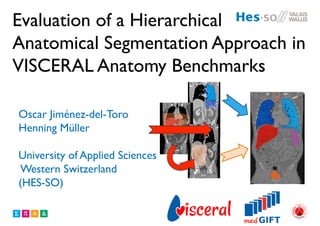
- 1. Evaluation of a Hierarchical Anatomical Segmentation Approach in VISCERAL Anatomy Benchmarks Oscar Jiménez-del-Toro Henning Müller University of Applied Sciences Western Switzerland (HES-SO)
- 2. 2 Overview • Motivation • VISCERAL • Method • Experimental Setup • Results Anatomy 1 Benchmark • Discussion • Conclusions
- 3. 3 Overview • Motivation • VISCERAL • Method • Experimental Setup • Results Anatomy 1 Benchmark • Discussion • Conclusions
- 4. • Anatomical segmentation is fundamental for further image analysis and Computer-Aided Diagnosis1 • Manual annotation and visual inspection is time consuming for radiologists • Accurate large scale data analysis techniques are needed 4 Motivation
- 5. 5 Overview • Motivation • VISCERAL • Method • Experimental Setup • Results Anatomy 1 Benchmark • Discussion • Conclusions
- 6. 6 Overview • Motivation • VISCERAL • Method • Experimental Setup • Results Anatomy 1 Benchmark • Discussion • Conclusions
- 7. VISual Concept Extraction challenge in RAdioLogy • EU funded project (2012-2015) – HES-SO, ETHZ, UHD, MUW, TUW, Gencat • Organize competitions on medical image analysis on big data o Anatomy benchmarks o Detection benchmark o Retrieval benchmark
- 8. VISCERAL Anatomy Benchmarks • All computations done in the cloud • Annotation by medical doctors • Automatic segmentation of anatomical structures (20) and landmark detection • CT and MR images (contrast-enhanced and non-enhanced)
- 9. 9 Overview • Motivation • VISCERAL • Method • Experimental Setup • Results Anatomy 1 Benchmark • Discussion • Conclusions
- 10. 10 Overview • Motivation • VISCERAL • Method • Experimental Setup • Results Anatomy 1 Benchmark • Discussion • Conclusions
- 11. Hierarchic Multi Atlas-Based Segmentation2 • Use multiple atlases for label estimation • Global and local alignment • Hierarchical selection of the registrations • Reuse registrations from the bigger structures (eg. liver) for the smaller ones • Label fusion
- 12. Image registration • Atlas = Patient volume + labels • Coordinate transformation increases spatial correlation between images3 • Multi-scale gaussian pyramid4
- 13. Affine alignment • Global rigid align • Independent local refinement for bigger structures (eg. liver, lungs) • Regions of interest based on the morphologically dilated initial estimations
- 14. Non-rigid alignment • Non-rigid b-spline • Multi-scale approach • Faster optimization due to better initial alignment
- 15. Label fusion • Majority voting threshold • Classification on a per-voxel basis • Local registration errors are reduced • Threshold optimization
- 16. Hierarchical Registration Affine Liver Right Kidney Urinary Bladder Global alignment Right Lung Left Lung 1st Lumbar Vertebra Gall-bladder Left Trachea Kidney Spleen Local Affine 2nd Local Affine B-spline non-rigid Label fusion
- 17. 17 Overview • Motivation • VISCERAL • Method • Experimental Setup • Results Anatomy 1 Benchmark • Discussion • Conclusions
- 18. 18 Overview • Motivation • VISCERAL • Method • Experimental Setup • Results Anatomy 1 Benchmark • Discussion • Conclusions
- 19. Experimental setup • VISCERAL Anatomy 1 • Testset :12 contrast-enhanced CT of the trunk • Applied to 10 anatomical structures • VISCERAL Anatomy 2 • Testset :10 contrast-enhanced CT of the trunk • 10 unenhanced whole body CT • Applied to ALL anatomical structures
- 20. 20 Overview • Motivation • VISCERAL • Method • Experimental Setup • Results Anatomy 1 Benchmark • Discussion • Conclusions
- 21. • Motivation • VISCERAL • Method • Experimental Setup • Results Anatomy 1 Benchmark • Discussion • Conclusions 21 Overview
- 22. Anatomy 1 Results DICE coefficient
- 23. Anatomy 1 Results DICE coefficient Top rank in benchmark
- 24. Anatomy 1 Results Average distance error
- 25. Anatomy 1 Results Average distance error Top rank in benchmark
- 26. Discussion Comparison with other participant methods: - SJ: Spanier et al. Rule-based approach with region growing for multiple organs - HJ: Huang et al. Multiple prior knowledge models and free-form deformation - W: Wang et al. Fast model bases level set method and hierarchical shape priors - K: Kazmig et al. Clustering and graph cut using shortest path constraint for spatial relations - GG: Gass et al. Multiple atlases via Markov random field registrations (DICE)
- 27. Discussion Comparison with other participant methods: - SJ: Spanier et al. Rule-based approach with region growing for multiple organs - HJ: Huang et al. Multiple prior knowledge models and free-form deformation - W: Wang et al. Fast model bases level set method and hierarchical shape priors - K: Kazmig et al. Clustering and graph cut using shortest path constraint for spatial relations - GG: Gass et al. Multiple atlases via Markov random field registrations (DICE)
- 28. Discussion • Competitive results compared with up to 5 segmentation methods in Anatomy1 • Similar to state-of-the-art methods for some organs: liver (0.89-0.96)5,6, kidneys (0.92-0.98)7,8 • Segments not only abdominal organs but can be implemented for any anatomical structure • Future work: Extend to method to other modalities
- 29. Conclusion • Straightforward automatic multi-structure segmentation method • Showed robustness in multiple structures particularly for ceCT • High overlap for the bigger structures (e.g. liver, lungs) and competitive overlap for smaller structures (e.g. gallbladder)
- 30. Thank you for your attention !!
- 31. References 1 K.Doi. Current status and future potential of computer-aided diagnosis in medical imaging. British Journal of Radiology, 78:3-19, 2005 2Jiménez del Toro et al., Multi-structure Atlas-Based Segmentation using Anatomical Regions of Interest. Proceedings of Medical Image Computing and Computer Assisted Intervention (MICCAI2013) MCV workshop, Nagoya, Japan, 2013 3Stefan Klein et al. Elastix: a toolbox for intensity-based medical image registration. IEEE Transactions on medical imaging, 29(1):196-205, 2010 4 Stefan Klein et al. Adaptive stochastic gradient descent optimisation for image registration. International Journal of Computer Vision, 81(3): 227-239, 2009
- 32. References 5Criminisi et al. Regression forests for efficient anatomy detection and localization in computed tomography scans. Medical Image Analysis, 17(8):1293-1303, 2013 6Okada et al. Abdominal multi-organ segmentation of CT images based on hierarchical spatial modeling of organ interrelations. Abdominal Imaging 2011, 7029:173-180, 2012 7Zhou et al. Automatic localization of solid organs on 3D CT images by a collaborative majority voting decision based on ensemble learning. Computerized Medical Imaging and Graphics, 36:304-313, 2012 8Wolz et al. Multi-organ abdominal CT segmentation using hierarchically weighted subject-specific atlases. Proceedings of Medical Image Computing and Computer Assisted Intervention (MICCAI2012), 7510:10-17, 2012