

Recommandé
Recommandé
Contenu connexe
Tendances
Tendances (20)
En vedette
En vedette (20)
Similaire à Stroke hyperacute treatment
Similaire à Stroke hyperacute treatment (20)
Plus de PS Deb
Plus de PS Deb (20)
Dernier
Dernier (20)
Stroke hyperacute treatment
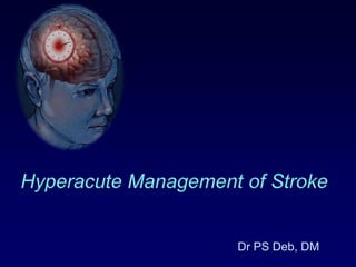
- 1. Hyperacute Management of Stroke Dr PS Deb, DM
- 2. Normal flow, normal function 50 Low flow, raised O2 extraction, normal function CBF (ml/100g brain) 20 Synaptic transmission failure 10 Membrane pump failure 0 3 4 5 1 2 Time in hours
- 3. Ischaemia - 02 ê glucose ê é lactate Anoxic depolarisation Ca2+ ié Hi é Free Fe2+ Glutamate Lipolysis NO synthase Proteolysis NA, Dopamine Free radicals Ischaemic Brain Injury
- 4. Aim of Rx: Ischemic Stroke Reperfusion Thrombolysis Mechanical disruption of clot Reducing the size of infarct Antiplatelate Anticoagulant Neuro-protection Blood pressure control Treat associated complication Raised ICT Seizure
- 5. Thrombolysis in acute stroke Within 3-4.5 hour of Stroke No occlusion Stop Medium Vessel Large Vessel IV rTPA/URK IA rTPA/URK C-D/ D-P mismatch
- 6. Challenge of thrombolysis Lack of awareness: Patient Physician Out of scope Operational issue Emergency team Imaging team Failure of medical thrombolysis Re-closure of opened vessel: (Stenting) Reperfusion injury with toxic edema and hemorrhage
- 7. Overcoming challange Education Patient Physician Alternative approach in failed cases Mechanical disruption of clot: Primary angioplasty Transcranialdoppler and thrombolysis Surgical emergency endarterectomy Stenting to prevent reclosure
- 8. Blood pressure control Systolic BP < 220 or diastolic < 120 Observe unless other end-organ involvement Systolic BP > 220 or diastolic >120 Labetalol 10-20 mg IV over 1-2 mins. May repeat or double every 10 mins. (300 mg/d) Diastolic BP > 140 Nitroprusside 0.5mcg/kg/min IV infusion *Aim for 10-15% reduction of BP Systolic BP > 185 or diastolic > 110 For thrombolysis Labetalol 10-20 mg IV over 1-2 mins. May repeat x 1 60 160 Mean systemic BP
- 9. Aspirin in Acute Stroke Aspirin 325mg Clopidogrel 75-150mg if Aspirin CI Early recurrent ischemic stroke – 7 fewer per 1,000 treated (p< 0.0001) Death from any cause – 4 fewer per 1,000 treated (p=0.05) Death or early recurrence of non-fatal stroke – 9 fewer per 1,000 treated (p=0.001) Death or dependency at discharge or six months – 13 fewer per 1,000 treated (p=0.007) Hazard : Hemorrhagic stroke or transformation – 2 more per 1,000 in ASA treated (p=0.06) Bath, 2001b [A]; Chinese Acute Stroke Trial Collaborative Group, 1997 [A]; International Stroke Trial Collaborative Group, 1997 [A]; Sandercock, 1993 [M]).
- 10. Anticoagulant in Acute Stroke Early use (<3Hr) may reduce mortality and morbidity. Hemorrhagic transformation is high Arterial dissection Cardioembolic infarct with or without AF Immediate for small infarct if re-embolization risk is high Delayed 2 weeks for all cases Heparin - 1000 units/hr. PTT 1.5 avoid bolus Enaoxaparin – 40mg SC BD Low dose Enoxaparin 40mg SC OD prophylactic for DVT
- 11. Other measures Statins Use after 2 days of onset (AHA) Continue in those already taking (NHS) Neuroprotection Citicolin Magnesium
- 14. The ICH volume on the 1-hour CT was 27 mL (bottom row). However, the 17-mL increase in ICH volume was not accompanied by any change in the 1-hour GCS (14) and NIH Stroke Scale (20) scores.Brott, T. et al. Sroke1997;28:1-5 Copyright ©1997 American Heart Association Early hemorrhage growth in patients with intracerebral hemorrhage.
- 15. Hematoma volume 30d outcome (Joseph P. Broderick et al Stroke 1993;24:987-993)
- 16. Approach to Treatment of ICH Stopping or slowing the initial bleeding Factor VIIa Blood pressure control Reducing raised ICT due to Hematoma and Edema Evacuation of hematoma Osmotherapy Neuroprotection Control of Seizure and other associated complication
- 17. A. Factor VIIa Trial Useful in Hemophilia with Ab to factor VII or IX Produce clotting by stimulating coagulation cascade in normal Used in cased of ICH secondary to coagulopathy There is mild reduction in progression of hematoma, morbidity and mortality (Phase II, III trial) INR may return to normal transiently it require repeated dose Routine use in Primary ICH remains investigational
- 18. B. Blood Pressure control Hypertension is common during early states of ICH -> Expansion, Peri-hematoma edema and re-bleeding A systolic BP above 140 to 150 mm Hg within 12 hours of ICH is associated with more than double the risk of subsequent death or dependency. Association of low BP and deterioration is not consistent like ischemic stroke. Blood pressure Antihypertensive Treatment in Acute Cerebral Hemorrhage (ATACH- I) INTensive Blood Pressure Reduction in Acute Cerebral Hemorrhage Trial (INTERACT- I)
- 19. Recommendation: AHA 2010 In patients presenting with a systolic BP of 150 to 220 mm Hg, acute lowering of systolic BP to 140 mm Hg is probably safe Class IIa; Level of Evidence: B
- 20. Raised ICT The Monro-Kellie doctrine
- 21. Problem secondary to raised ICT Early hematoma expansion and secondary edema-induced brain compression and consequent neuronal death; Cytotoxic (intracellular) and vasogenic (extracellular) edema resulting from disruption of the blood–brain barrier; Reductions in cerebral perfusion pressure (CPP) from mass effect and raised intracranial pressure (ICP); Brain herniation
- 22. Approach to raised ICT Head end elevation Surgical evacuation of hematoma Osmotherapy Hyperventilation Analgesia and sedation Neuromuscular block Barbiturate coma Hypothermia Corticosteroid CSF drainage Defroxamin
- 23. Head-of-Bed Elevation Elevation of the head of the bed to 30° improves jugular venous outflow and lowers ICP. The head should be midline, and head turning to either side should be avoided. In patients who are hypovolemic, elevation of the head of the bed may be associated with a fall in blood pressure and an overall fall in CPP.
- 24. Principals of Osmo-therapy Brain volume falls as long as there is an osmotic gradient between blood and brain. Short lived action few hours. Normal brain shrink (White/ Gray) Rebound edema. Dose not clear: 10 mOsm/L change in osmolality may be effective Chronic use not recommended as brain adapt
- 25. Mannitol Wise and Chater in 1962 Cleared from brain and CSF due to large molecular wt (182Dalton), less rebound phenomenon. Rebound phenomenon with increasing dose to sustain hyperosmolality of brain, as serum osmolality falls Dose: 1g/kg increases serum osmolality 20-30 mOsm/L for 3-4 hours. Higher dose 1.5-2gm/kg lower CSF pressure for longer time with increased rebound phenomenon. Lower dose 0.5gm/kg has less rebound
- 26. Mannitol Use in Acute Stroke Case Fatality at 30 Days and 1 Year In a tricenter, prospective study, 809 patient with ICH within 72 hours were analyzed 30-day and 1-year case fatality with respect to mannitol treatment in 2/3rd cases No recommendations can be made on the use of mannitol in acute stroke. Stroke. 2003;34:1730-1735.
- 27. Mannitol in ICH Randomized, controlled, double-blind study. Mannitol 20% 100ml q6h within 6 days of ictus for 5 days, tapered in next 2 days Mannitol did not seem to be beneficial in patients with ICH J Neurol Sci. 2005 Jul 15;234(1-2):41-5.
- 28. Effect of single mannitol bolus in intracerebral hemorrhage CT scan >3cm midline shift in ICH Randomized with bolus dose of Mannitol /Saline. Superior sagittal sinus to pontomesencephalic junction (SSS-PMJ) distance and edema hematoma complex were measured. Mannitol led to transient clinical improvement in five patients without significant reduction in superior sagittal sinus to pontomesencephalic junction (SSS-PMJ) distance at 30 and 60 min. U. K. Misra et al Department of Neurology, Sanjay Ghandi PGIMS, Lucknow, IndiaEur J Neurol. 2007 Oct;14(10):1118-23. Epub 2007 Aug 28.
- 29. Effect of mannitol on early enlargement of hematoma following hypertensive cerebral hemorrhage Hypertensive cerebral hemorrhage cases were randomized Group A 36 – Mannitol Group B 35 – furosemide Two follow-up CT were done Result: Enlargement of hematoma Group A : 33 3% patients(12 cases) Group B: 17 1% patients(6 cases) The inapt use of mannitol may be one reason of the early enlargement of hematoma following hypertensive cerebral hemorrhage Wang Minzhong,PangZaiying,FengYabo,et al
- 30. Hypertonic Saline Used in head injury and when Mannitol or Hyperventilation failed No trial on stroke patient Abrupt change in serum osmolality may leads Coma, Seizure and Subdural hematoma Pontine myelinolysis Volume expansion Cardiac failure Altered coagulation parameters -> bleeding Prolongation of PT, TT Decreased platelet aggregation Rebound phenomenon
- 31. Recommendation: Raised ICT Conservative An elevation of the head of the bed Analgesia and sedation Aggressive therapies Osmotic diuretics (mannitol and hypertonic saline solution), Drainage of CSF via ventricular catheter, Neuromuscular blockade, Hyperventilation, generally require concomitant monitoring of ICP and blood pressure with a goal to maintain CPP >70 mm Hg Class IIa, Level of Evidence B
- 32. Seizure and Antiepileptic drugs Incidence at onset -> 2 weeks: 3-17% Electrographic seizure 28-31% (on AET) One study prophylactic AE reduce clinical Sz. after Lobar hematoma Prospective population based studies did not show clinical Sz and worsened neurological outcome or mortality. Subclinical Sz.: one study showed prophylactic PHY increased disability and death at 90 days.
- 33. Recommendation: Seizure control Clinical seizures should be treated with antiepileptic drugs Class I; Level of Evidence: A Continuous EEG monitoring in patients with depressed mental status out of proportion to the degree of brain injury Class IIa; Level of Evidence: B Patients with a change in mental status who are found to have electrographic seizures on EEG should be treated with antiepileptic drugs Class I; Level of Evidence: C Prophylactic anticonvulsant medication should not be used Class III; Level of Evidence:B
- 34. Summary - ICH Rx Blood pressure control remains the mainstay of management in hemorrhagic stroke Osmotherapy has doubtful role and routine use is not indicated in minor bleed. Clinical and subclinical seizure with EEG abnormality should be treated. Surgical evacuation of hematoma and Supportive and critical care of patient during acute state is most important to reduce morbidity and mortality
Notes de l'éditeur
- Treatment of stroke has not really changed over years, however there is lots of variation in management of stroke. What I am going to do in next 15min is to tell you the physiologic basis of hyperacute stroke treatment and evidence to support them.
- Brain is about 2.3% of body weight, consumes one fifth of cardiac output and oxygen. Blood flow of the brain tissue is very high, about 50ml per 100gm. There is reflex increase in oxygen extraction with falling blood flow till it reaches critical level of 20ml when there is further fall to stop synaptic transmission to protect neuronal cell death. Any fall below 10ml over time leads to progressive death of nerve cell. Reopening of blocked circulation before there is substantial damage improve chance of recovery, however reperfusion after death of neuronal cell and blood vessel leads to severe reperfusion hemorrhage into the ischemic tissue.
- Part of brain surrounding the core of ischemic insult is call penumbra zone which has synaptic transmission failure with progressive cell death depends on the collateral circulation. There is cascade of biochemical event proceeds during ischemic insult resulting in accumulation of toxic substance like glutamate, free radical and lactate leading to entry of calcium into the cell which itself is suicidal to mitochondrial function. Once there is failure of sodium potassium pump, water enter into the cell causing rupture.
- Opening the thrombosed vessel ThrombolysisMechanical disruption of clotReducing the size of infarctAntiplatelateAnticoagulantNeuro-protectionBlood pressure controlTreat associated complicationRaised ICTSeizure
- CPatientWithin 3 hours of onsetNormal CT scan, MRI Diffusion/ perfusion or clinical mismatchBP <180/100 mmHgNo bleeding tendencyDrug/Dose 0.9mg /Kg. (max 90mg), 10% bolus, Rest 60 min. infusionRisk ICH in 6% of patientsPromise Reduced morbidity by 30%
- . ineligible for thrombolytic drug therapy or. who have failed to improve clinically or recanalise following intravenous thrombolysis.Mechanical clot disruption (including clot maceration by guidewire, clot snaring and balloon angioplasty) may achieve recanalisation in patients with persistent MCA or ICA occlusion after standard IV or IA rt-PA.123,124 Immediate recanalisation was achieved in 38% of patients and final recanalisation in 75% compared to rates of 6% and 72% for simple clot penetration by microcatheter.123 Bleeding risks did not appear to be increased.5.7.2 Transcranialdoppler and thrombolysis The use of continuous pulsed wave TCD ultrasound at 2 MHz in patients undergoing IV rt-PA thrombolysis for middle cerebral artery occlusions (diagnosed by ultrasound) within three hours of symptom onset is associated with higher rates of early recanalisation and a trend towards more favourable clinical outcomes compared to thrombolysis alone. Forty nine per cent of patients had complete recanalisation or dramatic recovery at two hours compared to 30% ofpatients receiving rt-PA alone (relative risk reduction, RRR=1.6; 95% CI 1.03 to 2.6).125 At three months 42% of patients receiving ultrasound had better functional outcome compared to 29% of the control group (p=0.2).125 Similar findings were reported for transcranialcolour coded sonography (TCCS) at identical frequency.126;; Augmentation of IV thrombolysis by continuous 2 MHz pulsed wave TCD ultrasound should be considered in the context of further clinical trials. Using lower frequencies of ultrasound causes less heating and gives better penetration, but there is an excess risk of inrtacerebral haemorrhage.127
- Treat Hypertension If Blood Pressure Greater than 185 Systolic or 110 DiastolicPatients with a systolic blood pressure (BP) greater than 185 mmHg or diastolic blood pressure greater than110 mmHg are excluded from this annotation only if the blood pressure remains elevated on consecutivemeasurements (Adams, 2007 [R]), and if aggressive treatment is required to lower the blood pressure intoan appropriate range (e.g., if more than a few doses of any medication is required or if nitroprusside dripis required).Guidelines for blood pressure management in this setting have been slowly evolving (Adams, 2007 [R];International Society of Hypertension Group, 2003 [R]; Powers, 1993 [R]; Stead, 2004 [M]; Strandgaard,1996 [R]).A full understanding of this issue requires understanding of the physiology. Cerebral blood flow (CBF)is regulated by the relationship between cerebral perfusion pressure (CPP) and cerebrovascular resistance(CVR) (CBF=CPP/CVR). CPP represents the difference between arterial blood pressure forcing the bloodinto the cerebral circulation and the venous back pressure. Under normal circumstances, the venous backpressure is negligible and CPP is equal to arterial blood pressure. Normally, changes in blood pressure (orCPP) over a wide range have little effect on CBF. This phenomenon, termed autoregulation, is mediatedvia changes in the CVR. An increase in CPP (or arterial blood pressure) produces vasoconstriction and adecrease produces vasodilatation. This autoregulation keeps the cerebral blood flow at a steady level overa range of 60-150 mmHg mean arterial pressure. In individuals with chronic hypertension, the range forautoregulation is shifted upwards so that they may be more tolerant of higher blood pressure and less tolerantof lower blood pressure (decreased cerebral blood flow).Acute ischemic stroke will cause a change in autoregulation in the ischemic zone by two mechanisms:First, when an artery is occluded, a central core of severe ischemia is produced. This is surrounded by azone with less reduction in blood flow termed the penumbra where perfusion is maintained by collateralcirculation. The blood vessels in the penumbra are maximally dilated, and for that reason blood flowthrough them may be completely dependent on blood pressure.Second, during the acute period, the phenomenon of autoregulation even outside of the penumbra canbe impaired in patients both with and without persistent arterial occlusion, changing the autoregulationcurve so that maintenance of blood flow is completely dependent on the blood pressure.These abnormalities in autoregulation may persist for days or weeks. There is evidence to suggest thatthere is slow improvement in disordered autoregulation in the acute period. But early on, lowering theblood pressure may reduce blood flow to critical levels in the ischemic region, potentially extendingthe area of infarct. This is supported by data from both animal and human studies (Christensen, 2002[D]; Powers, 1993 [R]).Although the potential dangers of lowering arterial blood pressure in patients with acute ischemic stroke areaccepted theory influencing practice, documentation of actual risk is based on a few published case reports(Britton, 1980 [D]; Grossman, 1996 [R]; Lavin, 1986 [D]). The theoretical adverse effects of overtreatmentare substantial. Whether carefully controlled treatment of hypertension in acute stroke might be beneficialhas not been adequately studied.A Cochrane review (2003) consisting of 34 randomized controlled trials and 5,368 patients examined theeffect of various drugs on blood pressure (BP) during the first 72 hours of acute ischemic stroke (AIS).Drugs shown to actually reduce BP included oral and IV calcium channel blockers, oral beta-blockers,glyceryltrinitrate, ACE inhibitors, prostacyclin (PGI2), and streptokinase. The effect of blood pressurereduction was not clear, likely due to the significant imbalances in baseline blood pressure between treatmentand control groups. Outcomes examined included early death and overall case fatality. The reviewconcluded that there is insufficient evidence to evaluate the effect of altering blood pressure on outcomeafter acute ischemic stroke. Another systematic review demonstrated increased mortality, early deterioration,and dependency associated with higher blood pressure in the acute stroke setting (Willmot, 2003 [M]).A recent placebo controlled trial in patients with stroke due to ischemia or hemorrhage assessed safety andoutcome efficacy of early BP reduction. Subjects were not on BP medications before the trial and had earlypost-stroke systolic blood pressure > 160 mmHg. Goal was reduction to 145-155 mmHg or by 15%. Thetrial was stopped when its funding ran out. It was underpowered to detect efficacy but suggested that mildBP reduction within 36 hours with labetolol or lisinopril was safe (Potter, 2009 [A]).The above review includes the IST trial (Leonardi-Bee, 2002 [A]), which demonstrated a U-shaped curvewhen BP was plotted against survival (i.e., increased mortality at lowest and highest pressure with lowestmortality at systolic pressure around 150 mmHg). Other investigators have reported a similar finding i.e., aU-shaped relationship with adverse outcomes in patient groups not treated with thrombolytic agents (Castillo,2004 [D]) and treated with tPA (Ahmed, 2009 [B]), providing grounds for the current consensus-basedguidelines to treat BP if it exceeds arbitrarily derived thresholds established according to thrombolysis status(see Table 3, "Approach to Elevated Blood Pressure in Acute Ischemic Stroke") (Adams, 2007 [R]). To beanticipated is more research to gather evidence about what the thresholds should actually be.Taking the above studies into consideration, the AHA issued a revised 2007 edition of "Guidelines for theEarly Management of Patients with Acute Ischemic Stroke" (Adams, 2007 [R]). In the absence of unambiguousdata, these consensus-based guidelines recommend the following measures for treatment of BP inpatients with acute ischemic stroke (AIS).Table 3.Approach to Elevated Blood Pressure in Acute Ischemic StrokeA. Not eligible for thrombolytic therapy Blood Pressure Level mm/Hg TreatmentSystolic BP < 220 or diastolic < 120 Observe unless other end-organ involvement, e.g., aortic dissection, acute myocardial infarction, pulmonaryedema, hypertensive encephalopathy. Treat other symptoms of stroke such as headache, pain, agitation, nausea and vomiting Treat other acute complications of stroke includinghypoxia, increased intracranial pressure, seizures or hypoglycemia.Systolic BP > 220 or diastolic BP >120 - Labetalol 10-20 mg IV over 1-2 mins. May repeat or double every 10 mins. (max. dose 300 mg in 24 hours)or- Nicardipine 5 mg/hr IV infusion as initial dose; titrate to desired effect by increasing 2.5 mg/hr every 5 mins. To maximum of 15 mg/hr.*Aim for 15% reduction of BPDiastolic BP > 140 Nitroprusside 0.5mcg/kg/min IV infusion as initial dose with continuous BP monitoring (max dose of 10 mcg/kg/min)*Aim for 10-15% reduction of BPB. Eligible for Thrombolytic Therapy Pretreatment Blood Pressure Level mmHgSystolic BP > 185 or diastolic BP > 110 - Labetalol 10-20 mg IV over 1-2 mins. May repeat x 1;or- Nitroglycerin ointment USP 2% 1-2 inches; or- Nicardipine infusion @ 5 mg/hr, titrate up by 2.5 mg/hrat 5-15 min intervals; max dose 15 mg/hr; when desiredBP attained, reduce to 3 mg/hr* If BP does not decline and remains > 185/100DO NOT administer tPADuring and After Treatment with rtPAMonitor BP Monitor BP every 15 minutes during treatment; followingtreatment, check BP every 15 mins for 2 hours, then every30 mins for 6 hrs, then every hour for 16 hours.Blood Pressure Level mmHgBP 180- 230/105-120 mmHg - Labetalol 10-20 mg IV over 1-2 mins, may repeat every10-20 mins (max dose 300 mg in 24 hours); or- Labetalol 10 mg IV followed by an infusion at2-8 mg/min (max dose 300 mg in 24 hours)BP > 230/121-140 mmHg - Labetalol 10-20 mg IV over 1-2 mins, may repeat every10-20 mins (max dose 300 mg in 24 hours); or- Labetalol 10 mg IV followed by an infusion at2-8 mg/min (max dose 300 mg in 24 hours); or- Nicardipine infusion 5 mg/hr, titrate to desired effect,may increase 2.5 mg/hr q 5- 15 mins; max dose of15 mg/hr.- If BP not controlled, consider nitroprusside infusion0.5 mcg/kg/min (max dose of 10 mcg/kg/min)Treat Blood Pressure If Greater than 220/120 mmHg or Mean Arterial Pressure Greaterthan 130 mmHgRecommendations – ischemic stroke, not a tPA candidate:• Admit to the intensive care unit or acute stroke care unit and perform cardiac monitoring.• Perform vital signs with neuro checks (not National Institutes of Health Stroke Scale).• Treat BP only if systolic blood pressure (SBP) is greater than 220 mmHg, diastolic blood pressure(DBP) is greater than 120 mmHg, and/or mean arterial pressure (MAP) is greater than 130 mmHg.• Use easily titrated agents, choosing those with the least effect on cerebrovasculature (labetolol,nitroglycerin ointment USP 2% or nicardipine). American Heart Association (AHA) recommendationssupport oral dosing if the patient has passed a bedside swallow test. If not, intravenous agentsshould be used.Dosing examples:labetalol oral 100-200 mg by mouth initially and every two hours as needed, upto 800 mg total in 24 hoursorlabetalol IV 10-20 mg IV over 1-2 min., repeat or double dose every 10-20 min.nitroglycerin ointment 1 to 2 inchesUSP 2%nicardipine 5 mg/hr IV infusion, titrate for BP control, increasing 2.5 mg/hrevery 5 min. to maximum of 15 mg/hrnitroprusside 0.5 mcg/kg/min IV; titrate for BP control as needed up to10 mcg/kg/min• Avoid agents that tend to cause precipitous drops in BP (e.g., sublingual calcium channel blockers).• Treat hypotension (IV fluids; treat congestive heart failure or arrhythmia and consider pressors).• Monitor BP and any corresponding neurological changes in the emergency department and first fewdays of hospitalization. Avoid overtreating BP.In patients with markedly increased blood pressure on presentation with acute stroke, measured reduction(e.g., 15% reduction targeted for the first 24 hours) is reasonable. The threshold for initiating such treatmentremains 220 mmHg systolic and/or 120 mmHg diastolic. This is despite preliminary evidence thatinitiating treatment at a lower level may be safe and beneficial (CHHIPS, 2008 [NA]). In patients who areon an antihypertensive medication program at the time of the ischemic stroke, these medications shouldgenerally be withheld for the initial 24 hours. They should be reinstated after 24 hours, assuming that oral ortube administration is possible and hypotension is not present (Adams, 2007 [R]). Many potential reasonsfor deviating from this general principle exist. For example, suspension of a beta-blocker in a patient withcoronary heart disease may be dangerous, and discontinuation of clonidine may cause rebound hypertension.• Continue antithrombotic therapy
- AspirinPatients who are not candidates for tPA should be given aspirin promptly in a dose of 325 mg (Adams, 2007 [R]) orally, rectally or by nasogastric tube and should be continued on a similar daily dose (Albers, 2004 [R]). Exceptions to this approach would be justified in those with contraindications to aspirin therapy (e.g., aspirin allergy, gastrointestinal hemorrhage). For patients with an aspirin allergy, 75 mg of clopidogrel may be reasonable. Intravenous or oral loading with 150-600 mg of clopidogrel establishes antiplatelet effect more rapidly; however, efficacy in this setting is unproven.Initiation of aspirin therapy should be withheld for 24 hours for patients who have received tPA. Although the benefits of aspirin therapy for long-term preventive therapy for stroke are well established, the use of aspirin to improve outcome in the acute treatment setting has also been demonstrated. Large randomized controlled trials have identified a small but measurable benefit with use of aspirin in the first 48 hours following ischemic stroke onset (Bath, 2001b [A]; Chinese Acute Stroke Trial Collaborative Group,1997 [A]; International Stroke Trial Collaborative Group, 1997 [A]; Sandercock, 1993 [M]).The studies together demonstrate benefit of small magnitude, but with statistical significance in the following outcome measures:• Early recurrent ischemic stroke – 7 fewer per 1,000 treated (p< 0.0001)• Death from any cause – 4 fewer per 1,000 treated (p=0.05)• Death or early recurrence of non-fatal stroke – 9 fewer per 1,000 treated (p=0.001)• Death or dependency at discharge or six months – 13 fewer per 1,000 treated (p=0.007)Also, the measured hazard appears to be small and statistically insignificant:• Hemorrhagic stroke or transformation – 2 more per 1,000 in ASA treated (p=0.06)Antiplatelet agents : SIGN 20085.2.1 aspirinA systematic review of twelve RCTs of over 40,000 patients showed that aspirin at 160 or 300 mg daily reduced death and disability, recurrent stroke, and improved the likelihood of full recovery in patients with ischaemic stroke.80 Two trials of aspirin (160 or 300 mg) given within 48 hours of stroke onset contributed 94% of the data analysed in the review. One was an open label trial and did not require a CT scan before randomisation. Exclusion criteria for these RCTs were vague and in all of the included trials mortality was lower than in the placebo arm (4-9%), suggesting that major strokes were under-represented.A Aspirin 300 mg daily should be commenced within 48 hours of ischaemic stroke and continued for at least 14 days.5.2.2 aspirin plus clopidogrelA small RCT of 110 patients with carotid stenosis showed that dual therapy with aspirin and clopidogrel significantly reduced the proportion of patients with embolic signals on transcranial Doppler ultrasound. The study was not powered to examine clinical end points.82 The FASTER trial randomised 392 patients with TIA or minor stroke to aspirin or aspirin plus clopidogrel for 90 days, commencing within 24 hours of symptom onset. A non-significant 3.8% absolute risk reduction (ARR) in total stroke was found for the combination therapy.25 Dual therapy with aspirin and clopidogrel given to patients within 48 hours of TIA or minor stroke was one component of several interventions in a prospective study, including early statins, antihypertensives and carotid endarterectomy. The contribution of combined aspirin and clopidogrel to the reduced stroke risk is not clear but there was no increase in the risk of intracerebral or other haemorrhage in the study group overall.24 Insufficient evidence exists to support the routine use of the combination of aspirin and clopidogrel for treating patients in the acute phase of stroke.
- 5.3 a nticoagulants SIGN 2008The administration of anticoagulants is contraindicated during the first 24 hours after IV thrombolytic therapy.83 Although recanalisation may be more successful, the risk of haemorrhage increases. Evidence on adjunctive anticoagulation is limited and neither the safety nor efficacy has been established.835.3.1 unfractionated heparinFixed dose unfractionated heparin (UFH) commenced within 48 hours of ischaemic stroke of any aetiology (including patients with atrial fibrillation) does not reduce the chances of death or dependence after 3-6 months and is not significantly superior to aspirin alone.84 A reduction of recurrent strokes (nine fewer for every 1,000 patients treated) was offset by nine extra symptomatic intracerebral haemorrhages for every 1,000 patients treated, and nine additional major extracranialhaemorrhages for every 1,000 patients treated.High dose UFH given within three hours of stroke onset was associated with a significant reduction in death or dependence. Patients eligible for very early heparin treatment are also likely to be candidates for thrombolysis.855.3.2 L ow Molecular Weight heparins and heparinoidsA non-significant trend towards reduction in death and dependence at 3-6 months was seen for low molecular weight heparins (LMWH) and heparinoids when compared to control, which was a mixture of placebo, aspirin, and unfractionated heparin (OR=0.85; 95% CI 0.66 to 1.08).86 In patients with atrial fibrillation, weight-adjusted high dose LMWH (dalteparin 100 iu/kg) was not superior to aspirin in preventing early stroke recurrence and carried a significantly higher risk of extracerebral haemorrhage.87Symptomatic and asymptomatic venous thromboembolic events are highly significantly reduced by all heparin treatments. Deep vein thrombosis (DVT) is avoided in 281 patients for every 1,000 treated and pulmonary thromboembolisms (PTE) are avoided in four patients per 1,000 treated.84 LMWH and heparinoids are associated with a significantly greater reduction in DVT than UFH without any additional hazard.86 Enoxaparin 40 mg once daily is superior to UFH 5,000 units twice daily and is not associated with any increase in bleeding complications or difference in mortality.885.3.3 heparin plus aspirinCombined treatment with aspirin and UFH reduced recurrent stroke by 10 fewer per 1,000 patients treated, although a significant increase in the number of symptomatic intracranial haemorrhages was also reported (10 more per 1,000 patients). For patients in atrial fibrillation (AF) with acute ischaemic stroke aspirin reduces the risk of further stroke by 21% giving a number needed to treat (NNT) to prevent one stroke of 100 over 2-4 weeks.89 Any benefit of UFH in this situation is offset by the increase in haemorrhagic stroke.895.3.4 WarfarinIn one trial 100 patients received warfarin within two weeks of stroke onset and none had haemorrhagic worsening.89 Large, disabling strokes were under-represented. An arbitrary time limit of two weeks is recommended for delaying warfarin treatment for AF following acute stroke.89The routine use of anticoagulants (UFH, LMWH, heparinoids, oral anticoagulants, direct thrombin inhibitors, fibrinogen-depleting agents) is not recommended for the treatmentof acute ischaemic stroke.. Anticoagulants are not recommended in patients with progressing stroke.A In patients at high risk of venous thromboembolic disease LMWH should be considered in preference to UFH.D Following administration of IV thrombolysis, heparin should not be given in any formfor 24 hours.C For patients in atrial fibrillation following stroke, anticoagulation with warfarin can be introduced early in patients with minor stroke or TIA, but should be deferred for two weeks after onset in those with major stroke.heparin or heparinoids. There is, as yet, insufficient evidence to decide whether specific subgroups of ischemic stroke (e.g., dissection, cardio-embolism with intra-cardiac clot) will benefit from therapeutic anticoagulation.• If a decision is made to use continuous heparin infusion, boluses should be avoided, and aPTT should be maintained in the 1.5-2 times baseline range.• Low-dose prophylactic parenteral anticoagulation (e.g., enoxaparin, 40 mg subcutaneously daily) is beneficial for prevention of deep vein thrombosis (DVT) or PE (pulmonaryembolism) in stroke patients with limited mobility.Considerations with Heparin UseIn contrast, to the proof of efficacy for aspirin, results from the International Stroke Trial provide powerful evidence against the routine use of any heparin regimen as intensive as the moderate-dose subcutaneous regimen utilized in this very large clinical trial (unfractionated heparin – 12,500 units subcutaneous twice daily) (International Stroke Trial Collaborative Group, 1997 [A]).The commonly cited indications of vertebrobasilar distribution ischemia or ischemic stroke in the setting of atrial fibrillation were analyzed separately and there was no measurable benefit in these specific subgroups.Similarly, the weight of available data regarding use of full-dose low-molecular-weight heparin for the acute treatment of stroke does not support their routine use for limiting disability or decreasing mortality in this setting (Publications Committee for the Trial of ORG 10172 in Acute Ischemic Stroke, 1998 [A]).In summary, the routine use of acute anticoagulation treatment with unfractionated heparin, low-molecularweight heparin, or heparinoid in acute ischemic stroke is not supported by the available evidence (International Stroke Trial Collaborative Group, 1997 [A]). This treatment does not appear to improve clinical outcome from the index stroke. There may be subgroups that benefit, but further studies of this problem are required for confirmation.Despite these discouraging results, the use of continuous heparin infusion in acute stroke has continued to be common in clinical practice (Albers, 2004 [R]; Berge, 2000 [A]; Coull, 2002 [M]; Diener, 2001 [A]).Given these data, if the decision is made to use full-dose continuous heparin infusion for a specific indication (e.g., large vessel atherothrombosis or dissection), physicians are strongly encouraged to discuss with their patients the lack of proof for this therapy and to detail the potential hazards.Heparin use for venous thromboembolism (VTE) prophylaxisFor patients at high risk for VTE where pharmacologic prophylaxis is contraindicated, intermittent pneumatic compression (IPC) should be used if the patient is confined to bed. Thigh-length graduated compression elastic stockings have been shown not to be effective (CLOTS Trials Collaboration, 2009 [A]). See also Annotation #38, "Other Post-Emergency Department Medical Management (First 24-48 Hours)“ (Kamphnisen, 2005 [R]).See the ICSI Venous Thromboembolism Prophylaxis guideline for more information.
- 5.8 P riorstatin therapyEvidence from retrospective studies suggests that in addition to reducing the risk of recurrent stroke, statin pre-treatment may improve stroke outcome.A small single centre RCT randomised 89 patients with ischaemic stroke, on prior statin therapy, to either atorvastatin 20 mg (initiated within 24 hours of symptom onset) or withdrawal of statin therapy for three days.128 Withdrawing statins increased the risk of death and dependency at 90 days (OR=4.66; 95% CI 1.46 to 14.91), and increased the risk of a poor clinical outcome (mRS>2; 60% versus 39%; p=0.043).Patients with ischaemic stroke on prior statin therapy should continue ;; treatment, via a nasogastric tube, if necessary.5.4 N europrotectantsMany agents have been studied for a potential neuroprotective effect in patients with stroke.There are systematic reviews of glutamate antagonists,92 calcium antagonists,93, 94 the free radicalscavenger tirilazad,95 vinca alkaloids,96 oral citicoline,97 the sodium antagonist lubeluzole,98corticosteroids,99 methylxanthine derivatives (pentoxiffyline, propentofylline and pentofylline),100aminophylline,101 and piracetam102 and phase III RCTs of the free radical spin trap NXY 059,1035HT1a agonist repinotan,104 neutrophilchemotaxis inhibitors enlimomab105 and UK-279,276,106magnesium,107 nalmefene,108 diazepam,109 chlomethiazole,110 cerebrolysin,111 and the free radicalscavenger edaravone.112These agents have widely differing pharmacological actions. For the majority of agents, there wasno evidence of benefit or harm. The exceptions are:Definite harm (increase in odds of death or dependence). tirilazad (OR=1.23; 95% CI 1.01 to 1.50)95. enlimomab (adjusted p=0.004 for worse distribution of Rankin grade).105Possible harm. aptiganel hydrochloride (mortality OR=1.32; 95% CI 0.91 to 1.93)92. selfotel (mortality OR=1.19; 95% CI 0.81 to 1.74)92. corticosteroids (OR=1.08; 95% CI 0.68 to 1.72)99. calcium antagonists (OR 1.07 0.97 to 1.18)94. gavestinel (mortality OR=1.12; 95% CI 0.95 to 1.32).92Possible benefit (reduced odds of death or dependence). citicoline (good outcome OR=1.38; 95% CI 1.10 to1.72)97. non-cortical stroke syndromes treated with magnesium (poor outcome OR=0.75; 95% CI0.58 to 0.97).107For other agents, there was insufficient evidence to define benefit or harm.;; Neuroprotectant agents should be used only within the context of randomised controlledtrials.
- Volume of Intracerebral HemorrhageA Powerful and Easy-to-Use Predictor of 30-Day MortalityJoseph P. Broderick, MD; Thomas G. Brott, MD; John E. Duldner, MD;Thomas Tomsick, MD; Gertrude Huster, MHSBackground and Purpose: The aim of this study was to determine the 30-day mortality and morbidity of intracerebral hemorrhage in a large metropolitan population and to determine the most important predictors of 30-day outcome.Methods: We reviewed the medical records and computed tomographic films for all cases of spontaneous intracerebral hemorrhage in Greater Cincinnati during 1988. Independent predictors of 30-day mortality were determined using univariate and multivariate statistical analyses.Results: The 30-day mortality for the 188 cases of intracerebral hemorrhage was 44%, with half of deaths occurring within the first 2 days of onset. Volume of intracerebral hemorrhage was the strongest predictor of 30-day mortality for all locations of intracerebral hemorrhage. Using three categories of parenchymal hemorrhage volume (0 to 29 cm3, 30 to 60 cm3, and 61 cm3 or more), calculated by a quick and easy-to-use ellipsoid method, and two categories of the Glasgow Coma Scale (9 or more and 8 or less), 30-daymortality was predicted correctly with a sensitivity of 96% and a specificity of 98%. Patients with a parenchymal hemorrhage volume of 60 cm3 or more on their initial computed tomogram and a Glasgow Coma Scale score of 8 or less had a predicted 30-day mortality of 91%. Patients with a volume of less than 30 cm3 and a Glasgow Coma Scale score of 9 or more had a predicted 30-day mortality of 19%o. Only one of the 71 patients with a volume of parenchymal hemorrhage of 30 cm3 or more could functionindependently at 30 days.Conclusions: Volume of intracerebral hemorrhage, in combination with the initial Glasgow Coma Scale score, is a powerful and easy-to-use predictor of 30-day mortality and morbidity in patients with spontaneous intracerebral hemorrhage. (Stroke 1993;24:987-993)KEY WoRDs * intracerebral hemorrhage * survival * tomo
- Overall Approach to Treatment of ICHPotential treatments of ICH include stopping or slowing the initial bleeding during the first hours after onset; Removing blood from the parenchyma or ventricles to eliminate both mechanical and chemical factors that cause brain injury; Management of complications of blood in the brain, including increased ICP and decreased cerebral perfusion; and good general supportive management of patients with severe brain injury. Good clinical practice includes management of airways, oxygenation, circulation, glucose level, fever, and nutrition, as well as prophylaxis for deep vein thrombosis. However, because of the lack of definitive randomized trials of either medical or surgical therapies for ICH, until recently, there has been great variability in the treatment of ICH worldwide.2004 Stroke June 2007Downloaded from stroke.ahajournals.org by on December 7, 2010
- Trials of Recombinant Activated Factor VIIrFVIIa is approved to treat bleeding in patients with hemophilia who have antibodies to factor VIII or IX, and it has been reported to reduce bleeding in patients without coagulopathy as well.38 Interaction of rFVIIa and tissue factor stimulates thrombin generation. rFVIIa also activates factor X on the surface of activated platelets, which leads to an enhanced thrombin burst at the site of injury.38,39 Thrombin converts fibrinogen into fibrin, which produces a stable clot. rFVIIa has a half-life of 2.6 hours, and the recommended dose for treatment of bleeding in patients with hemophilia is 90 g/kg intravenously every 3 hours.38 Two small dose-ranging pilot safety studies and a largerdose-finding phase II study focusing on decreasing the growth of ICHs have been published.33,34,40 In the 2 small dose-ranging studies, the overall thromboembolic and seriousadverse event rate in the 88 patients tested at escalating doses from 5 to 160 g/kg was low enough to encourage further testing.The second larger, randomized, dose-escalation trial included 399 patients with ICH diagnosed by CT within 3 hours after onset who were randomized to receive placebo (96patients) or rFVIIa 40 g/kg body weight (108 patients), 80 g/kg (92 patients), or 160 g/kg (103 patients) within 1 hour after the baseline scan. The primary outcome measure was the percent change in the volume of the ICH at 24 hours. Clinical outcomes were assessed at 90 days. Hematoma volume increased more in the placebo group than in the rFVIIagroups. The mean increase was 29% in the placebo group, compared with 16%, 14%, and 11% in the groups given rFVIIa 40, 80, and 160 g/kg, respectively (P0.01 forcomparison of the 3 rFVIIa groups with the placebo group).Growth in the volume of ICH was reduced by 3.3, 4.5, and 5.8 mL, respectively, in the 3 treatment groups versus that in the placebo group (P0.01). Sixty-nine percent of placebotreatedpatients died or were severely disabled (as defined by a modified Rankin Scale score of 4 to 6), compared with 55%, 49%, and 54% of the patients who were given rFVIIa 40, 80, and 160 g/kg, respectively (P0.004 for comparison of the 3 rFVIIa groups with the placebo group). The rate of death at 90 days was 29% for patients who received placebo versus 18% in the 3 rFVIIa groups combined (P0.02). Serious thromboembolic adverse events, mainly myocardial or cerebral infarction, occurred in 7% of rFVIIa-treated patients versus 2% of those given placebo (P0.12). In this moderatesizedphase II trial, treatment with rFVIIa within 4 hours after the onset of ICH limited the growth of the hematoma, reduced the mortality rate, and improved functional outcome at 90 days despite a small increase in the frequency of thromboembolic adverse events. A larger phase III randomized trial of rFVIIa has been completed, and presentation of the results will occur in May 2007 at the American Academy of Neurology Meeting in Boston, Mass.In addition, several case reports of the use of rFVIIa in the setting of warfarin-associated ICH have been published.41,42Although rFVIIa can reverse the elevated INR measurements rapidly, its use in this setting remains investigational. In addition, a normal INR after use of rFVIIa does not implycomplete normalization of the clotting system, and the INR may rise again after the initial rFVIIa dose.43
- 210 guideBlood PressureBlood Pressure and Outcome in ICHBlood pressure (BP) is frequently, and often markedly, elevated in patients with acute ICH; these elevations in BP are greater than that seen in patients with ischemic stroke.72,73Although BP generally falls spontaneously within several days after ICH, high BP persists in a substantial proportion of patients.72,73 Potential pathophysiologic mechanisms include stress activation of the neuroendocrine system (sympathetic nervous system, renin-angiotensin axis, or glucocorticoid system) and increased intracranial pressure. Hypertension theoretically could contribute to hydrostatic expansion of the hematoma, peri-hematoma edema, and rebleeding, all of which may contribute to adverse outcomes in ICH, although a clear association between hypertension within the first few hours after ICH and the risk of hematoma expansion (or eventual hematoma volume) has not been clearly demonstrated.25,74A systematic review75 and a recent large multisite study in China73 show that a measurement of systolic BP above 140 to 150 mm Hg within 12 hours of ICH is associated with more than double the risk of subsequent death or dependency.Compared with ischemic stroke, where consistent U- or J-shaped associations between BP levels and poor outcome have been shown,76 only 1 study of ICH has shown a pooroutcome at very low systolic BP levels (140 mm Hg).77 For both ischemic stroke and possibly ICH, a likely explanation for such association is reverse causation, whereby very lowBP levels occur disproportionately in more severe cases, so that although low BP levels may be associated with a high case fatality, it may not in itself be causal.Broderick et al Guidelines for Management of Spontaneous ICH in Adults 2005Downloaded from stroke.ahajournals.org by on December 7, 2010Recent Pilot Trial of Acute Blood Pressure ManagementThe optimal level of a patient’s blood pressure should be based on individual factors such as chronic hypertension, ICP, age, presumed cause of hemorrhage, and interval sinceonset. Theoretically, elevated blood pressure may increase the risk of ongoing bleeding from ruptured small arteries and arterioles during the first hours. Blood pressure is correlated with increased ICP and volume of hemorrhage. However, it has been difficult to determine whether elevated blood pressure is a cause of hemorrhage growth or an effect of increasing volumes of ICH and increased ICP. A prospective observational study of growth in the volume of ICH did not demonstrate a relationship between baseline blood pressure and subsequent growth of ICH, but frequent early use of hypertensive agents in that study may have obscured any relationship.15 Conversely, overaggressive treatment of blood pressure may decrease cerebral perfusion pressure (CPP) and theoretically worsen brain injury, particularly in the setting of increased ICP.Powers and colleagues44 studied 14 patients with acute supratentorial ICH 1 to 45 mL in size at 6 to 22 hours after onset. Cerebral blood flow (CBF) was measured withpositron emission tomography and [15O]water. After completion of the first CBF measurement, patients were randomized to receive either nicardipine or labetalol to reduce meanarterial pressure by 15%, and the CBF study was repeated.Mean arterial pressure was lowered by 16.75.4% from 14310 to 11911 mm Hg. No significant change was observed in either global CBF or periclot CBF. The authorsconcluded that in patients with small- to medium-sized acute ICHs, autoregulation of CBF was preserved with arterial blood pressure reductions in the range studied.Blood Pressure ManagementThe previous AHA recommendation for the management of blood pressure after ICH outlined the important concept of selecting a target blood pressure on the basis of individualpatient factors,6 such as baseline blood pressure, presumed cause of hemorrhage, age, and elevated ICP. The primary rationale for lowering the blood pressure is to avoid hemorrhagic expansion from potential sites of bleeding. This is especially true for hemorrhage resulting from a ruptured aneurysm or arteriovenous malformation, in which the risk of continued bleeding or rebleeding is presumed to be highest.However, in primary ICH, in which a specific large-vessel vasculopathy is not apparent, the risk of hemorrhagic expansion with mild blood pressure elevation may be lower andmust be balanced with the theoretical risks of inducing cerebral ischemia in the edematous region that surrounds the hemorrhage. This theoretical risk has been somewhatmuted by prospective observational studies in both animals and human beings44,46 that have dispelled the concept of major ischemia in the edematous tissue surrounding thehemorrhage. Nevertheless, some controversy persists on the basis of human MRI–apparent diffusion coefficient studies of the perihemorrhagic region,47 which indicate arim of tissue at risk for secondary ischemia in large hematomas with elevated ICP.Nonetheless, for primary ICH, little prospective evidence exists to support a specific blood pressure threshold. The previous recommendation was to maintain a systolic bloodpressure 180 mm Hg and/or mean arterial pressure 130 mm Hg. The evidence to support any specific recommendation can be briefly summarized as follows: (1) Isolatedsystolic blood pressure 210 mm Hg is not clearly related to hemorrhagic expansion or to neurological worsening.48 (2)Reduction in mean arterial pressure by 15% (mean 14210 to 11911 mm Hg) does not result in CBF reduction in humans as measured by positron emission tomography.44 (3)In one prospective observational study,49 reduction of systolic blood pressure to a target 160/90 mm Hg was associated with neurological deterioration in 7% of patients and withhemorrhagic expansion in 9% but was associated with a trend toward improved outcome in those patients in whom systolic blood pressure was lowered within 6 hours of hemorrhage onset. (4) Baseline blood pressure was not associated with growth of ICH in the largest prospective study of ICH growth and in the Recombinant Activated Factor VII Intracerebral Hemorrhage Trial.15,16,50 (5) Hemorrhagic enlargement occurs more frequently in patients with elevated systolic blood pressure, but it is not known whether this is an effect of increased growth of ICH with associated increases in ICP or a contributing cause to the growth of ICH.51 (6) A rapid decline in blood pressure during the acute hospitalization was associated with increased death rate in one retrospective study.52 (7) Experience in traumatic brain hemorrhage, as well as spontaneous ICH, supports preservation of the CPP 60 mm Hg.53–56 Thus, whether more aggressive control of blood pressure during the first hours after onset of ICH can decrease bleeding without compromising the perfusion of brain surrounding the ICH remains unknown. The Antihypertensive Treatment in Acute Cerebral Hemorrhage (ATACH) Pilot Study, begun in2005, has been funded by the National Institute of Neurological Disorders and Stroke to investigate the control of blood pressure in patients with ICH. This study will involve a3-dose–tiered trial of reducing systolic blood pressure to 3 predetermined levels: 170 to 200, 140 to 170, and 110 to 140 mm Hg. In addition, the phase III randomized international INTERACT study (Intensive Blood Pressure Reduction in Acute Cerebral Hemorrhage) is planned to begin in 2006. The goal of this study is to determine whether lowering high blood pressure levels after the start of ICH will reduce the chances of a person dying or surviving with a long-term disability. The study includes patients with acute stroke due to spontaneous ICH with at least 2 systolic blood pressure measurements of 150 mm Hg and 220 mm Hg recorded 2 or more minutes apart and who are able to commence a randomly assigned blood pressure–lowering regimen within 6 hours of ICH onset.AbstractObjective: To determine the feasibility and acute (i.e., within 72 hrs) safety of three levels of systolic blood pressure reduction in subjects with supratentorial intracerebral hemorrhage treated within 6 hrs after symptom onset.Design: A traditional phase I, dose-escalation, multicenter prospective study.Settings: Emergency departments and intensive care units.Patients: Patients with intracerebral hemorrhage with elevated systolic blood pressure ≥170 mm Hg who present to the emergency department within 6 hrs of symptom onset.Intervention: Intravenous nicardipine to reduce systolic blood pressure to a target of: (1) 170 to 200 mm Hg in the first cohort of patients; (2) 140 to 170 mm Hg in the second cohort; and (3) 110 to 140 mm Hg in the third cohort.Primary outcomes of interest were: (1) treatment feasibility (achieving and maintaining the systolic blood pressure goals for 18–24 hrs); (2) neurologic deterioration within 24 hrs; and (3) serious adverse events within 72 hrs.A total of 18, 20, and 22 patients were enrolled in the respective three tiers of systolic blood pressure treatment goals. Overall, 9 of 60 patients had treatment failures (all in the last tier). A total of seven subjects with neurologic deterioration were observed: one (6%), two (10%), and four (18%) in tier one, two, and three, respectively. Serious adverse events were observed in one subject (5%) in tier two and in three subjects (14%) in tier three. However, the safety stopping rule was not activated in any of the tiers. Three (17%), two (10%), and five (23%) subjects in tiers one, two, and three, respectively, died within 3 monthsConclusions: The observed proportions of neurologic deterioration and serious adverse events were below the prespecified safety thresholds, and the 3-month mortality rate was lower than expected in all systolic blood pressure tiers. The results form the basis of a larger randomized trial addressing the efficacy of systolic blood pressure reduction in patients with intracerebral hemorrhage.Effects of BP-Lowering TreatmentsThe strong observational data cited previously and sophisticated neuroimaging studies that fail to identify an ischemic penumbra in ICH78 formed the basis for the INTensive BloodPressure Reduction in Acute Cerebral Hemorrhage Trial (INTERACT) pilot study, published in 2008.79 INTERACT was an open-label, randomized, controlled trial undertaken in404 mainly Chinese patients who could be assessed, treated, and monitored within 6 hours of the onset of ICH; 203 were randomized to a treatment with locally available intravenous BP-lowering agents to target a low systolic BP goal of 140 mm Hg within 1 hour and maintained for at least the next 24 hours, and 201 were randomized to a more modest systolic BP target of 180 mm Hg, as recommended in an earlier AHA guideline.80 The study showed a trend toward lower relative and absolute growth in hematoma volumes from baseline to 24 hours in the intensive treatment group compared with the control group. In addition, there was no excess of neurological deterioration or other adverse events related to intensive BP lowering, nor were there any differences across several measures of clinical outcome, including disability and quality of life between groups, although the trial was not powered to detect such outcomes. The study provides an important proof of concept for early BP lowering in patients with ICH, but the data are insufficient to recommend a definitive policy.
- Recommendations 20101. Until ongoing clinical trials of BP intervention for ICH are completed, physicians must manage BP on the basis of the present incomplete efficacy evidence.Current suggested recommendations for target BP in various situations are listed in Table 6 and may be considered (Class IIb; Level of Evidence: C). (Unchanged from the previous guideline)2. In patients presenting with a systolic BP of 150 to 220 mm Hg, acute lowering of systolic BP to 140 mm Hg is probably safe (Class IIa; Level ofEvidence: B). (New recommendation)TABLE 2. Suggested Recommended Guidelines for Treating Elevated Blood Pressure in Spontaneous ICH1. If SBP is 200 mm Hg or MAP is 150 mm Hg, then consider aggressive reduction of blood pressure with continuous intravenous infusion, with frequent blood pressure monitoring every 5 minutes.2. If SBP is 180 mm Hg or MAP is 130 mm Hg and there is evidence of or suspicion of elevated ICP, then consider monitoring ICP and reducing blood pressure using intermittent or continuous intravenous medications to keep cerebral perfusion pressure 60 to 80 mm Hg.3. If SBP is 180 mm Hg or MAP is 130 mm Hg and there is not evidence of or suspicion of elevated ICP, then consider a modest reduction of blood pressure (eg, MAP of 110 mm Hg or target blood pressure of 160/90 mm Hg) using intermittent or continuous intravenous medications to control blood pressure, and clinically reexamine the patient every 15 minutes.SBP indicates systolic blood pressure; MAP, mean arterial pressure.The following algorithm adapted from guidelines for antihypertensive therapy in patients with acute stroke may be used in the first few hours of ICH (level of evidence V, grade C recommendation): If systolic BP s > 230 mmHg or diastolic BP > 140 mm Hg on 2 readings 5 minutes apart, institute nitroprusside. If systolic BP is 180 to 230 mmHg, diastolic BP 105 to 140mm Hg, or mean arterial BP >= 130 mm Hg on 2 readings 20 minutes apart, institute intravenous labetalol, esmolol, or enalapril. Avoid oral or sublingual nifedipine. If systolic BP is < 180 mmHg and diastolic BP is < 105mmHg, defer antihypertensive therapy unless concurrent coronary ischemia is suspected. Choice of medication depends on other medical contraindications (e.g., avoid labetalol in patients with asthma). If ICP monitoring is available, cerebral perfusion pressure should be kept at > 70 mmHg at all times. Any clinical deterioration in association with reduction of BP should prompt reconsideration of ongoing BP management strategy.
- Mechanism of raised intracranial pressureThe presence of hematoma initiates edema and neuronal damage in the surrounding parenchyma. Fluid begins to collect immediately around the hematoma resulting in edema which usually persists for five days,[5] but may last for two weeks. Early edema around the hematoma results from the release and accumulation of osmotically active serum protein from the clot.[6],[7] Cytotoxic and vasogenic edema follow, owing to failure of the sodium pump, death of neurons and disruption of the blood brain barrier.[8] Mass effect as a result of the volume of the hematoma, edema surrounding the hematoma and obstructive hydrocephalus may subsequently result in midline shift and herniation. This is the major cause of death during the first few days after ICH. The local increase in volume following ICH is initially accomodated by ventricular and subarachnoid spaces and later a marked progressive elevation of intracranial pressure (ICP) is seen, especially in patients with massive ICH. Localized mechanical damage and even transtentorialherniation may be seen in the absence of global increase in intracranial pressure.[9],[10] The major cause of mortality in ICH in acute stage is raised ICP. Various medical and surgical procedures have been undertaken to tide over this problem. In this review, the current status of osmotic therapy will be discussed.REVIEW ARTICLEYear : 2003 | Volume : 51 | Issue : 1 | Page : 104--109Current status of osmotherapy in intracerebral hemorrhage J Kalita, P Ranjan, UK Misra Department of Neurology, Sanjay Gandhi PGIMS, Lucknow, IndiaIntracerebral hemorrhage (ICH) is the most serious form of stroke, with more than two-thirds of the patients either dying or left permanently disabled from the condition. Despite considerable research effort, there is still no treatment of proven efficacy for ICH and the chances of surviving an ICH has failed to improve in recent decades. The brain damage from the initial hematoma is considered largely irreversible, which is because of the early time window of opportunity for treatment benefit and the modest potential effects of any medical therapy that limits hematoma growth. Knowledge has accumulated regarding the nature of secondary effects of the perihematomal edema in ICH, making it an attractive therapeutic target. The pathophysiology of ICH-related perihematomal edema is complex: a number of different mechanisms are involved from the initial hydrostatic pressure of the hematoma to the subsequent toxic effects of breakdown products resulting from coagulation cascade activation and erythrocyte lysis as part of the natural process of hematoma resolution. Although perihematomal edema and hematoma volumes are strongly correlated, there is less and conclusive evidence regarding the independent prognostic role of perihematomal edema per se. Patient management is primarily supportive and aimed at reducing resulting increases in intracranial pressure. No therapies have been shown to definitely influence outcome, and all are associated with some hazard. Further studies are required to clarify the relationship between perihematomal edema and outcome in ICH, and to translate the positive results of therapies identified in the laboratory into the clinical domain.Of the various pathological stroke types, intracerebral hemorrhage (ICH) is themost devastating and least treatable, causing death, disability, and long-term suffering in most of those affected.1 Despite considerable research effort, there is still a paucity of evidence regarding the efficacy of pharmacological and surgical interventions for ICH, and the chances of surviving ICH does not appear to have improved in recent decades.1,2 Although ICH constitutes about 10–15% of strokes in Western populations, its impact in terms of acute and long-term medical care costs as well as productivity loss is high, estimated at US$6 billion annually in the United States alone.3 In Asian populations, where rates of ICH are higher and people tend to be affected at younger ages, the impact of ICH is likely to be enormous. The heavy burden of ICH, coupled with the lack of proven therapies, underpins the need to develop novel strategies to improve outcomes. A better understanding of the underlying pathophysiological and pathochemicalsequelae of ICH should aid in this endeavor. The following processes have been shown to occur in ICH:early hematoma expansion and secondary edema-induced brain compression and consequent neuronal death; Cytotoxic (intracellular) and vasogenic (extracellular) edema resulting from disruption of the blood–brain barrier; Reductions in cerebral perfusion pressure (CPP) from mass effect and raised intracranial pressure (ICP); brain herniation; and finally, in those who survive, residual brain atrophy from the original lesion.4,5 Along with hematoma expansion, perihematomal edema is implicated in many of the fundamental processes driving the neuronal damage in ICH (Figure 1), although the prognostic significance of perihematomal edema on its own remains uncertain.6,7 However, while it is widely accepted that hematoma-induced brain damage is irreversible, the injury arising from perihematomal edema may be reversible, and thereby presents a potential therapeutic target for intervention.This review examines ICH-related perihematomal edema: pathophysiology and the factors influencing growth; its contribution to neurological deficits; and what potential therapeutic measures are currently available to limit its growth and enhance its resolution.
- ICH-related perihematomal edema growth is a progressive process, with multiple mechanisms implicated in its pathophysiology encompassing the hematologic, immunologic,and inflammatory pathways. Although the significance of perihematomal edema as a prognostic indicator per se is as yet uncertain, its secondary effects on ICP are clear and emphasize the consideration of treatment options that are largely supportive and aimed at reducing ICP and associated complications, most notably of brain herniation-related death. Further studies are therefore required to elucidate the prognostic significance of perihematomal edema in ICH and to translate the results the considerable experimental animal research into the clinical domain.Thus, treatment of the ICP resulting from perihematomaledema is primarily directed at the underlying cause, especially if there is hydrocephalus or mass effect from the hematoma. Because of limited data regarding ICP in ICH, management principles for elevated ICP are borrowed from traumatic brain injury guidelines that emphasize maintaining a CPP of 50–70 mmHg depending on the status of cerebral autoregulation.96 ICH patients with a Glasgow Coma Scale (GCS) score of 8 or less, those with clinical evidence of transtentorialherniation, or those with significant intraventricular hemorrhage or hydrocephalus may be considered for ICP monitoring and treatment.
- The role of ventriculostomy has never been studied prospectively, and its use has been associated with very high mortality68 and morbidity69 rates. When an intraventricularcatheter is used to monitor ICP, CSF drainage is an effective method for lowering ICP, particularly in the setting of hydrocephalus. When an intraventricular catheter is used tomonitor ICP, CSF drainage is an effective method for lowering ICP. This can be accomplished by intermittent drainage for short periods in response to elevations in ICP.The principal risks associated with ventriculostomy are infection and hemorrhage. Most studies report rates of bacterial colonization rather than symptomatic infection that rangefrom 0% to 19%.60,70 The incidence of ventriculostomyassociated bacterial meningitis varies between 6% and 22%.70,71Analgesia and SedationIntravenous sedation is needed in unstable patients who are intubated for maintenance of ventilation and control of airways, as well as for other procedures. Sedation should betitrated to minimize pain and increases in ICP, yet should enable evaluation of the patient’s clinical status. This is usually accomplished with intravenous propofol, etomidate,or midazolam for sedation and morphine or alfentanil for analgesia and antitussive effect.c. Barbiturate coma can result in respiratory and cardiovascular depression.91Neuromuscular BlockadeMuscle activity may further raise ICP by increasing intrathoracic pressure and obstructing cerebral venous outflow. If the patient is not responsive to analgesia and sedation alone,neuromuscular blockade is considered. However, the prophylactic use of neuromuscular blockade in patients without proven intracranial hypertension has not been shown toimprove outcome. It is associated with an increased risk of complications such as pneumonia and sepsis and can obscure seizure activity.b. Induced neuromuscular paralysis can reduce agitation and restlessness and allows use of assisted ventilation, but increases the risk of complications such as pneumonia and sepsis, and can obscure the degree of neurological deficit and any underlying seizure activityHyperventilationHyperventilation is one of the most effective methods available for the rapid reduction of ICP. The CO2 reactivity of intracerebral vessels is one of the normal mechanisms involvedin the regulation of CBF. Experimental studies using a pial window technique have clearly demonstrated that the action of CO2 on cerebral vessels is exerted via changes inextracellular fluid pH.74 Molecular CO2 and bicarbonate ions do not have independent vasoactivity on these vessels. As a result, hyperventilation consistently lowers ICP. Despite the effectiveness of hyperventilation in lowering ICP, broad and aggressive use of this treatment modality to substantially lower PCO2 levels has fallen out of favor, primarily because of the simultaneous effect on lowering CBF. Another characteristic of hyperventilation that limits its usefulness as a treatment modality for intracranial hypertension is the transient nature of its effect. Because the extracellular space of the brain rapidly accommodates to the pH change induced by hyperventilation, the effects on CBF and on ICP are short-lived. In fact, after a patient has been hyperventilated for 6 hours, rapid normalization of arterial PCO2 can cause a significant rebound increase in ICP. The target levels of CO2 for hyperventilation are 30 to 35 mm Hg. Lower levels of CO2 are not recommended.75Barbiturate ComaBarbiturates in high doses are effective in lowering refractory intracranial hypertension but ineffective or potentially harmful as a first-line or prophylactic treatment in patients withbrain injuries. High-dose barbiturate treatment acts by depressing cerebral metabolic activity. This results in a reduction in CBF, which is coupled to metabolism, and a fall inICP. The use of barbiturates in the treatment of refractory intracranial hypertension requires intensive monitoring and is associated with a significant risk of complications,76 the most common being hypotension. Cerebral electrical activity should ideally be monitored during high-dose barbiturate treatment, preferably on a continuous basis, with burst suppression activity providing a physiological end point for dose titration.d. Systemic hypothermia is potentially neuroprotective, often used following global anoxic brain damage after cardiac arrest, but is associated with a relatively high risk of cardiac,immunologic, hematologic, andmetabolic complications.93Corticosteroids, namely, dexamethasone, have traditionally been used for the treatment of cerebral edema, as they are purportedly beneficial in extracellular (vasogenic) edema through the inhibition of inflammatory mediator release, which limits blood–brain barrier permeability and suppresses the extracellular buildup of fluid.27 The approach has been regarded as predominantly preventative and thereby more efficacious in the early stages of edema formation.27 IronSystemic treatment with the iron chelatordeferoxamine ameliorates ICH-induced changes in markers of DNA damage, attenuates brain edema, and improves functional recoveryin rat models of ICH.107–111 A few studies have examined the role of iron in ICH patients and reported that high serum ferritin levels are associated with poor outcome after ICH112 and correlate with the perihematoma edema volume.113,114 Limiting iron-mediated toxicity is a promising therapeutic target in ICH. Besides chelating iron, deferoxamine exhibits other neuroprotective properties.115 It induces transcription of heme oxygenase-1 and inhibits hemoglobin-mediated glutamate excitotoxicity and hypoxia inducible factor prolylhydroxylases. 116–119 Further studies in this area are warranted, but no current therapeutic recommendation can be made at present.
- Broderick et al Guidelines for Management of Spontaneous ICH in Adults 2007Downloaded from stroke.ahajournals.org by on December 7, 2010Treatment of ICP Treatment of intracranial hypertension has evolved around patients with head injuries and may not apply to the specifics of patients with ICH. The “Lund protocol” assumes a disruption of the blood– brain barrier and recommends manipulations to decrease the hydrostatic and increase the osmotic forces that favor maintenance of fluid within the vascular compartment.62 The other primary approach, CPP-guided therapy, focuses on maintaining a CPP of 70 mm Hg to minimize reflex vasodilation or ischemia and has become a popular treatment for intracranial hypertension.55,56,63,64 However, cerebral ischemia and hypoxia may still occur with CPP-guided therapy, and concern remains that blood pressure elevation to maintain CPP may advance intracranial hypertension. A recent study on this matter concluded that the majority of patients did have increases in ICP when their mean arterial pressure was elevated therapeutically.65Despite long-standing debates, no controlled clinical trial has demonstrated the superiority of either approach. In today’s neurological critical care environment, various potenttreatments to combat intracranial hypertension are available, but these are far from perfect and are associated with serious adverse events. Nonselective hyperventilation may enhance secondary brain injury; mannitol can cause intravascular volume depletion, renal failure, and rebound intracranial hypertension; barbiturates are associated with cardiovascular and respiratory depression and prolonged coma; and cerebrospinal fluid (CSF) drainage via intraventricular catheter insertion may result in intracranial bleeding and infection and tissue shifts. Systemic cooling to 34°C can be effective in lowering refractory intracranial hypertension but is associated with a relatively high rate of complications, including pulmonary, infectious, coagulation, and electrolyte problems.66 A significant rebound in ICP also appears to occur when induced hypothermia is reversed.67The exact frequency of increased ICP in patients with ICH is not known. Many patients with smaller ICHs will likely not have increased ICP and require no measures to decrease ICP, as is the case for many patients with ischemic stroke. However, for those patients with clinical evidence of increased ICP, a balanced approach to ICP makes use of any ofthe approaches detailed below, with appropriate monitoring safeguards in a critical care unit. A balanced approach begins with simple and less aggressive measures, such as headpositioning, analgesia, and sedation, and then progresses to more aggressive measures as clinically indicated. In general, the more aggressive the measures, the more critical is the need to monitor ICP and CPP. No randomized clinical trial has demonstrated the efficacy of monitoring ICP and CPP in the setting of ICH.Head-of-Bed ElevationElevation of the head of the bed to 30° improves jugular venous outflow and lowers ICP. The head should be midline, and head turning to either side should be avoided. In patients who are hypovolemic, elevation of the head of the bed may be associated with a fall in blood pressure and an overall fall in CPP; therefore, care must be taken initially to exclude hypovolemia. The position of the arterial pressure transducer will also need to be adjusted to ensure reliable measurements of CPP.
- Osmotic TherapyThe most commonly used agent is mannitol, an intravascular osmotic agent that can draw fluid from both edematous and nonedematous brain tissue. In addition, it increases cardiac preload and CPP, thus decreasing ICP through cerebral autoregulation. Mannitol decreases blood viscosity, which results in reflex vasoconstriction and decreased cerebrovascular volume. The major problems associated with mannitol administration are hypovolemia and the induction of a hyperosmotic state. Target serum osmolality has often been recommended as 300 to 320 mOsm/kg, but definitive data on the effectiveness of specific thresholds are lacking. A few principles seem certain. First, brain volume falls as long as there is an osmotic gradient between blood and brain. Second, osmotic gradients obtained with hypertonic parenteral fluids are short-lived because each of the solutes reaches an equilibrium concentration in the brain after a delay of only a few hours. Third, the parts of the brain most likely to "shrink" are normal areas; thus, with focal vasogenic edema, the normal regions of the hemisphere shrink but edematous regions with increased capillary permeability do not. Fourth, a rebound in the severity of the edema may follow use of any hypertonic solution because the solute is not excluded from the edematous tissue; if tissue osmolality rises,the tissue water is increased. Finally, there is scant rationale for chronic use of hypertonic fluids, either orally or parenterally, because the brain adapts to sustained hyperosmolality with an increase in intracellular osmolality due to the solute and to idiogenicosmoles.There is some uncertainty about the size of an increase in plasma osmolality that causes a therapeutically significant decrease in brain volume and intracranial pressure in humans. Acute increases as small as 10 mOsm/L may be therapeutically effective. It should be emphasized that accurate dose-response relationships in different clinical situations have not been well defined with any of the hypertonic agents.Mechanism of osmotic agents in ICP reductionThe physiological principle underlying the ICP-lowering effect of various osmotic agents is the same. The solute must be relatively restricted in its entry across the blood brain barrier. An osmotic gradient is necessary to draw water from the brain cell to the site of higher osmolality, the plasma [Figure:1].[11],[12] Once the solute has reached equilibrium in both plasma and brain cell, the intracellular volume returns to its initial state and the intracranial pressure returns, to its initial level. Dimattio et al in 1975 showed in cats that a minimum of 10 per cent change in serum osmolality, i.e. about 30 mosm/L was needed to increase or decrease the water content of the brain.[13] However, smaller osmotic gradients have been shown to provide effective therapy in man. In a clinical study of Glycerol therapy, Rottenberg et al in 1977 reported that an osmotic gradient of just 10 mosm/L, was effective in reducing the intracranial pressure.[14] In addition to its complex effects on brain volume and metabolism, hyperosmolality in laboratory animals has also been shown to inhibit the formation of CSF within the ventricular system.[11],[13] The data on human beings regarding the quantitative importance of such an effect, however, is lacking.The progressive entry of the administered solute from the plasma into the damaged brain tissue as a result of altered blood brain barrier increases the osmolality of brain tissue. As a result, there is increased influx of water from plasma to this area leading to increasing brain edema.The solute also reaches equilibrium in CSF. It's exit from the CSF is slower than from the plasma. This results in greater osmolality of the CSF as compared to plasma, more water influx into the CSF, result in higher CSF pressure.Appearance of new idiogenicosmoles within brain cells increase intracellular osmolality and favor water retention.Defective blood brain barrier leads to rapid entry of solutes into extravascular brain tissue, thus obliterating the osmotic gradient. This results in influx of water into the brain tissue. This produces swelling of edematous brain and shrinkage of normal brain resulting in more midline shift and herniations.
- Mannitol: Mannitol has a molecular weight of 182 Dalton and was introduced in neurological practice by Wise and Chater in 1962.[43] It is relatively cleared from CSF and brain because of its higher molecular weight, which reduces the rebound phenomenon. Rebound however has been noted in clinical practice.[44] Rebound occurs when large dose of mannitol is given at a frequency that exceeds its urinary exertion rate. If sustained hyperosmolality of the brain is obtained, rebound is likely to occur when the plasma osmolality falls more rapidly than the brain. Mannitol in a dose of 1 gm/kg of body weight, given intravenously over a period of 10 to 15 minutes results in raised serum osmolality of approximately 20 to 30 mosm/L, which returns to normal level in about 3 hrs. A dose of 1.5 to 2 gm/kg lowers CSF pressure significantly for 3 to 8 hrs. Lower dosage of mannitol (0.5 gm/kg or less) is desirable to avoid excessive hyperosmolarity and rebound.[44] Miller and Leech reported 8 patients studied before and after the administration of mannitol (0.5 gm/kg). The ventricular CSF pressure was reduced in all 8 patients; the maximum reduction (35%) occurred 15 min after administration and it persisted for 45 min. The pressure volume response tested by injecting 1 ml saline in one second into the ventricular fluid showed maximum reduction at 15 minutes. It was concluded that not only mannitol influenced ICP, but also intracranial compliance. Thus an improvement in the neurological status may occur following osmotherapy in spite of little measurable effect on ICP.[45]Rheological effects of mannitolIntravenous infusion of mannitol causes influx of extravascular water into the circulation. This leads to acute expansion of plasma volume and increase in cerebral blood flow. This is counterbalanced by compensatory vasoconstriction leading to reduction in CBF, which also results in reduction of ICP. Mannitol results in 15% shrinkage of RBCs, improves deformation and cell wall flexibility thereby improves tissue oxygenation.[46],[47] This may be important for marginally perfused and hypoxic tissue.The side effects of mannitol are related to its mechanisms of action as osmotic diuretic and by intra vascular volume expansion. Water may move back into the extravascular space, but much will be lost via renal excretion. Thus hemodilution and positive effect on the viscosity of blood following mannitol are lost as the circulating blood volume decreases. This effect may not be obvious after a single dose of mannitol in the newly resuscitated and over hydrated patient, but as dehydration occurs, mannitol becomes less effective, later associated with rebound. Therefore, the diuresed fluid after mannitol administration, must be replaced ml for ml and the smallest effective dose of mannitol should be used. Inadequate fluid replacement may result in renal tubular toxicity. This toxicity is reversible but when high dose of mannitol is combined with vasopressor, aminoglycosides or other nephrotoxic drugs, especially in the presence of hypoxic or hypotensive episodes, devastating nephrotoxicity may occur.Controversies regarding mannitol therapyHematoma or large hemispheric infarction with surrounding edema act as space occupying lesion producing mass effect and brain herniation. Compartmentalized ICP difference is an important cause of neurological deterioration rather than a global increase in ICP.[48],[49] Every hypothetical mechanism of action of mannitol has a maximal effect in the normal hemisphere which can lead to potential aggravation of dangerous pressure difference because of differential lowering of compartmentalized ICP. It has been suggested that mannitol may aggravate midline shift and lead to neurological deterioration.[50] Garcia et al in their experimental study found that a high dose of mannitol led to 21% ICP reduction over the injured hemisphere and 26% reduction over the normal hemisphere.[51] Videen et al studied 6 patients who had acute middle cerebral arterial infarction and CT evidence of midline shift measured using the brain boundary shift on sequential T1weighted images acquired before and after a 1.5 gm/kg bolus infusion of mannitol. At 50 to 55 minutes after the baseline scan, total brain volume significantly decreased. The non-infarcted hemisphere shrank more compared to the infarcted hemisphere. However, the clinical implication of this study could not be ascertained.[52] Kauffman et al in an experimental study investigated the pharmacokinetics of mannitol administered for treatment of vasogenic cerebral edema. Control animals received no mannitol, while the treated group received either a single dose or five doses of 0.33 gm /kg of radiolabeled mannitol administered 4-hourly. Water content measurement showed that a single dose of mannitol failed to reduce cerebral water content or edema progression at 4 hrs, while multiple doses produced 3% increase in water content in edematous region. It was concluded that reversal of the osmotic concentration gradient between edematous brain and plasma develops following multiple mannitol injections, which was associated with exacerbation of vasogenic cerebral edema.[53]Manno et al evaluated the effect of a single large dose of mannitol on midline shift after large cerebral infarction in seven patients. The final average change in midline shift compared to the initial displacement was only 0.0 + 1 mm in horizontal and 0.25 + l.3 mm in vertical. Stroke scale improved in two, Glasgow coma scale score improved in three and pupillary light reactivity returned in two patients. No patient worsened. They concluded that a single large dose of mannitol used in patients with infarction did not alter midline shift or worsen neurological status.[54] Similarly, Paczynski et al, demonstrated in the rat model that repeated mannitol infusion reduced the water content of both edematous and normal brain tissue in contrast to earlier studies. Though midline shift was not directly measured in this model, the mean difference in water content between hemispheres was small when mannintol was used in a dose of 0.33 gm/kg. Large and repeated dosing of mannitol however, led to progressive increase in water loss from the normal hemisphere.[55]Despite the fact that mannitol has been widely used to decrease elevated ICP in both ischemic and hemorrhagic strokes, randomized clinical trials are very few. Most of the trials were confounded and a randomized clinical trial of mannitol in intracerebral hemorrhage is lacking.[56] Wang studied 44 cases of acute hemorrhagic stroke treated with FCMCK therapy and compared them with 44 cases treated with mannitol. The mortality rate of the FCMCK treated group was 4.5% which was significantly lower than that of the mannitol group (47.7%).[57] Gigliuto et al reviewed 20 patients with ICH admitted to intensive care units with raised ICP. They concluded that there was no significant therapeutic benefit using mannitol osmotherapy.[58] Based on these small clinical and experimental trials, no conclusion can be drawn. The American Heart Association, in their guidelines for the management of spontaneous intracerebral heamorrhage, recommend mannitol 20% (0.25 gm/kg every four hours) in patients with type B ICP waves, progressively increasing ICP and clinical deterioration due to mass effect.[59] However, enough evidence is lacking for the routine use of mannitol in ICH.
- Mannitol Use in Acute Stroke Case Fatality at 30 Days and 1 YearDánielBereczki, MD, PhD, DHAS; LászlóMihálka, MD, PhD; SzabolcsSzatmári, MD, DSci;KláraFekete, MD; David Di Cesar, MD; BélaFülesdi, MD, PhD;LászlóCsiba, MD, PhD, DHAS; IstvánFekete, MD, PhD(Stroke. 2003;34:1730-1735.)Background and Purpose—Mannitol is used worldwide to treat acute stroke, although its efficacy and safety have not beenproven by randomized trials.Methods—In a tricenter, prospective study, we analyzed the 30-day and 1-year case fatality with respect to mannitol treatment status in 805 patients consecutively admitted within 72 hours of stroke onset. Confounding factors were compared between treated and nontreated patients.Results—Two thirds of the patients received intravenous mannitol as part of their routine treatment (mean dose, 4722 g/d; mean duration, 63 days). The case fatality was 25% versus 16% (P0.006) at 30 days and 38% versus 25% (P0.001) at 1 year in the-mannitol treated and nontreated groups, respectively. Mannitol treatment effect was adjusted for age, stroke severity, fever in the first 3 days, and aspirin treatment (for ischemic strokes) in logistic regression models. Depending on the factors entered into the model, either no effect or harm could be attributed to mannitol. When the analysis was restricted to those admitted within 24 hours (n568), case fatality differed significantly only at 1 year (35% in treated and 26% in nontreated patients, P0.044). Although the prognostic scores of the Scandinavian Neurological Stroke Scale were similar in treated and nontreated patients, both in ischemic and hemorrhagic strokes, the patient groups differed in several factors that might also have influenced survival. Conclusions—Based on the results of this study, no recommendations can be made on the use of mannitol in acute stroke,and properly randomized, controlled trials should be performed to come to a final conclusion.
- Design:Randomized, controlled, double-blind study. Inclusion CriteriaPatients with CT proven primary supratentorial ICH within 6 days of ictus. Exclusion CriteriaPatients with renal and hepatic failure, hyperglycemia >250 mg%, past history of ICH, infratentorial hematoma, vascular malformation, tumor or anticoagulant bleed. Patient Involvement:All the patients were subjected to detailed neurological evaluation, and consciousness was assessed by Glasgow coma scale (GCS) and severity of stroke by Canadian Neurological Scale (CNS). Patients were then randomized into the treatment and the control groups. The study group received mannitol 20%, 100 ml every 4 h for 5 days, tapered in the next 2 days. The control group received sham infusionPrimary Outcome: One-month mortality. Secondary Outcome: Functional disability at 3 months assessed by Barthel index score, with a BI score below 12 as a poor outcome, partial BI=12–19, and complete BI=20Low dose mannitol did not seem to be beneficial in patients with ICHJ Neurol Sci. 2005 Jul 15;234(1-2):41-5.
- Effect of single mannitol bolus in intracerebral hemorrhageU. K. Misra1, J. Kalita1, A. Vajpayee1, R. V. Phadke2, A. Hadique2, V. Savlani2European Journal of NeurologyVolume 14, Issue 10, pages 1118–1123, October 2007Because of existing controversy about use of mannitol in intracerebral hemorrhage (ICH) this open exploratory trial with blinded outcome assessment of single mannitol bolus in ICH was undertaken. CT proven primary supratentorial ICH patients having midline shift of ≥3 mm were randomized into 20% mannitol (1.5 g/kg) and control groups. Clinical evaluation included Glasgow coma scale (GCS) score, Canadian Neurological scale (CNS) score, pupils, breathing, extensor posturing and contra-lateral pyramidal signs. On cranial MRI horizontal (HS), superior sagittal sinus to pontomesencephalic junction (SSS-PMJ) distance and edema hematoma complex were measured. Twelve patients each were in mannitol and control groups. The age, sex, GCS score, CNS score, pupillary asymmetry, contra-lateral pyramidal signs, HS and SSS-PMJ distance in mannitol and control groups did not differ significantly. Mannitol infusion resulted clinical improvement in five patients, which lasted for 30–60 min. HS and SSS-PMJ distance in mannitol and control groups did not change at 30 or 60 min from the baseline. The change in HS and SSS-PMJ distance were also not significantly different between the two groups both at 30 and 60 min. Mannitol led to transient clinical improvement in five patients without significant reduction in HS or SSS-PMJ distance at 30 and 60 min.Is Mannitol useful in ICH Annals of Indian Academy of Neurology, 2007 by J. Kalita, U. K. Misra, R. V. Phadke, A. Vajpayee Summary: Background: Mannitol is used and recommended in stroke by most physicians and academic bodies without any evidence. Our studies on low dose manniol (0.5 mg/kg) for 1 week in ICH did not reduce 1 month mortality nor it significantly lead to blood flow changes as evaluate by SPECT. Objective: Due to existing controversy about use of mannitol in intracerebral hemorrhage (ICH) this open exploratory trial with blinded outcome assessment of single mannitol bolus in ICH was undertaken. Design: CT proven primary supratentorial ICH patients having midline shift of &rt; 3 mm were randomized into 20% mannitol (1.5 g/kg) and control groups. Clinical evaluation included Glasgow coma scale (GCS) score, Canadian Neurological scale (CNS) score, pupils, breathing, extensor posturing and contra-lateral pyramidal signs. On cranial MRI horizontal (HS) and superior sagittal sinus to pontomesencephalic junction (SSS-PMJ) distance were measured. Results: Twelve patients each were in mannitol and control groups. The age, sex, GCS score, CNS score, pupillary asymmetry, contralateral pyramidal signs, and SSS-PMJ distance in mannitol and control groups did not differ significantly. Mannitol infusion resulted clinical improvement in 5 patients, which lasted for 30-60min. HS and SSS-PMJ distance in mannitol and control groups did not change at 30 or 60min from the baseline. The change in HS and SSS-PMJ distance were also not significantly different between the two groups both at 30 and 60 min. Conclusion: Mannitol led to transient clinical improvement in 5 patients without significant reduction in HS, SSS-PMJ distance or EHC at 30 and 60min.ABSTRACT FROM AUTHOR Copyright of Annals of Indian Academy of Neurology is the property of Medknow Publications &amp; Media Pvt. Ltd. and its content may not be copied or emailed to multiple sites or posted to a listserv without the copyright holder's express written permission. However, users may print, download, or email articles for individual use. This abstract may be abridged. No warranty is given about the accuracy of the copy. Users should refer to the original published version of the material for the full abstract.
- Effect of mannitol on early enlargement of hematoma following hypertensive cerebral hemorrhageWang Minzhong,PangZaiying,FengYabo,et alTo study the effect of mannitol on early enlargement of hematoma following hypertensive cerebral hemorrhage The emergency patients with hypertensive cerebral hemorrhage were devided into group A(36 cases)and group B(35 cases) The mannitol was used in group A and the furosemide was used in group B All patents were examined two times by cranial CT It was found that 33 3% patients(12 cases)in group A had enlargement of hematoma and 17 1% patients(6 cases)in group B had it So the inapt use of mannitol may be one reason of the early enlargement of hematoma following hypertensive cerebral hemorrhage
- Hypertonic saline: In 1919, Weed and Mc Kibben reported immediate shrinkage of brain parenchyma on gross visualization after intravenous injection of 30% saline solution in anaesthetized cats. Maximum shrinkage was observed 15-20 min after completion of injection.[25] Wilson et al in 1951 reported the effect of various hypertonic salt solutions on cisternal pressure in a group of dogs. The authors evaluated the effect of iso-osmolar doses of 1 M NaCl (5.8%), 1 M Na Lactate (11.2%) and 2/3 M Na succinate (18%) on ICP. The ICP decreased by approximately 10 cm of H2O after administration of hypertonic solutions. The cisternal pressure remained low for 2.5 to 4 hours.[26] Besides its osmotic effect, hypertonic saline also improved regional cerebral blood flow.[27],[28] Hypertonic saline restores normal resting membrane potential by normalizing intracellular concentration of Na and Cl.[29] Theoretically, membrane stabilization may help in preserving blood brain barrier; however, this remains unproven.[30]The potential complications of hypertonic saline are both neurological and systemic. Abrupt changes in serum osmolality and sodium concentration may result in coma and seizures.[31] Both subdural and intracerebral hemorrhages have been observed after abrupt changes in serum sodium.[32] Rapid correction of preexisting hyponatremia has been linked to central pontinemyelinolysis.[33] Prolonged or repeated administration can also result in rebound phenomenon.[34] Rapid volume expansion may precipitate congestive cardiac failure in patients with cardiac dysfunction.[35] Hypokalemia and hypochloremicacidemia can occur when large amounts of sodium chloride are infused without concomitant potassium replacement.[36] The prolongation of prothrombin and activated partial thromboplastin time with decreased platelet aggregation can lead to bleeding complication.[37] This is seen when hypertonic saline replaces normal plasma. It also results in phlebitis[38] and renal failure.[39]Quereshi et al compared equiosmolar dosage of mannintol, 3% NaCl and 23.4% NaCL in a canine model of intracerebral hemorrhage. ICP reduction was most prominent after 23.4% NaCl administration. After 2 hours of administration, only 3% NaCl had a sustained effect on ICP. The ICP in the mannitol group exceeded the pretreatment level. The cerebral perfusion pressure at 2 hours was significantly higher in the 3% NaCl treated group compared to mannitol. The water content in the affected white matter after 2 hrs was also lowest in the 3% NaCl group. The presence of a focal lesion and relative preservation of blood brain barrier may account for the favorable effect of hypertonic saline.[40]In a prospective study, Quershi et al did not find any beneficial effect of continuous infusion of 3% saline acetate in 14 patients who had suffered massive stroke.[41] Schwartz et al evaluated the effect of hypertonic saline in 6 stroke patients with increased intracranial pressure in whom mannitol was ineffective.[42] The maximum ICP reduction after 35 minutes of infusion was 9.9 mm of Hg; thereafter ICP began to rise. Cerebral perfusion pressure was increased although there was no effect on mean arterial blood pressure. They concluded that 75 ml of 10% hypertonic saline reduces ICP and increases cerebral perfusion pressure in stroke patients in whom mannitol was ineffective. All human studies on hypertonic saline for the treatment of cerebral edema and elevated ICP, are based on case reports, case series and small controlled groups. There is no large randomized double blind placebo controlled study on the use of hypertonic saline. A uniform concentration of hypertonic saline has not been studied, dose response curve is lacking and its safety and efficacy need further evaluation.
- Adults 2009Downloaded from stroke.ahajournals.org by on December 7, 2010Class II1. Treatment of elevated ICP should include a balanced and graded approach that begins with simple measures, such as elevation of the head of the bed andanalgesia and sedation. More aggressive therapies to decrease elevated ICP, such as osmotic diuretics (mannitol and hypertonic saline solution), drainage of CSF via ventricular catheter, neuromuscular blockade, and hyperventilation, generally require concomitant monitoring of ICP and blood pressure with a goal to maintain CPP >70 mm Hg (Class IIa, Level of Evidence B).3. Until ongoing clinical trials of blood pressure intervention for ICH are completed, physicians must manage blood pressure on the basis of the present incomplete evidence. Current suggested recommendations for target blood pressures in various situations and potential medications are listed in Tables 2 and 3 and may be considered (Class IIb, Level of Evidence C).4. Treatment with rFVIIa within the first 3 to 4 hours after onset to slow progression of bleeding has shown promise in one moderate-sized phase II trial; however, the efficacy and safety of this treatment must be confirmed in phase III trials before its use in patients with ICH can be recommended outside of a clinical trial (Class IIb, Level of Evidence B).5. A brief period of prophylactic antiepileptic therapy soon after ICH onset may reduce the risk of early seizures in patients with lobar hemorrhage (Class IIb,Level of Evidence C).Conclusion Over the years, some of the treatment options for raised ICP such as barbiturates or hyperventilation have lost their significance. Hypertonic solutions such as urea, hypertonic saline, glycerol and mannitol have been used to lower ICP for a long time. A large number of clinical and experimental studies demonstrated that a single dose of mannitol reduces elevated ICP transiently. The enthusiasm about mannitol for patients with ICH however is dampened by several factors: 1) Most of the clinical studies on mannitol are on patients with head injuries; 2) No randomized study on ICH patients has been undertaken, and the long-term beneficial effect of mannitol is unknown; and 3) There is evidence that repeated infusion of mannitol may even aggravate brain edema. Hypertonic saline infusion has been reported to reduce ICP when mannitol fails. However, this observation is based on small series and case reports. A properly designed randomized placebo controlled study is therefore needed to evaluate the role of mannitol and hypertonic saline in different concentrations, to address this issue. References1Dennis MS, Burn JP, Sandercock PA, BamfordJm, Wade DT, Warlow CP. Long term survival after first ever stroke: the Oxfordshire community stroke project. Stroke 1993;24:796-800.2Broderick JP, Brott T, Tomsick T, Huster G, Miller R. The risk of subarachnoid and intracerebral haemorrhage in blacks as compared to whites. N Eng J Med 1992;326:733-6.3Misra UK, Kalita J. Recurrent hypertensive intracerebral haemorrhage. Am J Med Sci 1995;1:156-7. 4Brott T, Broderick J, Kothari R, et al. Early haemorrhage growth in patients with intracerebral haemorrhage: incidence and time course. Stroke 1997;28:1-5.5Yang GB, Betz AL, Chenevert TL, Brunberg JA, Hoff JT. Experimental intracerebral haemorrhage relationship between brain oedema, blood flow and blood barin barrier permeability in rats. J Neurosurg 1994;81:93-102.6Zazulia AR, Diringer MN, Derdeyn CP, Powers WJ. Progreession of mass effect after intracerebral haemorrhage. Stroke 1999;30:1167-73.7Wagner KR, XiG, Hau Y, et al. Lobar intracerebral haemorrhage models in pigs: rapid oedema development in perihaematomal white matter. Stroke 1996;27:490-7.8Wagner KR, Xi G, Hau Y, Kleinholz M, et al. Early metabolic alterations in edematous perihaematomal brain region following experimental intracerebral haemorrhage. J Neurosurg 1998;88:1058-65.9Janny P, Papo I, Chazal J, Colnet G, Barretto LC. Intracranial hypertension and prognosis of spontaneous intracerebral haematomas:a correlative study of 60 patients. ActaNeuroChir (Wien) 1982;61:181-6.10Papo I, Janny P, Caruselli G, Colonet G, Luongo A. Intracranial pressure time course in primary intracerebral haemorrhage. Neurosurg 1979;4:504-11.11Hochwald, G M, Wald A, Malhan C. The sink action of CSF volume flow effect on brain water content. Arch Neurol 1976;33:339-44.12Rosenberg GA. Brain fluid and metabolism. New York: Oxford University Press; 1990. pp. 207.13Dimattio J, Hochwald GM, Malhan. C, Wald A. Effect of changes in serum osmolality on bulk flow of fluid into cerebral ventricles and on brain water content. P fluegers Arch 1975;359:253-64.14Rottenberg DA, Hurwitz BJ, Posner JB. The effect of oral glycerol on intraventricular pressure in man. Neurology 1977;27:600-8.15MC Dowell, ME, Wolf AV, Steer A. Osmotic volumes of distribution Idiogenic changes in osmotic pressure associated with the administration of hypertonic solution. Am J Physiol 1955;180:545-56.16Chan PH, Fishman RA. Elevetion of rat brain aminoacids, ammonia and ideogenicosmoles induces by hyperosmolality. Brain Res 1979;161:293-302.17Weed LH, MC Kibben PS. Pressure changes in the CSF following intravenous injection of solutions of various concentration. Am J Physiol 1919;48:512-30.18Bullock, LT, Gregerson MI, Kinney R. The use of hypertonic sucrose solution intravenously to reduce CSF pressure without a secondary rise. Am J Physiol 1935;112:82-96.19Reed DJ, Woodburg DM. Effect of urea and acetazolemide on brain volume and CSF pressure. J Physiol (Lond) 1962;164:265-73.20Cantore G, Guidetti B, Virno M. Oral glycerol for the reduction if intracranial pressure. J Neurosurg 1962;21:278-83.21Tourtellote WW, Reinglass JL, Newkirk TA. Cerebral dehydration action of glycerol. ClinPharamacolTherp 1972;13:159-71.22Mathew NT, Meyer JS, Rivera VM, et al. Double blind evaluation of glycerol therapy in acute cerebral infarction. Lancet 1972;2:1327-9.23Larsson, O, Marinovich N, Barber K. Double blind trial of glycerol therapy in early stroke Lancet 1976;1:832-4.24Yu, YL, Kumana CR, lauder IJ, Cheung YK, Chan FL, Kou MHV, et al. Treatment of Acute cerebral haemorrhage with intravenous glycerol-A double blind, Placebo controlled, Randomised trial. Stroke 1992;23:967-71.25Weed LH, McKibben PS. Experimental alteration of brain bulk. Am J Physiol 1999;48:531-58.26Wilson BJ, Jones RF, Coleman ST, et al. The effects of various hypertonic sodium salt solution on cisternal pressure. Surgery 1951;30:361-6.27Shackford SR, Zhuang J, Schrroker J. Intravenous fluid toxicity: effect on intracerebral pressure, cerebral blood flow, and cerebral oxygen delivery in focal brain injury. J Neurosurg 1992;76:91-8.28Shackford SR, Schmoker JD, Zhuang J. The effect of hypertonic resuscitation on pial arteriolar tone after brain injury and shock. J Trauma 1994;37:899-908.29Nakayama S, Kramer GC, Carlsen RC, et al. Infusion of very hypertonic saline to bleeding rats. Membrane potential and fluid shifts. J Surg Res 1985;38:180-6.30Gurmar W, Jonesson O, Merlotti G, et al. Head injury and haemorrhagicshock:studies of blood brain barrier and intracranial pressure after rescuscitation with normal saline solution, 3% saline solution and dextran 40. Surgery 1988;103:398-407.31Schell RM, Applegate RL II, Cole DJ. Salt, starch and water on the brain. J NeurosurgAnaesthesiol 1996:8:178-82.32Finberg L. Dangers to infants caused by changes in osmotor concentration. Paediatrics 1967;40:1031-4. 33Laureno R. experimental pontine and extrapontinemyelinolysis. Trans Am Neurol Assoc 1980;105:354-8. 34Qureshi AI, Suarez JI, Bhardwaj A. Malignant cerebral edema in patients with hypertensive intracerebral haemorrhage associates with hypertonic saline infusion. A rebound phenomenon? J NeurosurgAnaesthesiol 1998;10:188-92.35Suarez JI, Qureshi AI, Bhardwaj A, et al. Treatment of refractory intracranial hypertension with 23.4% saline, Crit Care Med 1998;26:1118-22. 36Shackford SR, Fortlage DA, Peters RM, et al. Serum osmolar and electrolytes changes associated with large infusion of hypertonic sodium lactate for intravascular volume expansion of patients undergoing aortic reconstruction. SurgGynecolObstet 1987;164:127-36. 37Reed RL II, Johnston TD, Chen Y, et al. Hypertonic saline alters plasma clotting times and platelet aggtregation. J Trauma 1991;31:8-14. 38Vasar MJ, Perry CA, Holocroft JW. Analysis of potential risk associated with 7.5% sodium chloride resuscitation of traumatic shock. Arch Surg 1990;125:1309-15.39Huang PP, Stucky FS, Dimick AR, et al. Hypertonic sodium resuscitation is associated with renal failure and death. Ann Surg 1995;221:543-54. 40Qureshi AD, Wilson DA, Traystman RJ. Treatment of elevated intracranial pressure in experimental intracerebral haemorrhage,Comparison between mannitol and hypertonic saline. Neurosurg 1999;44:1055-64.41Qureshi AI, Suuarez JI, Bhardwaj A, Misski M, Schnitzer MS, Hanley OF, et al. Use of hypertonic (3%) saline/acetate infusion in the treatment of cerebral edema. Effect on intracranial pressure and lateral displacement of the brain. Crit Care Med 1998:26:440-6.42Schwarz S, Georgiadis D, Aschoff A, Schwas S. Effect of hypertonic (10%) saline in patients with raised intracranial pressure after stroke. Stroke 2002;33:136-40.43Wise BL, Chater M. The nature of hypertonic mannitol solution in decreasing brains mass and lowering of cerebrospinal fluid pressure. J Neurosurg 1962;10:1038-43. 44Mc Graw CP, Alexander EP, Howard C. Effect of dose and dose schedule on the response of intracranial pressure to mannitol. SurgNeurol 1978;10:127-30.45Miller TD, Leech P. The effect of mannitol and steroid therapy on intracranial volume pressure relationship in human. J Neurosurg 1975;42:274-81.46Mendehow AD, Teasdale GM, Russel T, Floof J, Paterson J, Murnay GD. Effect of mannitol on cerebral perfusion pressure in human head injury. J Neurosurg 1985;63:43.47Muizelaar JP, Wei EP, Kontos HA, Becker DP. Mannitol causes compensatory vasoconstriction and vasodilatation in response to blood viscosity changes. J Neurosurg 1983;59:822.48Cardaso ER, Kupchak JA. Evaluation of intracranial pressure gradient by mean of transcranialdopplerultrasonography. ActaNeurochiro 1992;55:1-5.49Weaver DD, Winn HR, Jane JA. Differential intracranial pressure in patients with unilateral mass lesion. J Neurosurg 1982:56;660-5.50Frank JI. Large hemispheric infarction, deterioration, and intracranial pressure. Neurology 1995;45:1286-90.51Garcia BR, Pulido P, Cepille P. The immediate and long term effect of mannitol and glycerol: a comparative experimental study. ActaNeurochir (Wien) 1991;109:114-21.52Videen TU, Zazulia AR, Manno EM, Derdeyn CP, Adams RE, Diringer MN, et al. Mannitol bolus preferentially shrinks non infarcted brain in patients with ischaemic stroke. Neurology 2001;57:2120-2.53Kaufmann AM, Cardaso ER. Aggravation of vasogenic cerebral edema by multiple dose of mannitol. J Neurosurg. 1992;77:584-9.54Manno EM, Adamas RE, Derdeyn CP, Powers WJ, Diringer MN. The effect of mannitol on cerebral edema after large hemispheric cerebral infarction. Neurology 1999;52:583-7.55Paczynski PP, Hc YY, Diringer MN, Hsu EY. Multiple dose mannitol reduces brain water content in a rat model of cortical infarction. Stroke 1997;28:1437-44.56Bereezki D, Liu M, Prado GFD, Fekete I. Cochrane report, a systematic review of mannitol therapy for Acute ischaemic stroke and cerebral parenchymalhaemorrhage. Stroke 2000;31:2719-22.57Wang J. Medical treatment of acute haemorrhagic stroke. Observation of 44 cases with FCMCK therapy. ZhongguoyixuekexueyuanxueBao 1990;12:61-7.58Gigliuto CM, Stone KE, Algus M. The use of mannitol in intracerebral bleed in the medical ICU. N J Med 1991;88:48-51.59Broderick JP, Adams JR HP, Barsan U, et al. Guidelines for the management of spontaneous intracerebral haemorrhage-a statement for health care professional from a special writing group of the stroke council, American Heart Association. Stroke 1999;30:905-15.
- Seizures and Antiepileptic DrugsThe incidence of clinical seizures within the first 2 weeks after ICH has been reported to range from 2.7% to 17%, with the majority occurring at or near onset.96–100 Studies of continuous electroencephalography (EEG) have reported electrographic seizures in 28% to 31% of select cohorts of ICH patients, despite most having received prophylactic anticonvulsants.101,102 In a large, single-center study, prophylactic antiepileptic drugs did significantly reduce the number of clinical seizures after lobar ICH.98 However, in prospective and population-based studies, clinical seizures have not been associated with worsened neurological outcome or mortality.97,103,104 The clinical impact of subclinical seizures detected on EEG is also not clear. A recent analysis from the placebo arm of an ICH neuroprotectant study found that patients who received antiepileptic drugs (primarily phenytoin) without a documented seizure were significantly more likely to be dead or disabled at 90 days, after adjusting for other established predictors of ICH outcome.105 Another recent single-center observational study had similar findings, specifically for phenytoin.106 Thus only clinical seizures or electrographic seizures in patients with a change in mental status should be treated with antiepileptic drugs. Continuous EEG monitoring should be considered in ICH patients with depressed mental status out of proportion to the degree of brain injury. The utility of prophylactic anticonvulsant medication remains uncertain.Antiepileptic DrugsSeizures occur commonly after ICH and may be nonconvulsive. The frequency of observed seizures after ICH depends on the extent of monitoring. In a recently published largeclinical series of 761 subsequent patients, early seizures occurred in 4.2% of patients, and 8.1% had seizures within 30 days after onset. Lobar location was significantly associatedwith the occurrence of early seizures.81 In a cohort of ICH patients undergoing continuous electrophysiological monitoring in a neurocritical care unit, electrographic seizures occurred in 18 (28%) of 63 patients with ICH during the initial 72 hours after admission. Seizures were independently associated with increased midline shift after intraparenchymal hemorrhage.82 ICH-related seizures are often nonconvulsive and are associated with higher NIHSS scores, a midline shift, and a trend toward poor outcome.82Treatment of clinical seizures in ICH patients during the hospitalization should include intravenous medications to control seizures quickly, as for any hospitalized patient.Initial choice of medications includes benzodiazepines such as lorazepam or diazepam, followed directly by intravenous fos-phenytoin or phenytoin. The European Federation ofNeurological Societies guidelines provide a detailed step approach to address more refractory cases of status epilepticus.83 A brief period of antiepileptic therapy soon after ICH onset may reduce the risk of early seizures, particularly in patients with lobar hemorrhage.81 Choice of medication for prophylaxis should include one that can be administered intravenously as needed during the hospitalization and orally after discharge.
- RecommendationsSeizures and Antiepileptic Drugs1. Clinical seizures should be treated with antiepileptic drugs (Class I; Level of Evidence: A). (Revised from the previous guideline) Continuous EEG monitoringis probably indicated in ICH patients with depressed mental status out of proportion to the degree of brain injury (Class IIa; Level of Evidence: B). Patientswith a change in mental status who are found to have electrographic seizures on EEG should be treated with antiepileptic drugs (Class I; Level ofEvidence: C). Prophylactic anticonvulsant medication should not be used (Class III; Level of Evidence:B). (New recommendation)
- There is always conflict between intension, belief, evidence and intuition. If our intension are good our belief works but change your belief with evidence , which should not be followed blindly without support of intuition.