
Recommandé
Contenu connexe
Tendances
Tendances (20)
En vedette
Similaire à Cellular transport notes
Similaire à Cellular transport notes (20)
Dernier
Dernier (20)
Cellular transport notes
- 2. Cell membrane structure A phospholipidbilayer The basic structure is two layers of phospholipids. Remind yourself of the structure of a lipid molecule!!! – 3 fatty acid and a glycerol In phospholipid there are only two fatty acids, a negatively charged phosphate group replaces the third fatty acid.
- 4. The phosphate head of the molecule is polar, one end is slightly positive and the rest is slightly negative.
- 5. We all know that fats and water don’t mix,so when added to water phospholipids arrange themselves to avoid contact between their hydrophobic tails and the water. They may form a layer on the surface with their hydrophobic tails directed out of water, arrange themselves into spherical cluster called micelle.
- 6. Cells are filled with watery or aqueous cytoplasm and are surround by aqueous tissue fluid. The cell surface membrane phospholipids tend to adapt their most stable arrangement themselves, which is bilayers. This arrangement avoids the hydrophobic fatty acid tails having any contact with water on either side of the membrane but ensures the hydrophilic phosphate heads are in contact with water.
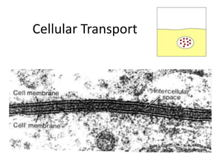
- 7. About Cell Membranes All cells have a cell membrane Functions: Controls what enters and exits the cell to maintain an internal balance called homeostasis Provides protection and support for the cell TEM picture of a real cell membrane.
- 8. About Cell Membranes (continued) Structure of cell membrane Lipid Bilayer -2 layers of phospholipids Phosphate head is polar (water loving) Fatty acid tails non-polar(water fearing) Proteins embedded in membrane Phospholipid Lipid Bilayer
- 9. Fluid Mosaic Model of the cell membrane Polar heads love water & dissolve. Membrane movement animation Non-polar tails hide from water. Carbohydrate cell markers Proteins
- 10. About Cell Membranes (continued) 4. Cell membranes have pores (holes) in it Selectively permeable: Allows some molecules in and keeps other molecules out The structure helps it be selective! Pores
- 11. Structure of the Cell Membrane Outside of cell Carbohydrate chains Proteins Lipid Bilayer Transport Protein Phospholipids Inside of cell (cytoplasm) Go to Section:
- 12. Evidence of fluid mosaic The most widely accepted membrane model made up until the early 1970s was a three layer protein lipid sandwich, based on the evidence of the electron micrographs in which the outer layers were thought to be proteins and the lighter region within was the lipid.
- 13. However, the protein lipid sandwich does not allow the hydrophilic phosphates head to be in contact with water, nor does it allow the hydrophobic amino acids on the outside of the membrane protein to be kept away from water. Experiment showed that there were two types of protein- those that could be dissociated from the membrane quite easily by increasing the ionic strength of the solution.
- 14. That could only be removed from the membrane by more drastic action such as adding detergents. This evidence supported the fluid mosaic model where some peripheral proteins are loosely attached on the outside surface of the membrane whilst integral membrane protein are fully embedded within the phospholipids, some even spanning both layers.
- 15. Additional evidence for integral membrane protein came from freeze fracture electron microscopy studies Frozen membrane sections were fractured along the weak point between the lipid layers, and the inner fractured surface coated in a heavy metal Scanning electron microscopy, which gives 3D , revealed a smooth mosaic like surface (the lipid tails) interspersed by much larger particles than the integral protein)
- 16. Finally, several experiment were carried out using labeled molecules that only attach to other specific molecules. In one experiment plant protein called lectins that bind to polysaccharides were labeled with ferritin (a protein with a core of ferric oxide) which is visible under the electron microscope.
- 17. When mixed with membrane samples, the lectins only bound to the outer surface of the membrane, and never to the inner. This showed that membranes are asymmetric ; the outside surface of the membrane is different to the inside, and once again did not support the protein sandwich model, which would be the same on both sides.
- 18. Another experiment involved fusing mouse cells with human cells. Before the cell fused a specific membrane protein was labelled in each cell type.
- 19. The mouse membrane protein was given a green fluorescent label, and the human membrane protein was given red fluorescent label. Immediately after fusing, the coloured remained in their respective halves, but after 40 min at 37⁰C there was complete intermixing of the protein.
- 20. The only way the proteins could have intermixed was to have diffused through the membranes, showing the components were indeed fluid.
- 21. Cell membrane Roles of carbohydrate Cell cell recognition- ability of a cell to determine if other cells it encounters are alike or different from it. Cell adhesion- bind with each other, causing adhesion between the cells. This enables cells to orientate and form a tissue.
- 22. LIPID Cholesterol- increasing the fluidity of cell membranes, by disrupting the close and orderly packing of phospholipids molecules in cell membrane.
- 23. PROTEIN Protein are required to facilitate diffusion, osmosis and active transport Facilitated diffusion and osmosis; where the protein span the cell surface membrane and form a hydrophilic channel/ carrier that is specific for particular molecule Active transport where the protein able to hydrolyse ATP as an energy source to pump substances across cell membrane Membrane protein may function as receptor sites. Receptor protein has a binding site for a hormone; such as insulin to allow external signal to trigger/ initiate reactions within cell.
- 24. *EXPLAIN WHY THE CELL MEMBRANE IS DESCRIBED AS HAVING A LFUID MOSAIC STRUCTURE (2m) It is a fluid because the phospholipids move around the membrane And it is mosaic because the membrane contain different size and shape of protein; pattern looks like tiles of mosaic.s protein/ glycoprotiens
- 25. The more phospholipids containing unsaturated fatty acids there are present in the membrane, the more fluid it is. The kinks in the hydrocarbon tails of the unsaturated phospholipids prevent them from packing closely together, so more movement is possible. Cholesterol reduces the fluidity of the membrane by preventing movement of the phospholipids.
- 26. ENDOCYTOSIS- materials are taken into the cell This is subdivide based in the type and size of material being transported. Occurs mainly in animal cells, as plants have rigid cell walls Mechanism; the cell forms pseudopodia that engulf macromolecules- the pseudopodia fuse, and the membrane pinches off, forming an internal vesicle-the vesicle fuses with the lysosomes-release