SlideShare une entreprise Scribd logo
1  sur  11
Télécharger pour lire hors ligne
CASE OF THE WEEK
                      PROFESSOR YASSER METWALLY
CLINICAL PICTURE

CLINICAL PICTURE:

8 years old male patient presented clinically with right sided hemiplegia since birth, Lennox Gastaut syndrome (with a
history suggestive of infantile spasm during the first few months of life) and mental subnormality. The vision in the left
eye was completely lost since birth and the vision in the right eye was markedly defective. The child showed evidence of
growth and developmental delay.

RADIOLOGICAL FINDINGS

RADIOLOGICAL FINDINGS:


Figure 1. Postcontrast CT scan images (A,B) and Precontrast MRI T1 images (C). Notice the cleft that extends through
the entire thickness of cerebral hemisphere from the ventricular surface (ependyma) to the periphery (pial surface) of the
brain, apparently the cleft is lined by gray matter (Open lip schizencephaly). The schizencephalic cleft is appreciated at
the parasylvian regions and is seen communicating with an encephalocele externally. The optic nerves are markedly
hypoplastic bilaterally. The septum pellucidum is markedly deficient, hypoplastic and can not be appreciated in the
presented images. The hypoplastic septum pellucidum resulted in a box- like appearance of the frontal horns. It is
difficult to see the pituitary stalk in the presented images. The cerebral cortex is lissencephalic and pachygyric and the
corpus callosum is markedly thinned anteriorly. The schizencephalic cleft walls are separated (open-lip or type II
schizencephaly).
Figure 2. Septo-optic dysplasia. Precontrast MRI T1 images. Notice the cleft that extends through the entire thickness of
cerebral hemisphere from the ventricular surface (ependyma) to the periphery (pial surface) of the brain, apparently the
cleft is lined by gray matter (Open lip schizencephaly). The schizencephalic cleft is appreciated at the parasylvian regions
and is seen communicating with an encephalocele externally. The optic nerves are markedly hypoplastic bilaterally. The
septum pellucidum is markedly deficient, hypoplastic and can not be appreciated in the presented images. The
hypoplastic septum pellucidum resulted in a box- like appearance of the frontal horns. It is difficult to see the pituitary
stalk in the presented images. The cerebral cortex is lissencephalic and pachygyric and the corpus callosum is markedly
thinned anteriorly (B,C). The schizencephalic cleft walls are separated (open-lip or type II schizencephaly).


Figure 3. Septo-optic dysplasia. Precontrast MRI T1 images. Notice the cleft that extends through the entire thickness of
cerebral hemisphere from the ventricular surface (ependyma) to the periphery (pial surface) of the brain, apparently the
cleft is lined by gray matter (Open lip schizencephaly). The schizencephalic cleft is appreciated at the parasylvian regions
and is seen communicating with an encephalocele externally. The optic nerves (A) and optic chiasma (B) are markedly
hypoplastic bilaterally. The septum pellucidum is markedly deficient, hypoplastic and can not be appreciated in the
presented images. The hypoplastic septum pellucidum resulted in a box- like appearance of the frontal horns. It is
difficult to see the pituitary stalk in the presented images. The cerebral cortex is lissencephalic and pachygyric and the
corpus callosum is markedly thinned anteriorly (A). The schizencephalic cleft walls are separated (open-lip or type II
schizencephaly). The pituitary gland appeared vacuolated (B)
Figure 4. Septo-optic dysplasia. Precontrast MRI T1 images. Notice the cleft that extends through the entire thickness of
cerebral hemisphere from the ventricular surface (ependyma) to the periphery (pial surface) of the brain, apparently the
cleft is lined by gray matter (Open lip schizencephaly). The schizencephalic cleft is appreciated at the parasylvian regions
and is seen communicating with an encephalocele externally. The optic nerves (A) and optic chiasma (B) are markedly
hypoplastic bilaterally. The septum pellucidum is markedly deficient, hypoplastic and can not be appreciated in the
presented images. The hypoplastic septum pellucidum resulted in a box- like appearance of the frontal horns. It is
difficult to see the pituitary stalk in the presented images. The cerebral cortex is lissencephalic and pachygyric. The
schizencephalic cleft walls are separated (open-lip or type II schizencephaly). The pituitary gland appeared vacuolated
(B)

The encephalocele and the skull wall expansion that are present at the opening of the open-lip schizencephaly (in the
presented case) is believed to result from CSF pulsations from the lateral ventricles transmitted through the cleft. [45]

 Case summary
      Open-lip or type II schizencephaly
      Deficient and hypoplastic septum pellucidum
      Dysplatic cerebral cortex (The cerebral cortex is lissencephalic and
       pachygyric)
      The pituitary gland appeared vacuolated
      Hypoplastic optic nerves and optic chiasma
      Hypoplastic corpus callosum
      Encephalocele

DIAGNOSIS:

DIAGNOSIS: SEPTO-OPTIC DYSPLASIA WITH OPEN LIP SCHIZENCEPHALY

DISCUSSION

DISCUSSION:

      Septo-optic dysplasia (de Morsier syndrome)

Septo-optic dysplasia (de Morsier syndrome) is a disorder characterized by the absence of the septum pellucidum, optic
nerve hypoplasia, and hypothalamic dysfunction. It may be associated with agenesis of the corpus callosum. This disorder
should be considered in any patient who exhibits at least two of the above abnormalities and perhaps even solely
hypothalamic dysfunction [2]. Septo-optic dysplasia also appears to involve prosencephalic cleavage and development of
anterior telencephalic structures [3]. About 50% of patients with septo-optic dysplasia have schizencephaly [1].

Patients may present with visual disturbance, seizures, mental retardation, hemiparesis (especially if associated with
schizencephaly), quadriparesis, or hypothalamic dysfunction. Endocrine abnormalities may include growth hormone,
thyroid hormone, or antidiuretic hormone function or levels. The consideration of septo-optic dysplasia necessitates an
evaluation of the hypothalamic-pituitary axis because as many as 60% of the children with this disorder might exhibit
evidence of a disturbance of endocrine function [4]. This evaluation can include thyroid function studies and electrolytes;
these patients are at high risk for growth retardation.

The recent identification of patients with this condition that harbor mutations in the transcriptional regulator gene
HESX1, suggest that the mechanism of this disorder is likely genetic and a patterning or segmental abnormality [5]. Even
though the genetic abnormality has been identified for a minority of patients, there exists the possibility that this may not
represent an entirely genetic disorder because associations have been made with young maternal age, diabetes, the use of
anticonvulsants, phencyclidine, cocaine, and alcohol [6,45]

            Pathology of septo-optic dysplasia

Several congenital malformations might be associated with macroscopic malformations of the septal nuclei. Most of them
involve the midline structures, including the septum pellucidum. For example, septo-optic dysplasia is a syndrome
characterized by the absence of the septum pellucidum and optic nerve hypoplasia It may also be associated with
pituitary insufficiency resulting in growth and developmental delay. The septal nuclei abnormalities are often an element
of a larger cerebral malformation, such as holoprosencephaly, schizencephaly, or porencephaly. In most cases, other
midline structures such as the optic nerves, hypothalamus, and septum pellucidum are involved. Absence of the septum
pellucidum may reach 80% to 90% in conditions such as schizencephaly. Focal heterotopias or cortical dysplasias may be
associated with epileptic seizures. Severe mental retardation and intractable seizures are typical features of the more
common larger cerebral malformations and of the syndromes associated with chromosomal anomalies. In some cases of
corpus callosum agenesis, it is possible to discern a few axon bundles in an anteroposterior orientation that attempted to
cross posteriorly. These are called Probst's bundles. If the corpus callosum is absent, the subcallosal interhemispheric
tracts such as the anterior, posterior, and hippocampal commissure might be enlarged. The presence of Probst's bundles
is considered by some to indicate that the agenesis of the corpus callosum is due to a primary cause and not the result of
secondary injury such as hypoxia. These primary causes include chromosomal abnormalities, single gene defects, and
metabolic defects. Cysts and lipomas may be associated with absence of the corpus callosum. [45]

Large occiput encephaloceles may include significant parts of the brain, inducing hypoplasia of the anterior and middle
cranial fossa. The brain in the encephalocele could show ischemia or polymicrogyria or be normal. The hypoplasia of the
anterior and middle cranial fossa may stretch the septal nuclei, fornix, and septum pellucidum, among other structures.
In contrast, transsphenoidal and sphenoethmoidal encephaloceles may directly involve the septal nuclei because they may
be included in the herniated brain. [45]

            Neuroendocrine disorders in septo-optic dysplasia

Syndromes that may be associated with pituitary-hypothalamic dysfunction include septo-optic dysplasia, Kallmann's
syndrome, and empty sella syndrome. In addition, other developmental anomalies may be associated with pituitary-
hypothalamic defects, including agenesis of the corpus callosum, holoprosencephaly, and basal cephaloceles. [45]

Septo-optic dysplasia, a disorder of ventral induction, is considered a mild form of holoprosencephaly. It is a disorder of
abnormal induction of the midline mesoderm occurring at the same time as the development of the optic vesicles. Failure
of differentiation of the mesoderm into the optic stalk results in aplasia of the stalk. [15] Septo-optic dysplasia is
characterized by absence or hypoplasia of the septum pellucidum associated with hypoplasia of the optic nerves as first
described by DeMorsier. [12] Associated hypothalamic-pituitary dysfunction occurs in two thirds of patients. [17,18,19]
Patients may have hypopituitarism that ranges from panhypopituitarism to growth hormone deficiency, thyroid-
stimulating hormone deficiency, elevated prolactin, or ACTH or ADH deficiency. [16] Visual symptoms include
nystagmus, amblyopia, or hemianopsia. This syndrome may result from in utero injury or genetic abnormalities. [9]
Environmental factors implicated are maternal diabetes, quinidine ingestion, anticonvulsants, alcohol or drug abuse, and
cytomegalovirus. [17,45] There may be associated anomalies of neuronal migration, such as schizencephaly and gray
matter heterotopias.

On imaging, there is absence or hypoplasia of the septum pellucidum resulting in a box- like appearance of the frontal
horns. [8,13,14] Hypoplasia of the optic disks is often diagnosed by an ophthalmologist; however, optic nerve, optic tract,
or optic chiasm hypoplasia may be seen in 50% of patients on imaging. [8] Other associated abnormalities include
absence of the fornix and callosal dysgenesis. [11,45] On MR imaging, three patterns of involvement have been described
in septo-optic dysplasia. [8,11.45]

  1. One group has neuronal migration anomalies such as schizencephaly and gray matter heterotopias, hypothalamic-
     pituitary dysfunction, and partial absence of the septum pellucidum. [45]

  2. The other group has complete absence of the septum pellucidum with cerebral white matter hypoplasia. This group
     may be associated with hypoplasia of the genu of the corpus callosum or hypoplasia of the anterior falx cerebri.

  3. Another group of these patients with ectopia of the posterior pituitary has also been described. [11]

Other anomalies that may be associated with complete or partial absence of the septum pellucidum include
holoprosencephaly, hydranencephaly, basilar cephaloceles, dysgenesis of the corpus callosum, chronic hydrocephalus,
Chiari II malformation, and schizencephaly. [7,10,20]

             Optic nerve hypoplasia in septo-optic dysplasia

Optic nerve hypoplasia, a specific clinical entity, is an increasingly common cause of infantile blindness in the United
States. It is characterized by a small optic disc surface area and thin optic nerve caused by a congenital nonprogressive
decrease in the number of optic nerve axons. Unilateral and bilateral optic nerve hypoplasia occurs with roughly equal
frequency. Various pathophysiologic theories have been proposed, including failure of axons to normally develop; a
destructive developmental event, such as a vascular insult; exaggerated apoptosis; or a genetic disorder. [45]

All patients with optic nerve hypoplasia show visual field defects; localized defects as well as generalized constriction are
seen. The loss of visual acuity is variable depending on the degree of involvement with axons from the macula, the center
of sight. [45]

Optic nerve hypoplasia is recognized as part of a spectrum of optic nerve, central nervous system (CNS), and
hypothalamic-pituitary axis congenital anomalies [21]. From the ophthalmologist's perspective, optic nerve hypoplasia
can be assigned into five sometimes overlapping groups: isolated unilateral or bilateral optic nerve hypoplasia, with
absence of the septum pellucidum (septo-optic dysplasia), with posterior pituitary ectopia or absence of the pituitary
stalk, with hemispheric migration anomalies, or with intrauterine and/or perinatal hemispheric injuries [22]. Optic nerve
hypoplasia is also associated with a variety of ocular and systemic disorders, including aniridia, albinism, maternal
diabetes, fetal alcohol syndrome, and other maternal ingestions [23,24,25].

 Groups with optic nerve hypoplasia
      Isolated unilateral or bilateral optic nerve hypoplasia
      Optic nerve hypoplasia with absence of the septum pellucidum
       (septo-optic dysplasia)
      Optic nerve hypoplasia with posterior pituitary ectopia or absence
       of the pituitary stalk
      Optic nerve hypoplasia with hemispheric migration anomalies
      Optic nerve hypoplasia with intrauterine and/or perinatal
       hemispheric injuries

             Schizencephaly in septo-optic dysplasia

Schizencephaly is the most complete form of migrational disorders. It is thought to be caused by a complete agenesis of a
section of the cerebral tissue, which results in clefts that extend through the entire thickness of cerebral hemisphere. At
the margin of the cleft the pial membrane and ependymal lining of the ventricle lie adjacent to each other and form a pial
ependymal seam [29] Schizencephaly probably occurs as the end result of a variety of insults occuring at a critical time
and in a critical location during brain development. No specific inciting or unusual prenatal events are described. The
lesion is most likely related to multiple etiologies including genetic, toxic, metabolic, vascular or infectious disease.
Familial cases have been reported. [30] Septooptic dysplasia, also called as De Morsier disease is a syndrome consisting of
blindness, hypoplastic optic nerves and absence of septum pellucidum in females. [29] It is known that Schizencephaly
and septo-optic dysplasia frequently coexists. [31] Associated anomalies are heterotopias, Dandy Walker malformation,
hydrocephalus and polymicrogyria. [28] Cleft may be unilateral or bilateral, symmetrical or asymmetrical. Commonly
located near pre/post central gyrus [26.27]] The cleft in Schizencephaly are lined either totally or in part by gray matter
and extend from pial surface to ependyma of the lateral ventricle. The clefts can be located anywhere but commonly
occur in parasylvian regions. The cavity formed in open lip type varies in size from small to large and may communicate
with lateral ventricle. The ventricular system may be enlarged, particularly in-patients with open lip form of
Schizencephaly. [30]

Absence of septum pellucidum, dysgenesis of corpus callosum are often associated with open lip Schizencephaly [28,29]
Differential diagnosis includes subarachnoid cysts and porencephaly. Using CT diagnosis of Schizencephaly is sometimes
difficult particularly in case of type I. CT may show a slight outpouching at the ependymal surface of cleft and a full
thickness cleft may be difficult to identify on CT Scan .Secondary findings like hydrocephalus, heterotopia,
polymicrogyria, subdural hygromas and arachnoid cysts can be identified. [30] MRI gives most detailed and precise
definition of anatomy and anomaly. For anatomy MRI T1 images suffice [28] MRI is the modality of choice. MRI better
delineates the gray matter lining the cleft, which is pathognomonic finding in Schizencephaly.MRI also provides superb
cortical anatomy detail and multiplanar capacity. Primary findings related to the cleft and secondary findings associated
with Schizencephaly are identified using MRI.The ability of MRI pulse sequence to differentiate gray matter and white
matter permit demonstration of gray matter heterotopias in the subcortical white matter beneath the cleft, abnormalities
involving the cortex (pachygyria and polymicrogyria) and other secondary findings are also identified. Homolateral
absence of sylvian vasculature, small medullary pyramids low position of fornix and thinning of the corpus callosum are
findings related to absent cerebral cortex and are better demonstrated by MRI than with other studies.

Sonography can be done in neonatal period in-patients in whom this anomaly is suspected. In Schizencephaly type I, a
hyperechoic line extends from the parasylvian region to the anterior portion of lateral ventricle. The hyperechoic line
represents the cortex lining the fused cleft. This type of anomaly is difficult to detect with ultrasound and requires high
index of suspicion and highly skilled operator. In Schizencephaly type II an anechoic band or cavity, representing the
fluid filled cleft extends from the cortical surface of lateral ventricle. The meeting of the closed lip portion or apex of the
cleft with the margin of ventricle may be identified as a ventricular diverticulum or dimple. The size of the caudate,
thalamus and lenticular nuclei (subcortical gray matter structures) is decreased. Other associated anomalies such as
ventricular enlargement may also be identified. [30]

This case represents a typical association of septo-optic dysplasia with open lip Schizencephaly with all cardinal
neurological cardinal features on one hand and a rare form of unilateral open lip cleft on the other. It highlights the
importance of MRI in diagnosis of such a condition.

SCHIZENCEPHALY

Schizencephaly is an uncommon disorder of neuronal migrational characterized by a cerebrospinal fluid–filled cleft,
which is lined by gray matter. The cleft extends across the entire cerebral hemisphere, from the ventricular surface
(ependyma) to the periphery (pial surface) of the brain.

The clefts may be unilateral or bilateral and may be closed (fused lips), as in schizencephaly type I, or separated (open
lips), as in schizencephaly type II. Presentation and outcome are variable, but patients typically present with seizures,
hemiparesis, and developmental deficits. Usually, the severity of symptoms is related to the amount of brain affected by
the abnormality.

      Aetiology and types of schizencephaly

Several theories have been proposed to explain the etiology of schizencephaly, although none is universally accepted. The
leading theory indicates that schizencephaly results from an early, focal destruction of the germinal matrix and
surrounding brain before the hemispheres are fully formed. Schizencephaly probably occurs as the end result of a variety
of insults occurring at a critical time and in a critical location during brain development. No specific inciting or unusual
prenatal events have been identified, and reported cases are sporadic. The lesion is most likely related to multiple
etiologies, including genetic, toxic, metabolic, vascular, or infectious causes. Familial cases of schizencephaly have been
reported. Schizencephaly is uncommon; to our knowledge, there are no documented geographical differences in its
occurrence. Severity of the symptoms depends on the amount of brain involved. Affected patients typically have seizures,
hemiparesis, variable developmental delay, and blindness. Patients also have variable degrees of mental retardation.
Patients with open-lip schizencephaly die at an earlier age than patients with the closed-lip form. Usually, death results
from failure to thrive, chronic infections, and respiratory problems. Patients with closed-lip schizencephaly may not
present clinically until later in infancy or early childhood and may live to early adulthood.

Schizencephaly is divided into 2 types, which have prognostic significance. In closed-lip or type I schizencephaly, the cleft
walls are in apposition. In open-lip or type II schizencephaly, the cleft walls are separated. Schizencephaly type II occurs
more commonly than type I.

The clefts in schizencephaly are lined either totally or in part by gray matter and extend from the pial surface to the
ependyma of the lateral ventricle. The clefts can be located anywhere, but they commonly occur in the parasylvian
regions. The clefts can be unilateral or bilateral, and can be either symmetric or asymmetric. The cavity formed in the
open-lip type varies in size from small to large and may communicate with the lateral ventricle. The ventricular system
may be enlarged, particularly in patients with the open-lip form of schizencephaly.

Gray-matter heterotopia (collections of gray matter in abnormal locations), polymicrogyria, and arachnoid cysts can be
associated with schizencephaly. Heterotopias and polymicrogyria typically line the clefts. Microcephaly has been noted in
some patients. The septum pellucidum is absent in 80-90% of patients, and schizencephaly may coexist with septo-optic
dysplasia.

      Clinical picture of schizencephaly

Clinical features of schizencephaly are highly variable. Patients with unilateral clefts with fused lips may have mild
hemiparesis and seizures but otherwise have normal development. When the cleft is open, patients present with mild-to-
moderate developmental delay and hemiparesis; severity is related to the extent of cortex involved in the defect.

Patients with bilateral clefts present with severe mental deficits and severe motor anomalies including spastic
quadriparesis. Frequently, these patients present with blindness, which is often associated with optic nerve hypoplasia.
Language development is more likely to be normal in patients with unilateral schizencephaly compared to patients with
bilateral clefts.

Intractable seizures frequently are noted in schizencephaly. Several types of seizures have been reported including
generalized tonic-clonic, partial motor, and sensory seizures. Infantile spasms have been seen in a few children. Reports
have found no correlation between the subtype of schizencephaly and the occurrence or type of seizure disorder.
Identification of gray matter lining the cleft is the pathognomonic finding in differentiating schizencephaly from
porencephaly; this is best demonstrated on MRIs.

The more complete information obtained by MRI enables a more accurate prediction of neurologic outcome.


                                                                      Figure 5. Open-lip schizencephaly with cortical
                                                                      dysplasia


Figure 6. Bilateral open lip schizencephaly

      Neuroimaging of schizencephaly

             CT scan

Using CT, the diagnosis of schizencephaly is sometimes difficult, particularly type I, or closed lip schizencephaly. CT
scans of closed-lip schizencephaly may show only a slight outpouching at the ependymal surface of the cleft, and a full-
thickness cleft may be difficult to identify on CT scan. The cleft is partially or totally lined by gray matter and extends
from the lateral ventricle to the pial surface of the cerebral hemisphere. Secondary findings that can be identified on CT
scan include hydrocephalus, heterotopia, polymicrogyria, subdural hygromas, and arachnoid cysts.

            MRI

MRI is the modality of choice for evaluating patients with schizencephaly. MRI better delineates the gray matter lining
the cleft, which is the pathognomonic finding in schizencephaly. MRI also provides superb cortical anatomy detail and
multiplanar capability. Primary findings related to the cleft and secondary findings associated with schizencephaly are
identified using MRI.

The ability of MRI pulse sequences to differentiate gray matter and white matter permits demonstration of gray-matter
heterotopias in the subcortical white matter beneath the cleft, abnormalities involving the cortex (eg, pachygyria or
polymicrogyria), and other secondary findings also identified by using CT scans. Homolateral absence of the sylvian
vasculature, small medullary pyramids, a low position of the fornix, and thinning of the corpus callosum are findings
related to absent cerebral cortex and are better demonstrated by MRI than with other studies.


Figure 7. A, Schizencephaly. Axial T2-weighted MRI in unilateral closed-lip (type I) schizencephaly. The cleft is lined by
gray matter and extends from the pial surface to the lateral ventricle. B, Schizencephaly. Axial T2-weighted (left) and
coronal T1-weighted (right) MRIs in bilateral closed-lip (type I) schizencephaly. A ventricular diverticulum defines the
meeting of the closed-lip portion of the clefts with the margin of the ventricles. The septum pellucidum is absent, and the
clefts are lined by gray matter and extend from the pial surface to the lateral ventricle. C, Schizencephaly. Axial T2-
weighted MRI demonstrates a small open-lip schizencephaly. The septum pellucidum is absent. D, Schizencephaly. Axial
T2-weighted MRI in unilateral open-lip (type II) schizencephaly. The septum pellucidum is absent, and a large
cerebrospinal fluid–filled cleft extends from the lateral ventricle to the cortical surface. The cleft is lined by gray matter.
E, Schizencephaly. Coronal sonograms with a corresponding coronal T1-weighted MRI of open-lip bilateral
schizencephaly. Extensive bilateral schizencephalic defects with large CSF-filled clefts extend from the lateral ventricles
to the cortical surface.


SUMMARY


SUMMARY

Schizencephaly is the term describing gray matter-lined clefts that extend through the entire hemisphere from the lateral
ventricles to the cortical surface, characterized pathologically by a pial-ependymal continuity [28,29]. The gray matter
lining these clefts is dysplastic and has irregular inner and outer surfaces, identical to that of polymicrogyria. The clefts
can be unilateral or bilateral and are most commonly located near the pre- and postcentral gyri, locations almost
identical to those in which polymicrogyria occurs [22,23]. It is probable that schizencephaly represents an extreme
variant of cortical dysplasia, in which the infolding of cortex extends all the way into the lateral ventricle [22,23]. The lips
of the cleft may be fused, in which case the walls are directly apposed, obliterating the CSF space within the cleft at that
point. When the lips are separated, CSF fills the cleft from the lateral ventricle to the subarachnoid space surrounding
the hemispheres [23,28-30]. As in polymicrogyria, large vessels are often seen in the cleft between the lips of the
schizencephaly.

The severity of the clinical symptoms is related to the amount of involved brain [23, 31,32]. Patients with a unilateral cleft
with fused lips typically present with mild hemiparesis or epilepsy but are otherwise developmentally normal. Patients
with unilateral clefts with separated lips more commonly present with hemiparesis and a mild to moderate developmental
delay, depending on the location of the cleft within the brain. Patients with bilateral clefts tend to be severely retarded,
with refractory seizures beginning at a very early age and severe motor anomalies, and they may appear to be blind.
Optic nerve hypoplasia is seen in up to one-third of affected patients [8,30, 33,34].

Routine spin-echo MR obtained in at least two planes is usually adequate for imaging of schizencephalies. These studies
show full-thickness clefts, lined by gray matter with an irregular inner surface, that extend through the hemisphere from
the ventricle to the surface of the brain. The gray matter lining the cleft often extends into the ventricle as a
subependymal heterotopia [6,8,23,30,32]. A dimple is usually seen in the wall of the lateral ventricle where it
communicates with the cleft. The dimple provides a helpful sign of continuity of the cleft with the ventricle when the lips
of the cleft are fused. The gyral pattern of the cortex adjacent to the cleft is usually abnormal, with features characteristic
of polymicrogyria. Polymicrogyria may also be present in the hemisphere contralateral to a unilateral schizencephaly
[22,23]. Therefore, the contralateral hemisphere should always be scrutinized.

The calvarium is often expanded over the opening of an open-lip schizencephaly. The expansion is believed to result from
CSF pulsations from the lateral ventricles transmitted through the cleft. When plagiocephaly is severe, insertion of a
ventriculoperitoneal shunt may help to dampen the pulsations and reverse the cranial asymmetry.


      Addendum

            A new version of this PDF file (with a new case) is uploaded in my web site every week (every Saturday and
             remains available till Friday.)

            To download the current version follow the link quot;http://pdf.yassermetwally.com/case.pdfquot;.
            You can also download the current version from my web site at quot;http://yassermetwally.comquot;.
            To download the software version of the publication (crow.exe) follow the link:
             http://neurology.yassermetwally.com/crow.zip
            The case is also presented as a short case in PDF format, to download the short case follow the link:
             http://pdf.yassermetwally.com/short.pdf
            At the end of each year, all the publications are compiled on a single CD-ROM, please contact the author to know
             more details.
            Screen resolution is better set at 1024*768 pixel screen area for optimum display.
            For an archive of the previously reported cases go to www.yassermetwally.net, then under pages in the right panel,
             scroll down and click on the text entry quot;downloadable case records in PDF formatquot;


REFERENCES

References

[1] Aicardi J, Goutieres F. The syndrome of absence of the septum pellucidum with porencephalies and other
developmental defects. Neuropediatrics 1981;12:319-29.

[2] Brickman JM, Clements M, Tyrell R, et al. Molecular effects of novel mutations in Hesxl/HESXI associated with
human pituitary disorders. Development 2001;128:5189-99.

[3] Ouvier R, Billson F. Optic nerve hypoplasia: a review. J Child Neurol 1986;1:181-8.

[4] Costin G, Murphree AL. Hypothalamic-pituitary function in children with optic nerve hypoplasia. Am J Dis Child
1985;139:249-54.

[5] Dattani MT, Martinez-Barbera JP, Thomas PQ, et al. Mutations in the homeobox gene HESXI/Hesxl associated with
septo-optic dysplasia in human and mouse. Nat Genet 1998; 19:125-33.
[6] Patel H, Tze WJ, Crichton JU, et al. Optic nerve hypoplasia with hypopituitarism: septo- optic dysplasia with
hypopituitarism. Am J Dis Child 1975;129:175-80.

[7] Aicardi J, Goutieres F: The syndrome of absence of the septum pellucidum with porencephalies and other
developmental defects. Neuropediatrics 12:319-329, 1981

[8] Barkovich A, Fram E, Norman D: Septooptic dysplasia: MR imaging. Radiology 171:189-192,1989

[9] Barkovich AJ: Pediatric Neuroimaging, ed 2. New York, Raven Press, 1995, pp 236-237

[10] Barkovich Aj, Norman D: Absence of the septum pellucidum: A useful sign in the diagnosis of congenital brain
malformations. AJR Am j Roentgenol 152:353- 360,1989

[11] Brodsky M, Glasier C: Optic nerve hypoplasia: Clinical significance of associated central nervous system
abnormalities on magnetic resonance imaging. Arch Ophthalmol 111:66-74,1993

{12] DeMorsier G: Etudes sur les dysraphics cranio-encephaliques 111. Agenese du septum lucidum avec malformation
du tractus optique. La dysplasie septo- optic. Schweiz Arch Neurol Neurochir Psychiatry 77:267-292,1956

[13] Fitz C: Holoprosencephaly and related entities. Neuroradiology 25:3-238, 1983

[14] Fitz CR: Holoprosencephaly and septo-optic dysplasia. Neuroimaging Clin North Am 4:263-281, 1994

[15] Hale BR, Rice P: Septo-optic dysplasia: Clinical and embryological aspects. Dev Med Child Neurol 16: 812-817,1974

[16] Izenberg N, Rosenblum M, Parks JS: The endocrine spectrum of septo-optic dysplasia. Clin Pediatr (Phila) 23:632-
636, 1984

[17] Morishima A, Aranoff G: Syndrome of septooptic- pituitary dysplasia: The clinical spectrum. Brain Dev 8:233-
239,1986

[18] Skarf B, Hoyt C: Optic nerve hypoplasia in children: Association with anomalies of the endocrine and CNS. Arch
Ophthalmol 102:62-67,1984

[19] Stanhope R, Preece M, Brook C: Hypoplastic optic nerves and pituitary dysfunction: A spectrum of anatomical and
endocrine abnormalities. Arch Dis Child 59:111-114,1984

[20] Harwood-Nash D, Fitz C: Neuroradiology in Infants and Children. St. Louis, CV Mosby, 1976, pp 484-486

[21] Campbell CL. Septo-optic dysplasia: a literature review. Optometry. 2003;74(7):417–426.

[22]Brodsky MC, Glasier CM. Optic nerve hypoplasia. Clinical significance of associated central nervous system
abnormalities on magnetic resonance imaging. Arch Ophthalmol. 1993;111(1):66–74.

[23]Brown GC. Optic nerve hypoplasia and colobomatous defects. J Pediatr Ophthalmol Strabismus. 1982;19(2):90–93.

[24]Chan T, Bowell R, O'Keefe M, et al.. Ocular manifestations in fetal alcohol syndrome. Br J Ophthalmol. 1991;75
(9):524–526.

[25]Mansour AM, Bitar FF, Traboulsi EI, et al.. Ocular pathology in congenital heart disease. Eye. 2004;19(1):29–34.

[26] S.K.Sethi, R.S.Solanki Radiological Quiz. Indian J. Radiology and Imaging, 2004,14:1:95-96.

[27] D.A Shah, G.D.Rathod, Radiological Quiz Ind J Radiology and Imaging, 2005,15:1:127-128.

[28] Yutaka Sato, Simon C.S.Kao, and Wilbur L.Smith, RCNA, March 1991, Vol. 29,No 2 :179-194,

[29] C.Raybaud Destructive lesions of Brain Neuroradiology 1983; 25:265-291.

[30] Ken R.Close Schizencephaly www emedicine.com/radio/topic622.htm last updated April 7, 2004

[31] AJNR, Mar/Apr, 1988:9:297-302.:MR imaging of Schizencephaly; A.James Barkovich, David Norman.

[32] Barkovich AJ, Norman D: MR imaging of schizencephaly. AJR Am J Roentgenol 1988 Jun; 150(6): 1391-6.
[33] Barkovich AJ: Schizencephaly. In: Pediatric Neuroimaging. 2nd ed. Philadelphia: Lippincott-Raven; 1996:219-25.

[34] Barth PG: Schizencephaly and nonlissencephalic cortical dysplasias. AJNR Am J Neuroradiol 1992 Jan-Feb; 13(1):
104-6.

[35] Bird CR, Gilles FH: Type I schizencephaly: CT and neuropathologic findings. AJNR Am J Neuroradiol 1987 May-
Jun; 8(3): 451-4.

[36] Byrd SE, Osborn RE, Bohan TP, Naidich TP: The CT and MR evaluation of migrational disorders of the brain. Part
II. Schizencephaly, heterotopia and polymicrogyria. Pediatr Radiol 1989; 19(4): 219-22.

[37] Chamberlain MC, Press GA, Bejar RF: Neonatal schizencephaly: comparison of brain imaging. Pediatr Neurol 1990
Nov-Dec; 6(6): 382-7.

[38] Denis D, Chateil JF, Brun M, et al: Schizencephaly: clinical and imaging features in 30 infantile cases. Brain Dev
2000 Dec; 22(8): 475-83.

[39] DiPietro MA, Brody BA, Kuban K, Cole FS: Schizencephaly: rare cerebral malformation demonstrated by
sonography. AJNR Am J Neuroradiol 1984 Mar-Apr; 5(2): 196-8.

[40] Komarniski CA, Cyr DR, Mack LA, Weinberger E: Prenatal diagnosis of schizencephaly. J Ultrasound Med 1990
May; 9(5): 305-7.

[41] Lee SH, Rao K, Zimmerman RA: Schizencephaly. In: Cranial MRI and CT. 4th ed. New York: McGraw-Hill; 1999:
163-5.

[42] Miller GM, Stears JC, Guggenheim MA, Wilkening GN: Schizencephaly: a clinical and CT study. Neurology 1984
Aug; 34(8): 997-1001.

[43] Osborne AG: Schizencephaly. In: Diagnostic Neuroradiology. St Louis: Mosby-Year Book; 1994:52-6.

[44] Packard AM, Miller VS, Delgado MR: Schizencephaly: correlations of clinical and radiologic features. Neurology
1997 May; 48(5): 1427-34.

[45] Metwally, MYM: Textbook of neuroimaging, A CD-ROM publication, (Metwally, MYM editor) WEB-CD agency
for electronic publication, version 9.2a April 2008

Contenu connexe

Tendances

Approach to neuroregression
Approach to neuroregressionApproach to neuroregression
Approach to neuroregressiondrswarupa
 
approaches to pale disc
approaches to pale discapproaches to pale disc
approaches to pale discPrajakta Matey
 
Pediatric epilepsy syndromes
Pediatric epilepsy syndromesPediatric epilepsy syndromes
Pediatric epilepsy syndromesNeurologyKota
 
AION Anterior Ischemic Optic Neuropathy
AION Anterior Ischemic Optic NeuropathyAION Anterior Ischemic Optic Neuropathy
AION Anterior Ischemic Optic NeuropathyNoor Munirah Aab
 
Neurocutaneous syndrome
Neurocutaneous syndromeNeurocutaneous syndrome
Neurocutaneous syndromeNeurologyKota
 
Approach to neurodegenerative disorders new praman
Approach to neurodegenerative disorders new pramanApproach to neurodegenerative disorders new praman
Approach to neurodegenerative disorders new pramanDr Praman Kushwah
 
Optic neuritis
Optic neuritisOptic neuritis
Optic neuritisfaqar2003
 
NEURODEGENERATIVE DISORDER OF CHILDHOOD
NEURODEGENERATIVE DISORDER OF CHILDHOODNEURODEGENERATIVE DISORDER OF CHILDHOOD
NEURODEGENERATIVE DISORDER OF CHILDHOODSamiul Hussain
 
Vogt Koyanagi Harada Disease
Vogt Koyanagi Harada DiseaseVogt Koyanagi Harada Disease
Vogt Koyanagi Harada DiseaseGauree Gattani
 
Benign intracranial hypertension by Dr.Syed Alam Zeb
Benign intracranial hypertension by Dr.Syed Alam ZebBenign intracranial hypertension by Dr.Syed Alam Zeb
Benign intracranial hypertension by Dr.Syed Alam ZebSyed Alam Zeb
 
Approach to a patient with bilateral vision loss
Approach to a patient with bilateral vision lossApproach to a patient with bilateral vision loss
Approach to a patient with bilateral vision lossNeurologyKota
 
Retinopathy of prematurity
Retinopathy of prematurityRetinopathy of prematurity
Retinopathy of prematurityBarun Garg
 
Differential Diagnosis of Disc Edema
Differential Diagnosis of Disc EdemaDifferential Diagnosis of Disc Edema
Differential Diagnosis of Disc EdemaSahil Thakur
 
Supranuclear control of gaze
Supranuclear control of gazeSupranuclear control of gaze
Supranuclear control of gazeDr. Arghya Deb
 

Tendances (20)

Transient Visual Loss
Transient Visual LossTransient Visual Loss
Transient Visual Loss
 
Approach to neuroregression
Approach to neuroregressionApproach to neuroregression
Approach to neuroregression
 
approaches to pale disc
approaches to pale discapproaches to pale disc
approaches to pale disc
 
Pediatric epilepsy syndromes
Pediatric epilepsy syndromesPediatric epilepsy syndromes
Pediatric epilepsy syndromes
 
AION Anterior Ischemic Optic Neuropathy
AION Anterior Ischemic Optic NeuropathyAION Anterior Ischemic Optic Neuropathy
AION Anterior Ischemic Optic Neuropathy
 
Gaze palcy
Gaze palcy Gaze palcy
Gaze palcy
 
Neurocutaneous syndrome
Neurocutaneous syndromeNeurocutaneous syndrome
Neurocutaneous syndrome
 
Approach to neurodegenerative disorders new praman
Approach to neurodegenerative disorders new pramanApproach to neurodegenerative disorders new praman
Approach to neurodegenerative disorders new praman
 
Gaze palsy
Gaze palsyGaze palsy
Gaze palsy
 
Optic neuritis
Optic neuritisOptic neuritis
Optic neuritis
 
Optic atrophy (b)
Optic atrophy (b)Optic atrophy (b)
Optic atrophy (b)
 
NEURODEGENERATIVE DISORDER OF CHILDHOOD
NEURODEGENERATIVE DISORDER OF CHILDHOODNEURODEGENERATIVE DISORDER OF CHILDHOOD
NEURODEGENERATIVE DISORDER OF CHILDHOOD
 
Vogt Koyanagi Harada Disease
Vogt Koyanagi Harada DiseaseVogt Koyanagi Harada Disease
Vogt Koyanagi Harada Disease
 
orbital cellulitis.pptx
orbital cellulitis.pptxorbital cellulitis.pptx
orbital cellulitis.pptx
 
Benign intracranial hypertension by Dr.Syed Alam Zeb
Benign intracranial hypertension by Dr.Syed Alam ZebBenign intracranial hypertension by Dr.Syed Alam Zeb
Benign intracranial hypertension by Dr.Syed Alam Zeb
 
Approach to a patient with bilateral vision loss
Approach to a patient with bilateral vision lossApproach to a patient with bilateral vision loss
Approach to a patient with bilateral vision loss
 
Retinopathy of prematurity
Retinopathy of prematurityRetinopathy of prematurity
Retinopathy of prematurity
 
Differential Diagnosis of Disc Edema
Differential Diagnosis of Disc EdemaDifferential Diagnosis of Disc Edema
Differential Diagnosis of Disc Edema
 
Retinopathy of prematurity
Retinopathy of prematurityRetinopathy of prematurity
Retinopathy of prematurity
 
Supranuclear control of gaze
Supranuclear control of gazeSupranuclear control of gaze
Supranuclear control of gaze
 

Similaire à Case record... Septo-optic dysplasia

Imaging of congenital cns lesions
Imaging of congenital cns lesionsImaging of congenital cns lesions
Imaging of congenital cns lesionsGobardhan Thapa
 
Imaging of congenital CNS lesions
Imaging of congenital CNS lesionsImaging of congenital CNS lesions
Imaging of congenital CNS lesionsMilan Silwal
 
Congenital CNS lesions
Congenital CNS lesionsCongenital CNS lesions
Congenital CNS lesionsMilan Silwal
 
Presentation1.pptx, congenital malformation of the brain.
Presentation1.pptx, congenital malformation of the brain.Presentation1.pptx, congenital malformation of the brain.
Presentation1.pptx, congenital malformation of the brain.Abdellah Nazeer
 
Imaging in holoprosencephaly and related disorders
Imaging in holoprosencephaly and related disordersImaging in holoprosencephaly and related disorders
Imaging in holoprosencephaly and related disordersKhalaf Saba
 
1.schizencephaly 2.holoprosencephaly 3.porencephaly
1.schizencephaly 2.holoprosencephaly 3.porencephaly1.schizencephaly 2.holoprosencephaly 3.porencephaly
1.schizencephaly 2.holoprosencephaly 3.porencephalyairwave12
 
Neuronal migration disorders
Neuronal migration disordersNeuronal migration disorders
Neuronal migration disordersAmr Hassan
 
Radiological findings of congenital anomalies of the spine and spinal cord
Radiological findings of congenital anomalies of the spine and spinal cordRadiological findings of congenital anomalies of the spine and spinal cord
Radiological findings of congenital anomalies of the spine and spinal cordDr. Armaan Singh
 
Radiological pathology of brain developmental disorders
Radiological pathology of brain developmental disordersRadiological pathology of brain developmental disorders
Radiological pathology of brain developmental disordersProfessor Yasser Metwally
 
Pediatric posterior head region epilepsy
Pediatric posterior head region epilepsyPediatric posterior head region epilepsy
Pediatric posterior head region epilepsyPramod Krishnan
 
Ultrasound image gallery
Ultrasound image galleryUltrasound image gallery
Ultrasound image galleryRitesh Mahajan
 
Imaging in developmental malformations of the brain
Imaging in developmental malformations of the brainImaging in developmental malformations of the brain
Imaging in developmental malformations of the brainChakradhar Reddy
 

Similaire à Case record... Septo-optic dysplasia (20)

Imaging of congenital cns lesions
Imaging of congenital cns lesionsImaging of congenital cns lesions
Imaging of congenital cns lesions
 
Imaging of congenital CNS lesions
Imaging of congenital CNS lesionsImaging of congenital CNS lesions
Imaging of congenital CNS lesions
 
Congenital CNS lesions
Congenital CNS lesionsCongenital CNS lesions
Congenital CNS lesions
 
Presentation1.pptx, congenital malformation of the brain.
Presentation1.pptx, congenital malformation of the brain.Presentation1.pptx, congenital malformation of the brain.
Presentation1.pptx, congenital malformation of the brain.
 
Imaging in holoprosencephaly and related disorders
Imaging in holoprosencephaly and related disordersImaging in holoprosencephaly and related disorders
Imaging in holoprosencephaly and related disorders
 
Case record...Cerebral palsy
Case record...Cerebral palsyCase record...Cerebral palsy
Case record...Cerebral palsy
 
1.schizencephaly 2.holoprosencephaly 3.porencephaly
1.schizencephaly 2.holoprosencephaly 3.porencephaly1.schizencephaly 2.holoprosencephaly 3.porencephaly
1.schizencephaly 2.holoprosencephaly 3.porencephaly
 
Case record...Cortical dysplasia
Case record...Cortical dysplasiaCase record...Cortical dysplasia
Case record...Cortical dysplasia
 
Neuronal migration disorders
Neuronal migration disordersNeuronal migration disorders
Neuronal migration disorders
 
Encephaloceles
EncephalocelesEncephaloceles
Encephaloceles
 
Caser record... Rasmussen's encephalitis
Caser record... Rasmussen's encephalitisCaser record... Rasmussen's encephalitis
Caser record... Rasmussen's encephalitis
 
Brain anomalies
Brain anomaliesBrain anomalies
Brain anomalies
 
CHARGE SYNDROME.pptx
CHARGE SYNDROME.pptxCHARGE SYNDROME.pptx
CHARGE SYNDROME.pptx
 
Radiological findings of congenital anomalies of the spine and spinal cord
Radiological findings of congenital anomalies of the spine and spinal cordRadiological findings of congenital anomalies of the spine and spinal cord
Radiological findings of congenital anomalies of the spine and spinal cord
 
Case record...Aicardi syndrome
Case record...Aicardi syndromeCase record...Aicardi syndrome
Case record...Aicardi syndrome
 
Radiological pathology of brain developmental disorders
Radiological pathology of brain developmental disordersRadiological pathology of brain developmental disorders
Radiological pathology of brain developmental disorders
 
Neural tube defects
Neural tube defectsNeural tube defects
Neural tube defects
 
Pediatric posterior head region epilepsy
Pediatric posterior head region epilepsyPediatric posterior head region epilepsy
Pediatric posterior head region epilepsy
 
Ultrasound image gallery
Ultrasound image galleryUltrasound image gallery
Ultrasound image gallery
 
Imaging in developmental malformations of the brain
Imaging in developmental malformations of the brainImaging in developmental malformations of the brain
Imaging in developmental malformations of the brain
 

Plus de Professor Yasser Metwally

The Snake, the Scorpion, the turtle in Egypt
The Snake, the Scorpion, the turtle in EgyptThe Snake, the Scorpion, the turtle in Egypt
The Snake, the Scorpion, the turtle in EgyptProfessor Yasser Metwally
 
Radiological pathology of epileptic disorders
Radiological pathology of epileptic disordersRadiological pathology of epileptic disorders
Radiological pathology of epileptic disordersProfessor Yasser Metwally
 
Radiological pathology of cerebrovascular disorders
Radiological pathology of cerebrovascular disordersRadiological pathology of cerebrovascular disorders
Radiological pathology of cerebrovascular disordersProfessor Yasser Metwally
 
Radiological pathology of spontaneous cerebral hemorrhage
Radiological pathology of spontaneous cerebral hemorrhageRadiological pathology of spontaneous cerebral hemorrhage
Radiological pathology of spontaneous cerebral hemorrhageProfessor Yasser Metwally
 
Radiological pathology of cerebral amyloid angiography
Radiological pathology of cerebral amyloid angiographyRadiological pathology of cerebral amyloid angiography
Radiological pathology of cerebral amyloid angiographyProfessor Yasser Metwally
 
Radiological pathology of cerebral microbleeds
Radiological pathology of cerebral microbleedsRadiological pathology of cerebral microbleeds
Radiological pathology of cerebral microbleedsProfessor Yasser Metwally
 
Issues in radiological pathology: Radiological pathology of watershed infarct...
Issues in radiological pathology: Radiological pathology of watershed infarct...Issues in radiological pathology: Radiological pathology of watershed infarct...
Issues in radiological pathology: Radiological pathology of watershed infarct...Professor Yasser Metwally
 
Radiological pathology of cortical laminar necrosis
Radiological pathology of cortical laminar necrosisRadiological pathology of cortical laminar necrosis
Radiological pathology of cortical laminar necrosisProfessor Yasser Metwally
 

Plus de Professor Yasser Metwally (20)

The Egyptian Zoo in Cairo 2015
The Egyptian Zoo in Cairo 2015The Egyptian Zoo in Cairo 2015
The Egyptian Zoo in Cairo 2015
 
End of the great nile river in Ras Elbar
End of the great nile river in Ras ElbarEnd of the great nile river in Ras Elbar
End of the great nile river in Ras Elbar
 
The Lion and The tiger in Egypt
The Lion and The tiger in EgyptThe Lion and The tiger in Egypt
The Lion and The tiger in Egypt
 
The monkeys in Egypt
The monkeys in EgyptThe monkeys in Egypt
The monkeys in Egypt
 
The Snake, the Scorpion, the turtle in Egypt
The Snake, the Scorpion, the turtle in EgyptThe Snake, the Scorpion, the turtle in Egypt
The Snake, the Scorpion, the turtle in Egypt
 
The Egyptian Parrot
The Egyptian ParrotThe Egyptian Parrot
The Egyptian Parrot
 
The Egyptian Deer
The Egyptian DeerThe Egyptian Deer
The Egyptian Deer
 
The Egyptian Pelican
The Egyptian PelicanThe Egyptian Pelican
The Egyptian Pelican
 
The Flamingo bird in Egypt
The Flamingo bird in EgyptThe Flamingo bird in Egypt
The Flamingo bird in Egypt
 
Egyptian Cats
Egyptian CatsEgyptian Cats
Egyptian Cats
 
Radiological pathology of epileptic disorders
Radiological pathology of epileptic disordersRadiological pathology of epileptic disorders
Radiological pathology of epileptic disorders
 
Radiological pathology of cerebrovascular disorders
Radiological pathology of cerebrovascular disordersRadiological pathology of cerebrovascular disorders
Radiological pathology of cerebrovascular disorders
 
Radiological pathology of spontaneous cerebral hemorrhage
Radiological pathology of spontaneous cerebral hemorrhageRadiological pathology of spontaneous cerebral hemorrhage
Radiological pathology of spontaneous cerebral hemorrhage
 
Radiological pathology of cerebral amyloid angiography
Radiological pathology of cerebral amyloid angiographyRadiological pathology of cerebral amyloid angiography
Radiological pathology of cerebral amyloid angiography
 
Radiological pathology of cerebral microbleeds
Radiological pathology of cerebral microbleedsRadiological pathology of cerebral microbleeds
Radiological pathology of cerebral microbleeds
 
The Egyptian Zoo in Cairo
The Egyptian Zoo in CairoThe Egyptian Zoo in Cairo
The Egyptian Zoo in Cairo
 
Progressive multifocal leukoencephalopathy
Progressive multifocal leukoencephalopathyProgressive multifocal leukoencephalopathy
Progressive multifocal leukoencephalopathy
 
Progressive multifocal leukoencephalopathy
Progressive multifocal leukoencephalopathyProgressive multifocal leukoencephalopathy
Progressive multifocal leukoencephalopathy
 
Issues in radiological pathology: Radiological pathology of watershed infarct...
Issues in radiological pathology: Radiological pathology of watershed infarct...Issues in radiological pathology: Radiological pathology of watershed infarct...
Issues in radiological pathology: Radiological pathology of watershed infarct...
 
Radiological pathology of cortical laminar necrosis
Radiological pathology of cortical laminar necrosisRadiological pathology of cortical laminar necrosis
Radiological pathology of cortical laminar necrosis
 

Dernier

psychiatric nursing HISTORY COLLECTION .docx
psychiatric  nursing HISTORY  COLLECTION  .docxpsychiatric  nursing HISTORY  COLLECTION  .docx
psychiatric nursing HISTORY COLLECTION .docxPoojaSen20
 
Sociology 101 Demonstration of Learning Exhibit
Sociology 101 Demonstration of Learning ExhibitSociology 101 Demonstration of Learning Exhibit
Sociology 101 Demonstration of Learning Exhibitjbellavia9
 
Measures of Dispersion and Variability: Range, QD, AD and SD
Measures of Dispersion and Variability: Range, QD, AD and SDMeasures of Dispersion and Variability: Range, QD, AD and SD
Measures of Dispersion and Variability: Range, QD, AD and SDThiyagu K
 
Unit-IV; Professional Sales Representative (PSR).pptx
Unit-IV; Professional Sales Representative (PSR).pptxUnit-IV; Professional Sales Representative (PSR).pptx
Unit-IV; Professional Sales Representative (PSR).pptxVishalSingh1417
 
Nutritional Needs Presentation - HLTH 104
Nutritional Needs Presentation - HLTH 104Nutritional Needs Presentation - HLTH 104
Nutritional Needs Presentation - HLTH 104misteraugie
 
ComPTIA Overview | Comptia Security+ Book SY0-701
ComPTIA Overview | Comptia Security+ Book SY0-701ComPTIA Overview | Comptia Security+ Book SY0-701
ComPTIA Overview | Comptia Security+ Book SY0-701bronxfugly43
 
General Principles of Intellectual Property: Concepts of Intellectual Proper...
General Principles of Intellectual Property: Concepts of Intellectual  Proper...General Principles of Intellectual Property: Concepts of Intellectual  Proper...
General Principles of Intellectual Property: Concepts of Intellectual Proper...Poonam Aher Patil
 
Ecological Succession. ( ECOSYSTEM, B. Pharmacy, 1st Year, Sem-II, Environmen...
Ecological Succession. ( ECOSYSTEM, B. Pharmacy, 1st Year, Sem-II, Environmen...Ecological Succession. ( ECOSYSTEM, B. Pharmacy, 1st Year, Sem-II, Environmen...
Ecological Succession. ( ECOSYSTEM, B. Pharmacy, 1st Year, Sem-II, Environmen...Shubhangi Sonawane
 
Unit-IV- Pharma. Marketing Channels.pptx
Unit-IV- Pharma. Marketing Channels.pptxUnit-IV- Pharma. Marketing Channels.pptx
Unit-IV- Pharma. Marketing Channels.pptxVishalSingh1417
 
Food Chain and Food Web (Ecosystem) EVS, B. Pharmacy 1st Year, Sem-II
Food Chain and Food Web (Ecosystem) EVS, B. Pharmacy 1st Year, Sem-IIFood Chain and Food Web (Ecosystem) EVS, B. Pharmacy 1st Year, Sem-II
Food Chain and Food Web (Ecosystem) EVS, B. Pharmacy 1st Year, Sem-IIShubhangi Sonawane
 
The basics of sentences session 2pptx copy.pptx
The basics of sentences session 2pptx copy.pptxThe basics of sentences session 2pptx copy.pptx
The basics of sentences session 2pptx copy.pptxheathfieldcps1
 
Web & Social Media Analytics Previous Year Question Paper.pdf
Web & Social Media Analytics Previous Year Question Paper.pdfWeb & Social Media Analytics Previous Year Question Paper.pdf
Web & Social Media Analytics Previous Year Question Paper.pdfJayanti Pande
 
Basic Civil Engineering first year Notes- Chapter 4 Building.pptx
Basic Civil Engineering first year Notes- Chapter 4 Building.pptxBasic Civil Engineering first year Notes- Chapter 4 Building.pptx
Basic Civil Engineering first year Notes- Chapter 4 Building.pptxDenish Jangid
 
PROCESS RECORDING FORMAT.docx
PROCESS      RECORDING        FORMAT.docxPROCESS      RECORDING        FORMAT.docx
PROCESS RECORDING FORMAT.docxPoojaSen20
 
2024-NATIONAL-LEARNING-CAMP-AND-OTHER.pptx
2024-NATIONAL-LEARNING-CAMP-AND-OTHER.pptx2024-NATIONAL-LEARNING-CAMP-AND-OTHER.pptx
2024-NATIONAL-LEARNING-CAMP-AND-OTHER.pptxMaritesTamaniVerdade
 
ICT Role in 21st Century Education & its Challenges.pptx
ICT Role in 21st Century Education & its Challenges.pptxICT Role in 21st Century Education & its Challenges.pptx
ICT Role in 21st Century Education & its Challenges.pptxAreebaZafar22
 
1029-Danh muc Sach Giao Khoa khoi 6.pdf
1029-Danh muc Sach Giao Khoa khoi  6.pdf1029-Danh muc Sach Giao Khoa khoi  6.pdf
1029-Danh muc Sach Giao Khoa khoi 6.pdfQucHHunhnh
 
Class 11th Physics NEET formula sheet pdf
Class 11th Physics NEET formula sheet pdfClass 11th Physics NEET formula sheet pdf
Class 11th Physics NEET formula sheet pdfAyushMahapatra5
 
Energy Resources. ( B. Pharmacy, 1st Year, Sem-II) Natural Resources
Energy Resources. ( B. Pharmacy, 1st Year, Sem-II) Natural ResourcesEnergy Resources. ( B. Pharmacy, 1st Year, Sem-II) Natural Resources
Energy Resources. ( B. Pharmacy, 1st Year, Sem-II) Natural ResourcesShubhangi Sonawane
 

Dernier (20)

psychiatric nursing HISTORY COLLECTION .docx
psychiatric  nursing HISTORY  COLLECTION  .docxpsychiatric  nursing HISTORY  COLLECTION  .docx
psychiatric nursing HISTORY COLLECTION .docx
 
Asian American Pacific Islander Month DDSD 2024.pptx
Asian American Pacific Islander Month DDSD 2024.pptxAsian American Pacific Islander Month DDSD 2024.pptx
Asian American Pacific Islander Month DDSD 2024.pptx
 
Sociology 101 Demonstration of Learning Exhibit
Sociology 101 Demonstration of Learning ExhibitSociology 101 Demonstration of Learning Exhibit
Sociology 101 Demonstration of Learning Exhibit
 
Measures of Dispersion and Variability: Range, QD, AD and SD
Measures of Dispersion and Variability: Range, QD, AD and SDMeasures of Dispersion and Variability: Range, QD, AD and SD
Measures of Dispersion and Variability: Range, QD, AD and SD
 
Unit-IV; Professional Sales Representative (PSR).pptx
Unit-IV; Professional Sales Representative (PSR).pptxUnit-IV; Professional Sales Representative (PSR).pptx
Unit-IV; Professional Sales Representative (PSR).pptx
 
Nutritional Needs Presentation - HLTH 104
Nutritional Needs Presentation - HLTH 104Nutritional Needs Presentation - HLTH 104
Nutritional Needs Presentation - HLTH 104
 
ComPTIA Overview | Comptia Security+ Book SY0-701
ComPTIA Overview | Comptia Security+ Book SY0-701ComPTIA Overview | Comptia Security+ Book SY0-701
ComPTIA Overview | Comptia Security+ Book SY0-701
 
General Principles of Intellectual Property: Concepts of Intellectual Proper...
General Principles of Intellectual Property: Concepts of Intellectual  Proper...General Principles of Intellectual Property: Concepts of Intellectual  Proper...
General Principles of Intellectual Property: Concepts of Intellectual Proper...
 
Ecological Succession. ( ECOSYSTEM, B. Pharmacy, 1st Year, Sem-II, Environmen...
Ecological Succession. ( ECOSYSTEM, B. Pharmacy, 1st Year, Sem-II, Environmen...Ecological Succession. ( ECOSYSTEM, B. Pharmacy, 1st Year, Sem-II, Environmen...
Ecological Succession. ( ECOSYSTEM, B. Pharmacy, 1st Year, Sem-II, Environmen...
 
Unit-IV- Pharma. Marketing Channels.pptx
Unit-IV- Pharma. Marketing Channels.pptxUnit-IV- Pharma. Marketing Channels.pptx
Unit-IV- Pharma. Marketing Channels.pptx
 
Food Chain and Food Web (Ecosystem) EVS, B. Pharmacy 1st Year, Sem-II
Food Chain and Food Web (Ecosystem) EVS, B. Pharmacy 1st Year, Sem-IIFood Chain and Food Web (Ecosystem) EVS, B. Pharmacy 1st Year, Sem-II
Food Chain and Food Web (Ecosystem) EVS, B. Pharmacy 1st Year, Sem-II
 
The basics of sentences session 2pptx copy.pptx
The basics of sentences session 2pptx copy.pptxThe basics of sentences session 2pptx copy.pptx
The basics of sentences session 2pptx copy.pptx
 
Web & Social Media Analytics Previous Year Question Paper.pdf
Web & Social Media Analytics Previous Year Question Paper.pdfWeb & Social Media Analytics Previous Year Question Paper.pdf
Web & Social Media Analytics Previous Year Question Paper.pdf
 
Basic Civil Engineering first year Notes- Chapter 4 Building.pptx
Basic Civil Engineering first year Notes- Chapter 4 Building.pptxBasic Civil Engineering first year Notes- Chapter 4 Building.pptx
Basic Civil Engineering first year Notes- Chapter 4 Building.pptx
 
PROCESS RECORDING FORMAT.docx
PROCESS      RECORDING        FORMAT.docxPROCESS      RECORDING        FORMAT.docx
PROCESS RECORDING FORMAT.docx
 
2024-NATIONAL-LEARNING-CAMP-AND-OTHER.pptx
2024-NATIONAL-LEARNING-CAMP-AND-OTHER.pptx2024-NATIONAL-LEARNING-CAMP-AND-OTHER.pptx
2024-NATIONAL-LEARNING-CAMP-AND-OTHER.pptx
 
ICT Role in 21st Century Education & its Challenges.pptx
ICT Role in 21st Century Education & its Challenges.pptxICT Role in 21st Century Education & its Challenges.pptx
ICT Role in 21st Century Education & its Challenges.pptx
 
1029-Danh muc Sach Giao Khoa khoi 6.pdf
1029-Danh muc Sach Giao Khoa khoi  6.pdf1029-Danh muc Sach Giao Khoa khoi  6.pdf
1029-Danh muc Sach Giao Khoa khoi 6.pdf
 
Class 11th Physics NEET formula sheet pdf
Class 11th Physics NEET formula sheet pdfClass 11th Physics NEET formula sheet pdf
Class 11th Physics NEET formula sheet pdf
 
Energy Resources. ( B. Pharmacy, 1st Year, Sem-II) Natural Resources
Energy Resources. ( B. Pharmacy, 1st Year, Sem-II) Natural ResourcesEnergy Resources. ( B. Pharmacy, 1st Year, Sem-II) Natural Resources
Energy Resources. ( B. Pharmacy, 1st Year, Sem-II) Natural Resources
 

Case record... Septo-optic dysplasia

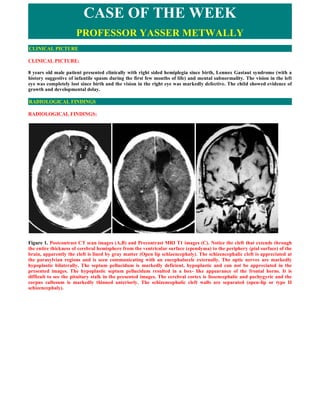
  • 1. CASE OF THE WEEK PROFESSOR YASSER METWALLY CLINICAL PICTURE CLINICAL PICTURE: 8 years old male patient presented clinically with right sided hemiplegia since birth, Lennox Gastaut syndrome (with a history suggestive of infantile spasm during the first few months of life) and mental subnormality. The vision in the left eye was completely lost since birth and the vision in the right eye was markedly defective. The child showed evidence of growth and developmental delay. RADIOLOGICAL FINDINGS RADIOLOGICAL FINDINGS: Figure 1. Postcontrast CT scan images (A,B) and Precontrast MRI T1 images (C). Notice the cleft that extends through the entire thickness of cerebral hemisphere from the ventricular surface (ependyma) to the periphery (pial surface) of the brain, apparently the cleft is lined by gray matter (Open lip schizencephaly). The schizencephalic cleft is appreciated at the parasylvian regions and is seen communicating with an encephalocele externally. The optic nerves are markedly hypoplastic bilaterally. The septum pellucidum is markedly deficient, hypoplastic and can not be appreciated in the presented images. The hypoplastic septum pellucidum resulted in a box- like appearance of the frontal horns. It is difficult to see the pituitary stalk in the presented images. The cerebral cortex is lissencephalic and pachygyric and the corpus callosum is markedly thinned anteriorly. The schizencephalic cleft walls are separated (open-lip or type II schizencephaly).
  • 2. Figure 2. Septo-optic dysplasia. Precontrast MRI T1 images. Notice the cleft that extends through the entire thickness of cerebral hemisphere from the ventricular surface (ependyma) to the periphery (pial surface) of the brain, apparently the cleft is lined by gray matter (Open lip schizencephaly). The schizencephalic cleft is appreciated at the parasylvian regions and is seen communicating with an encephalocele externally. The optic nerves are markedly hypoplastic bilaterally. The septum pellucidum is markedly deficient, hypoplastic and can not be appreciated in the presented images. The hypoplastic septum pellucidum resulted in a box- like appearance of the frontal horns. It is difficult to see the pituitary stalk in the presented images. The cerebral cortex is lissencephalic and pachygyric and the corpus callosum is markedly thinned anteriorly (B,C). The schizencephalic cleft walls are separated (open-lip or type II schizencephaly). Figure 3. Septo-optic dysplasia. Precontrast MRI T1 images. Notice the cleft that extends through the entire thickness of cerebral hemisphere from the ventricular surface (ependyma) to the periphery (pial surface) of the brain, apparently the cleft is lined by gray matter (Open lip schizencephaly). The schizencephalic cleft is appreciated at the parasylvian regions and is seen communicating with an encephalocele externally. The optic nerves (A) and optic chiasma (B) are markedly hypoplastic bilaterally. The septum pellucidum is markedly deficient, hypoplastic and can not be appreciated in the presented images. The hypoplastic septum pellucidum resulted in a box- like appearance of the frontal horns. It is difficult to see the pituitary stalk in the presented images. The cerebral cortex is lissencephalic and pachygyric and the corpus callosum is markedly thinned anteriorly (A). The schizencephalic cleft walls are separated (open-lip or type II schizencephaly). The pituitary gland appeared vacuolated (B)
  • 3. Figure 4. Septo-optic dysplasia. Precontrast MRI T1 images. Notice the cleft that extends through the entire thickness of cerebral hemisphere from the ventricular surface (ependyma) to the periphery (pial surface) of the brain, apparently the cleft is lined by gray matter (Open lip schizencephaly). The schizencephalic cleft is appreciated at the parasylvian regions and is seen communicating with an encephalocele externally. The optic nerves (A) and optic chiasma (B) are markedly hypoplastic bilaterally. The septum pellucidum is markedly deficient, hypoplastic and can not be appreciated in the presented images. The hypoplastic septum pellucidum resulted in a box- like appearance of the frontal horns. It is difficult to see the pituitary stalk in the presented images. The cerebral cortex is lissencephalic and pachygyric. The schizencephalic cleft walls are separated (open-lip or type II schizencephaly). The pituitary gland appeared vacuolated (B) The encephalocele and the skull wall expansion that are present at the opening of the open-lip schizencephaly (in the presented case) is believed to result from CSF pulsations from the lateral ventricles transmitted through the cleft. [45] Case summary  Open-lip or type II schizencephaly  Deficient and hypoplastic septum pellucidum  Dysplatic cerebral cortex (The cerebral cortex is lissencephalic and pachygyric)  The pituitary gland appeared vacuolated  Hypoplastic optic nerves and optic chiasma  Hypoplastic corpus callosum  Encephalocele DIAGNOSIS: DIAGNOSIS: SEPTO-OPTIC DYSPLASIA WITH OPEN LIP SCHIZENCEPHALY DISCUSSION DISCUSSION:  Septo-optic dysplasia (de Morsier syndrome) Septo-optic dysplasia (de Morsier syndrome) is a disorder characterized by the absence of the septum pellucidum, optic nerve hypoplasia, and hypothalamic dysfunction. It may be associated with agenesis of the corpus callosum. This disorder should be considered in any patient who exhibits at least two of the above abnormalities and perhaps even solely hypothalamic dysfunction [2]. Septo-optic dysplasia also appears to involve prosencephalic cleavage and development of anterior telencephalic structures [3]. About 50% of patients with septo-optic dysplasia have schizencephaly [1]. Patients may present with visual disturbance, seizures, mental retardation, hemiparesis (especially if associated with schizencephaly), quadriparesis, or hypothalamic dysfunction. Endocrine abnormalities may include growth hormone, thyroid hormone, or antidiuretic hormone function or levels. The consideration of septo-optic dysplasia necessitates an evaluation of the hypothalamic-pituitary axis because as many as 60% of the children with this disorder might exhibit
  • 4. evidence of a disturbance of endocrine function [4]. This evaluation can include thyroid function studies and electrolytes; these patients are at high risk for growth retardation. The recent identification of patients with this condition that harbor mutations in the transcriptional regulator gene HESX1, suggest that the mechanism of this disorder is likely genetic and a patterning or segmental abnormality [5]. Even though the genetic abnormality has been identified for a minority of patients, there exists the possibility that this may not represent an entirely genetic disorder because associations have been made with young maternal age, diabetes, the use of anticonvulsants, phencyclidine, cocaine, and alcohol [6,45]  Pathology of septo-optic dysplasia Several congenital malformations might be associated with macroscopic malformations of the septal nuclei. Most of them involve the midline structures, including the septum pellucidum. For example, septo-optic dysplasia is a syndrome characterized by the absence of the septum pellucidum and optic nerve hypoplasia It may also be associated with pituitary insufficiency resulting in growth and developmental delay. The septal nuclei abnormalities are often an element of a larger cerebral malformation, such as holoprosencephaly, schizencephaly, or porencephaly. In most cases, other midline structures such as the optic nerves, hypothalamus, and septum pellucidum are involved. Absence of the septum pellucidum may reach 80% to 90% in conditions such as schizencephaly. Focal heterotopias or cortical dysplasias may be associated with epileptic seizures. Severe mental retardation and intractable seizures are typical features of the more common larger cerebral malformations and of the syndromes associated with chromosomal anomalies. In some cases of corpus callosum agenesis, it is possible to discern a few axon bundles in an anteroposterior orientation that attempted to cross posteriorly. These are called Probst's bundles. If the corpus callosum is absent, the subcallosal interhemispheric tracts such as the anterior, posterior, and hippocampal commissure might be enlarged. The presence of Probst's bundles is considered by some to indicate that the agenesis of the corpus callosum is due to a primary cause and not the result of secondary injury such as hypoxia. These primary causes include chromosomal abnormalities, single gene defects, and metabolic defects. Cysts and lipomas may be associated with absence of the corpus callosum. [45] Large occiput encephaloceles may include significant parts of the brain, inducing hypoplasia of the anterior and middle cranial fossa. The brain in the encephalocele could show ischemia or polymicrogyria or be normal. The hypoplasia of the anterior and middle cranial fossa may stretch the septal nuclei, fornix, and septum pellucidum, among other structures. In contrast, transsphenoidal and sphenoethmoidal encephaloceles may directly involve the septal nuclei because they may be included in the herniated brain. [45]  Neuroendocrine disorders in septo-optic dysplasia Syndromes that may be associated with pituitary-hypothalamic dysfunction include septo-optic dysplasia, Kallmann's syndrome, and empty sella syndrome. In addition, other developmental anomalies may be associated with pituitary- hypothalamic defects, including agenesis of the corpus callosum, holoprosencephaly, and basal cephaloceles. [45] Septo-optic dysplasia, a disorder of ventral induction, is considered a mild form of holoprosencephaly. It is a disorder of abnormal induction of the midline mesoderm occurring at the same time as the development of the optic vesicles. Failure of differentiation of the mesoderm into the optic stalk results in aplasia of the stalk. [15] Septo-optic dysplasia is characterized by absence or hypoplasia of the septum pellucidum associated with hypoplasia of the optic nerves as first described by DeMorsier. [12] Associated hypothalamic-pituitary dysfunction occurs in two thirds of patients. [17,18,19] Patients may have hypopituitarism that ranges from panhypopituitarism to growth hormone deficiency, thyroid- stimulating hormone deficiency, elevated prolactin, or ACTH or ADH deficiency. [16] Visual symptoms include nystagmus, amblyopia, or hemianopsia. This syndrome may result from in utero injury or genetic abnormalities. [9] Environmental factors implicated are maternal diabetes, quinidine ingestion, anticonvulsants, alcohol or drug abuse, and cytomegalovirus. [17,45] There may be associated anomalies of neuronal migration, such as schizencephaly and gray matter heterotopias. On imaging, there is absence or hypoplasia of the septum pellucidum resulting in a box- like appearance of the frontal horns. [8,13,14] Hypoplasia of the optic disks is often diagnosed by an ophthalmologist; however, optic nerve, optic tract, or optic chiasm hypoplasia may be seen in 50% of patients on imaging. [8] Other associated abnormalities include absence of the fornix and callosal dysgenesis. [11,45] On MR imaging, three patterns of involvement have been described in septo-optic dysplasia. [8,11.45] 1. One group has neuronal migration anomalies such as schizencephaly and gray matter heterotopias, hypothalamic- pituitary dysfunction, and partial absence of the septum pellucidum. [45] 2. The other group has complete absence of the septum pellucidum with cerebral white matter hypoplasia. This group may be associated with hypoplasia of the genu of the corpus callosum or hypoplasia of the anterior falx cerebri. 3. Another group of these patients with ectopia of the posterior pituitary has also been described. [11] Other anomalies that may be associated with complete or partial absence of the septum pellucidum include holoprosencephaly, hydranencephaly, basilar cephaloceles, dysgenesis of the corpus callosum, chronic hydrocephalus,
  • 5. Chiari II malformation, and schizencephaly. [7,10,20]  Optic nerve hypoplasia in septo-optic dysplasia Optic nerve hypoplasia, a specific clinical entity, is an increasingly common cause of infantile blindness in the United States. It is characterized by a small optic disc surface area and thin optic nerve caused by a congenital nonprogressive decrease in the number of optic nerve axons. Unilateral and bilateral optic nerve hypoplasia occurs with roughly equal frequency. Various pathophysiologic theories have been proposed, including failure of axons to normally develop; a destructive developmental event, such as a vascular insult; exaggerated apoptosis; or a genetic disorder. [45] All patients with optic nerve hypoplasia show visual field defects; localized defects as well as generalized constriction are seen. The loss of visual acuity is variable depending on the degree of involvement with axons from the macula, the center of sight. [45] Optic nerve hypoplasia is recognized as part of a spectrum of optic nerve, central nervous system (CNS), and hypothalamic-pituitary axis congenital anomalies [21]. From the ophthalmologist's perspective, optic nerve hypoplasia can be assigned into five sometimes overlapping groups: isolated unilateral or bilateral optic nerve hypoplasia, with absence of the septum pellucidum (septo-optic dysplasia), with posterior pituitary ectopia or absence of the pituitary stalk, with hemispheric migration anomalies, or with intrauterine and/or perinatal hemispheric injuries [22]. Optic nerve hypoplasia is also associated with a variety of ocular and systemic disorders, including aniridia, albinism, maternal diabetes, fetal alcohol syndrome, and other maternal ingestions [23,24,25]. Groups with optic nerve hypoplasia  Isolated unilateral or bilateral optic nerve hypoplasia  Optic nerve hypoplasia with absence of the septum pellucidum (septo-optic dysplasia)  Optic nerve hypoplasia with posterior pituitary ectopia or absence of the pituitary stalk  Optic nerve hypoplasia with hemispheric migration anomalies  Optic nerve hypoplasia with intrauterine and/or perinatal hemispheric injuries  Schizencephaly in septo-optic dysplasia Schizencephaly is the most complete form of migrational disorders. It is thought to be caused by a complete agenesis of a section of the cerebral tissue, which results in clefts that extend through the entire thickness of cerebral hemisphere. At the margin of the cleft the pial membrane and ependymal lining of the ventricle lie adjacent to each other and form a pial ependymal seam [29] Schizencephaly probably occurs as the end result of a variety of insults occuring at a critical time and in a critical location during brain development. No specific inciting or unusual prenatal events are described. The lesion is most likely related to multiple etiologies including genetic, toxic, metabolic, vascular or infectious disease. Familial cases have been reported. [30] Septooptic dysplasia, also called as De Morsier disease is a syndrome consisting of blindness, hypoplastic optic nerves and absence of septum pellucidum in females. [29] It is known that Schizencephaly and septo-optic dysplasia frequently coexists. [31] Associated anomalies are heterotopias, Dandy Walker malformation, hydrocephalus and polymicrogyria. [28] Cleft may be unilateral or bilateral, symmetrical or asymmetrical. Commonly located near pre/post central gyrus [26.27]] The cleft in Schizencephaly are lined either totally or in part by gray matter and extend from pial surface to ependyma of the lateral ventricle. The clefts can be located anywhere but commonly occur in parasylvian regions. The cavity formed in open lip type varies in size from small to large and may communicate with lateral ventricle. The ventricular system may be enlarged, particularly in-patients with open lip form of Schizencephaly. [30] Absence of septum pellucidum, dysgenesis of corpus callosum are often associated with open lip Schizencephaly [28,29] Differential diagnosis includes subarachnoid cysts and porencephaly. Using CT diagnosis of Schizencephaly is sometimes difficult particularly in case of type I. CT may show a slight outpouching at the ependymal surface of cleft and a full thickness cleft may be difficult to identify on CT Scan .Secondary findings like hydrocephalus, heterotopia, polymicrogyria, subdural hygromas and arachnoid cysts can be identified. [30] MRI gives most detailed and precise definition of anatomy and anomaly. For anatomy MRI T1 images suffice [28] MRI is the modality of choice. MRI better delineates the gray matter lining the cleft, which is pathognomonic finding in Schizencephaly.MRI also provides superb cortical anatomy detail and multiplanar capacity. Primary findings related to the cleft and secondary findings associated with Schizencephaly are identified using MRI.The ability of MRI pulse sequence to differentiate gray matter and white matter permit demonstration of gray matter heterotopias in the subcortical white matter beneath the cleft, abnormalities involving the cortex (pachygyria and polymicrogyria) and other secondary findings are also identified. Homolateral absence of sylvian vasculature, small medullary pyramids low position of fornix and thinning of the corpus callosum are findings related to absent cerebral cortex and are better demonstrated by MRI than with other studies. Sonography can be done in neonatal period in-patients in whom this anomaly is suspected. In Schizencephaly type I, a
  • 6. hyperechoic line extends from the parasylvian region to the anterior portion of lateral ventricle. The hyperechoic line represents the cortex lining the fused cleft. This type of anomaly is difficult to detect with ultrasound and requires high index of suspicion and highly skilled operator. In Schizencephaly type II an anechoic band or cavity, representing the fluid filled cleft extends from the cortical surface of lateral ventricle. The meeting of the closed lip portion or apex of the cleft with the margin of ventricle may be identified as a ventricular diverticulum or dimple. The size of the caudate, thalamus and lenticular nuclei (subcortical gray matter structures) is decreased. Other associated anomalies such as ventricular enlargement may also be identified. [30] This case represents a typical association of septo-optic dysplasia with open lip Schizencephaly with all cardinal neurological cardinal features on one hand and a rare form of unilateral open lip cleft on the other. It highlights the importance of MRI in diagnosis of such a condition. SCHIZENCEPHALY Schizencephaly is an uncommon disorder of neuronal migrational characterized by a cerebrospinal fluid–filled cleft, which is lined by gray matter. The cleft extends across the entire cerebral hemisphere, from the ventricular surface (ependyma) to the periphery (pial surface) of the brain. The clefts may be unilateral or bilateral and may be closed (fused lips), as in schizencephaly type I, or separated (open lips), as in schizencephaly type II. Presentation and outcome are variable, but patients typically present with seizures, hemiparesis, and developmental deficits. Usually, the severity of symptoms is related to the amount of brain affected by the abnormality.  Aetiology and types of schizencephaly Several theories have been proposed to explain the etiology of schizencephaly, although none is universally accepted. The leading theory indicates that schizencephaly results from an early, focal destruction of the germinal matrix and surrounding brain before the hemispheres are fully formed. Schizencephaly probably occurs as the end result of a variety of insults occurring at a critical time and in a critical location during brain development. No specific inciting or unusual prenatal events have been identified, and reported cases are sporadic. The lesion is most likely related to multiple etiologies, including genetic, toxic, metabolic, vascular, or infectious causes. Familial cases of schizencephaly have been reported. Schizencephaly is uncommon; to our knowledge, there are no documented geographical differences in its occurrence. Severity of the symptoms depends on the amount of brain involved. Affected patients typically have seizures, hemiparesis, variable developmental delay, and blindness. Patients also have variable degrees of mental retardation. Patients with open-lip schizencephaly die at an earlier age than patients with the closed-lip form. Usually, death results from failure to thrive, chronic infections, and respiratory problems. Patients with closed-lip schizencephaly may not present clinically until later in infancy or early childhood and may live to early adulthood. Schizencephaly is divided into 2 types, which have prognostic significance. In closed-lip or type I schizencephaly, the cleft walls are in apposition. In open-lip or type II schizencephaly, the cleft walls are separated. Schizencephaly type II occurs more commonly than type I. The clefts in schizencephaly are lined either totally or in part by gray matter and extend from the pial surface to the ependyma of the lateral ventricle. The clefts can be located anywhere, but they commonly occur in the parasylvian regions. The clefts can be unilateral or bilateral, and can be either symmetric or asymmetric. The cavity formed in the open-lip type varies in size from small to large and may communicate with the lateral ventricle. The ventricular system may be enlarged, particularly in patients with the open-lip form of schizencephaly. Gray-matter heterotopia (collections of gray matter in abnormal locations), polymicrogyria, and arachnoid cysts can be associated with schizencephaly. Heterotopias and polymicrogyria typically line the clefts. Microcephaly has been noted in some patients. The septum pellucidum is absent in 80-90% of patients, and schizencephaly may coexist with septo-optic dysplasia.  Clinical picture of schizencephaly Clinical features of schizencephaly are highly variable. Patients with unilateral clefts with fused lips may have mild hemiparesis and seizures but otherwise have normal development. When the cleft is open, patients present with mild-to- moderate developmental delay and hemiparesis; severity is related to the extent of cortex involved in the defect. Patients with bilateral clefts present with severe mental deficits and severe motor anomalies including spastic quadriparesis. Frequently, these patients present with blindness, which is often associated with optic nerve hypoplasia. Language development is more likely to be normal in patients with unilateral schizencephaly compared to patients with bilateral clefts. Intractable seizures frequently are noted in schizencephaly. Several types of seizures have been reported including generalized tonic-clonic, partial motor, and sensory seizures. Infantile spasms have been seen in a few children. Reports
  • 7. have found no correlation between the subtype of schizencephaly and the occurrence or type of seizure disorder. Identification of gray matter lining the cleft is the pathognomonic finding in differentiating schizencephaly from porencephaly; this is best demonstrated on MRIs. The more complete information obtained by MRI enables a more accurate prediction of neurologic outcome. Figure 5. Open-lip schizencephaly with cortical dysplasia Figure 6. Bilateral open lip schizencephaly  Neuroimaging of schizencephaly  CT scan Using CT, the diagnosis of schizencephaly is sometimes difficult, particularly type I, or closed lip schizencephaly. CT scans of closed-lip schizencephaly may show only a slight outpouching at the ependymal surface of the cleft, and a full- thickness cleft may be difficult to identify on CT scan. The cleft is partially or totally lined by gray matter and extends from the lateral ventricle to the pial surface of the cerebral hemisphere. Secondary findings that can be identified on CT
  • 8. scan include hydrocephalus, heterotopia, polymicrogyria, subdural hygromas, and arachnoid cysts.  MRI MRI is the modality of choice for evaluating patients with schizencephaly. MRI better delineates the gray matter lining the cleft, which is the pathognomonic finding in schizencephaly. MRI also provides superb cortical anatomy detail and multiplanar capability. Primary findings related to the cleft and secondary findings associated with schizencephaly are identified using MRI. The ability of MRI pulse sequences to differentiate gray matter and white matter permits demonstration of gray-matter heterotopias in the subcortical white matter beneath the cleft, abnormalities involving the cortex (eg, pachygyria or polymicrogyria), and other secondary findings also identified by using CT scans. Homolateral absence of the sylvian vasculature, small medullary pyramids, a low position of the fornix, and thinning of the corpus callosum are findings related to absent cerebral cortex and are better demonstrated by MRI than with other studies. Figure 7. A, Schizencephaly. Axial T2-weighted MRI in unilateral closed-lip (type I) schizencephaly. The cleft is lined by gray matter and extends from the pial surface to the lateral ventricle. B, Schizencephaly. Axial T2-weighted (left) and coronal T1-weighted (right) MRIs in bilateral closed-lip (type I) schizencephaly. A ventricular diverticulum defines the meeting of the closed-lip portion of the clefts with the margin of the ventricles. The septum pellucidum is absent, and the clefts are lined by gray matter and extend from the pial surface to the lateral ventricle. C, Schizencephaly. Axial T2- weighted MRI demonstrates a small open-lip schizencephaly. The septum pellucidum is absent. D, Schizencephaly. Axial T2-weighted MRI in unilateral open-lip (type II) schizencephaly. The septum pellucidum is absent, and a large cerebrospinal fluid–filled cleft extends from the lateral ventricle to the cortical surface. The cleft is lined by gray matter. E, Schizencephaly. Coronal sonograms with a corresponding coronal T1-weighted MRI of open-lip bilateral schizencephaly. Extensive bilateral schizencephalic defects with large CSF-filled clefts extend from the lateral ventricles to the cortical surface. SUMMARY SUMMARY Schizencephaly is the term describing gray matter-lined clefts that extend through the entire hemisphere from the lateral ventricles to the cortical surface, characterized pathologically by a pial-ependymal continuity [28,29]. The gray matter lining these clefts is dysplastic and has irregular inner and outer surfaces, identical to that of polymicrogyria. The clefts can be unilateral or bilateral and are most commonly located near the pre- and postcentral gyri, locations almost
  • 9. identical to those in which polymicrogyria occurs [22,23]. It is probable that schizencephaly represents an extreme variant of cortical dysplasia, in which the infolding of cortex extends all the way into the lateral ventricle [22,23]. The lips of the cleft may be fused, in which case the walls are directly apposed, obliterating the CSF space within the cleft at that point. When the lips are separated, CSF fills the cleft from the lateral ventricle to the subarachnoid space surrounding the hemispheres [23,28-30]. As in polymicrogyria, large vessels are often seen in the cleft between the lips of the schizencephaly. The severity of the clinical symptoms is related to the amount of involved brain [23, 31,32]. Patients with a unilateral cleft with fused lips typically present with mild hemiparesis or epilepsy but are otherwise developmentally normal. Patients with unilateral clefts with separated lips more commonly present with hemiparesis and a mild to moderate developmental delay, depending on the location of the cleft within the brain. Patients with bilateral clefts tend to be severely retarded, with refractory seizures beginning at a very early age and severe motor anomalies, and they may appear to be blind. Optic nerve hypoplasia is seen in up to one-third of affected patients [8,30, 33,34]. Routine spin-echo MR obtained in at least two planes is usually adequate for imaging of schizencephalies. These studies show full-thickness clefts, lined by gray matter with an irregular inner surface, that extend through the hemisphere from the ventricle to the surface of the brain. The gray matter lining the cleft often extends into the ventricle as a subependymal heterotopia [6,8,23,30,32]. A dimple is usually seen in the wall of the lateral ventricle where it communicates with the cleft. The dimple provides a helpful sign of continuity of the cleft with the ventricle when the lips of the cleft are fused. The gyral pattern of the cortex adjacent to the cleft is usually abnormal, with features characteristic of polymicrogyria. Polymicrogyria may also be present in the hemisphere contralateral to a unilateral schizencephaly [22,23]. Therefore, the contralateral hemisphere should always be scrutinized. The calvarium is often expanded over the opening of an open-lip schizencephaly. The expansion is believed to result from CSF pulsations from the lateral ventricles transmitted through the cleft. When plagiocephaly is severe, insertion of a ventriculoperitoneal shunt may help to dampen the pulsations and reverse the cranial asymmetry.  Addendum  A new version of this PDF file (with a new case) is uploaded in my web site every week (every Saturday and remains available till Friday.)  To download the current version follow the link quot;http://pdf.yassermetwally.com/case.pdfquot;.  You can also download the current version from my web site at quot;http://yassermetwally.comquot;.  To download the software version of the publication (crow.exe) follow the link: http://neurology.yassermetwally.com/crow.zip  The case is also presented as a short case in PDF format, to download the short case follow the link: http://pdf.yassermetwally.com/short.pdf  At the end of each year, all the publications are compiled on a single CD-ROM, please contact the author to know more details.  Screen resolution is better set at 1024*768 pixel screen area for optimum display.  For an archive of the previously reported cases go to www.yassermetwally.net, then under pages in the right panel, scroll down and click on the text entry quot;downloadable case records in PDF formatquot; REFERENCES References [1] Aicardi J, Goutieres F. The syndrome of absence of the septum pellucidum with porencephalies and other developmental defects. Neuropediatrics 1981;12:319-29. [2] Brickman JM, Clements M, Tyrell R, et al. Molecular effects of novel mutations in Hesxl/HESXI associated with human pituitary disorders. Development 2001;128:5189-99. [3] Ouvier R, Billson F. Optic nerve hypoplasia: a review. J Child Neurol 1986;1:181-8. [4] Costin G, Murphree AL. Hypothalamic-pituitary function in children with optic nerve hypoplasia. Am J Dis Child 1985;139:249-54. [5] Dattani MT, Martinez-Barbera JP, Thomas PQ, et al. Mutations in the homeobox gene HESXI/Hesxl associated with septo-optic dysplasia in human and mouse. Nat Genet 1998; 19:125-33.
  • 10. [6] Patel H, Tze WJ, Crichton JU, et al. Optic nerve hypoplasia with hypopituitarism: septo- optic dysplasia with hypopituitarism. Am J Dis Child 1975;129:175-80. [7] Aicardi J, Goutieres F: The syndrome of absence of the septum pellucidum with porencephalies and other developmental defects. Neuropediatrics 12:319-329, 1981 [8] Barkovich A, Fram E, Norman D: Septooptic dysplasia: MR imaging. Radiology 171:189-192,1989 [9] Barkovich AJ: Pediatric Neuroimaging, ed 2. New York, Raven Press, 1995, pp 236-237 [10] Barkovich Aj, Norman D: Absence of the septum pellucidum: A useful sign in the diagnosis of congenital brain malformations. AJR Am j Roentgenol 152:353- 360,1989 [11] Brodsky M, Glasier C: Optic nerve hypoplasia: Clinical significance of associated central nervous system abnormalities on magnetic resonance imaging. Arch Ophthalmol 111:66-74,1993 {12] DeMorsier G: Etudes sur les dysraphics cranio-encephaliques 111. Agenese du septum lucidum avec malformation du tractus optique. La dysplasie septo- optic. Schweiz Arch Neurol Neurochir Psychiatry 77:267-292,1956 [13] Fitz C: Holoprosencephaly and related entities. Neuroradiology 25:3-238, 1983 [14] Fitz CR: Holoprosencephaly and septo-optic dysplasia. Neuroimaging Clin North Am 4:263-281, 1994 [15] Hale BR, Rice P: Septo-optic dysplasia: Clinical and embryological aspects. Dev Med Child Neurol 16: 812-817,1974 [16] Izenberg N, Rosenblum M, Parks JS: The endocrine spectrum of septo-optic dysplasia. Clin Pediatr (Phila) 23:632- 636, 1984 [17] Morishima A, Aranoff G: Syndrome of septooptic- pituitary dysplasia: The clinical spectrum. Brain Dev 8:233- 239,1986 [18] Skarf B, Hoyt C: Optic nerve hypoplasia in children: Association with anomalies of the endocrine and CNS. Arch Ophthalmol 102:62-67,1984 [19] Stanhope R, Preece M, Brook C: Hypoplastic optic nerves and pituitary dysfunction: A spectrum of anatomical and endocrine abnormalities. Arch Dis Child 59:111-114,1984 [20] Harwood-Nash D, Fitz C: Neuroradiology in Infants and Children. St. Louis, CV Mosby, 1976, pp 484-486 [21] Campbell CL. Septo-optic dysplasia: a literature review. Optometry. 2003;74(7):417–426. [22]Brodsky MC, Glasier CM. Optic nerve hypoplasia. Clinical significance of associated central nervous system abnormalities on magnetic resonance imaging. Arch Ophthalmol. 1993;111(1):66–74. [23]Brown GC. Optic nerve hypoplasia and colobomatous defects. J Pediatr Ophthalmol Strabismus. 1982;19(2):90–93. [24]Chan T, Bowell R, O'Keefe M, et al.. Ocular manifestations in fetal alcohol syndrome. Br J Ophthalmol. 1991;75 (9):524–526. [25]Mansour AM, Bitar FF, Traboulsi EI, et al.. Ocular pathology in congenital heart disease. Eye. 2004;19(1):29–34. [26] S.K.Sethi, R.S.Solanki Radiological Quiz. Indian J. Radiology and Imaging, 2004,14:1:95-96. [27] D.A Shah, G.D.Rathod, Radiological Quiz Ind J Radiology and Imaging, 2005,15:1:127-128. [28] Yutaka Sato, Simon C.S.Kao, and Wilbur L.Smith, RCNA, March 1991, Vol. 29,No 2 :179-194, [29] C.Raybaud Destructive lesions of Brain Neuroradiology 1983; 25:265-291. [30] Ken R.Close Schizencephaly www emedicine.com/radio/topic622.htm last updated April 7, 2004 [31] AJNR, Mar/Apr, 1988:9:297-302.:MR imaging of Schizencephaly; A.James Barkovich, David Norman. [32] Barkovich AJ, Norman D: MR imaging of schizencephaly. AJR Am J Roentgenol 1988 Jun; 150(6): 1391-6.
  • 11. [33] Barkovich AJ: Schizencephaly. In: Pediatric Neuroimaging. 2nd ed. Philadelphia: Lippincott-Raven; 1996:219-25. [34] Barth PG: Schizencephaly and nonlissencephalic cortical dysplasias. AJNR Am J Neuroradiol 1992 Jan-Feb; 13(1): 104-6. [35] Bird CR, Gilles FH: Type I schizencephaly: CT and neuropathologic findings. AJNR Am J Neuroradiol 1987 May- Jun; 8(3): 451-4. [36] Byrd SE, Osborn RE, Bohan TP, Naidich TP: The CT and MR evaluation of migrational disorders of the brain. Part II. Schizencephaly, heterotopia and polymicrogyria. Pediatr Radiol 1989; 19(4): 219-22. [37] Chamberlain MC, Press GA, Bejar RF: Neonatal schizencephaly: comparison of brain imaging. Pediatr Neurol 1990 Nov-Dec; 6(6): 382-7. [38] Denis D, Chateil JF, Brun M, et al: Schizencephaly: clinical and imaging features in 30 infantile cases. Brain Dev 2000 Dec; 22(8): 475-83. [39] DiPietro MA, Brody BA, Kuban K, Cole FS: Schizencephaly: rare cerebral malformation demonstrated by sonography. AJNR Am J Neuroradiol 1984 Mar-Apr; 5(2): 196-8. [40] Komarniski CA, Cyr DR, Mack LA, Weinberger E: Prenatal diagnosis of schizencephaly. J Ultrasound Med 1990 May; 9(5): 305-7. [41] Lee SH, Rao K, Zimmerman RA: Schizencephaly. In: Cranial MRI and CT. 4th ed. New York: McGraw-Hill; 1999: 163-5. [42] Miller GM, Stears JC, Guggenheim MA, Wilkening GN: Schizencephaly: a clinical and CT study. Neurology 1984 Aug; 34(8): 997-1001. [43] Osborne AG: Schizencephaly. In: Diagnostic Neuroradiology. St Louis: Mosby-Year Book; 1994:52-6. [44] Packard AM, Miller VS, Delgado MR: Schizencephaly: correlations of clinical and radiologic features. Neurology 1997 May; 48(5): 1427-34. [45] Metwally, MYM: Textbook of neuroimaging, A CD-ROM publication, (Metwally, MYM editor) WEB-CD agency for electronic publication, version 9.2a April 2008