Cardiovascular System
•
11 j'aime•5,844 vues

The human heart heart length, width, and thickness are 12 cm, 8.5 cm, and 6 cm, respectively. In addition, the mean weight of the heart is 280-340 g in males and 230-280 g in females.

Recommandé
Contenu connexe
Tendances
Tendances (20)
Similaire à Cardiovascular System
Similaire à Cardiovascular System (20)
Plus de Abhay Rajpoot
Plus de Abhay Rajpoot (20)
Dernier
Dernier (20)
Cardiovascular System
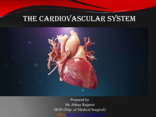
- 1. The Cardiovascular System Prepared by Mr.Abhay Rajpoot HOD (Dep. of Medical Surgical) abhayrajpoot5591@gmail.com
- 2. The Cardiovascular System Cardiovascularsystem: organ system that distributes blood to all parts of thebody Major function – transportation, using blood as thetransport vehicle
- 3. The Cardiovascular System This system carries oxygen, nutrients, cell wastes, hormones and other substances vital for body homeostasis to and formcells The force to move blood around the body is provided by the pumping heart and blood pressure
- 5. The Heart The human heart heart length, width, and thickness are 12 cm, 8.5 cm, and 6 cm, respectively. In addition, the mean weight of the heart is 280-340 g in males and 230-280 g in females. It is enclosed within the inferior mediastinum, the medial cavity of the thorax, and flanked on each side by the lungs
- 6. The Heart The pointed apex is directed toward the left hipand restsat about the fifth intercostal space The broad aspect, or base, points towardthe right shoulder and lies beneath the second rib
- 7. The Heart The heart is enclosed by a double-walled sac called the pericardium The superficial loosely fitted part is called the fibrous pericardium Protects and anchors theheart
- 8. The Heart
- 9. The Heart Deep to the fibrous pericardium is the slippery, two-layer serouspericardium The parietal layer lines the interior of the fibrous pericardium
- 10. The Heart The parietal layerattaches to the large arteries leaving the heart and then makes a U-turn and continues inferiorly overthe heart surfaceas thevisceral layer, or epicardium
- 11. The Heart A slippery lubricating fluid is produced by the serous pericardial membranes which allows the heart to beat easily in a relative frictionless environment
- 12. The Heart Inflammation of the pericardium, pericarditis,often results in a decrease in the serous fluid The cause the pericardial layers to stick, forming painful adhesions that interfere with heartmovements
- 13. The Heart The heartwallsarecomposed of three layers: 1. outerepicardium 2. myocardium 3. endocardium
- 14. The Heart The myocardium consists of thick bundles of the cardiac muscle twisted into ring like arrangements This is the layer ofthe heart that actually contracts Reinforced by dense, fibrous connectivetissue (“heart skeleton”)
- 15. The Heart The endocardium is a thin, glistening sheetof endothelium that lines the heartchambers Continuouswith the liningsof the blood vessels leaving and entering theheart
- 16. The Heart The heart has four hollowchambers: 2 atria – receivingchambers 2 ventricles – fillingchambers
- 17. The Heart Blood flows intothe atria under low pressure from the veins, andcontinues into theventricles
- 18. The Heart The ventricles are thick- walled discharging chambers They are the pumpsof the heart When they contract, blood is propelled out of the heart and into circulation The right ventricle forms most of the heart’s anterior surface The left ventricle forms theapex
- 20. The Heart The septum that divides the heart longitudinally is the interventricular septum or theinteratrial septum based on the chambers itseparates
- 21. The Heart The heart functionsas a doublepump The right sideworksas the pulmonarycircuit pump Receives relatively oxygen-poor blood from the veins of the body through the large superior and inferior vena cava
- 22. The Heart
- 23. The Heart The blood then pumps out through the pulmonary trunk which splits into the left and right pulmonary arteries The pulmonary arteries carry blood to thelungs, where oxygen is picked up and carbon dioxideis unloaded Oxygen-rich blood drains from the lungs and is returned tothe leftsideof the heart through the four pulmonaryveins
- 24. The Heart This circuit is call pulmonarycirculation Itsonly function is tocarry blood to the lungs forgas exchange and then return it to the heart
- 26. The Heart Blood returned to the leftside of the heart is pumped out of the heart into the aorta The systemicarteries branch from theaorta tosupply the body tissues withblood Oxygen-poor blood circulates from the tissues back to the right atrium via the systemic veins, which empty their blood into either the superior or inferior vena cava
- 27. The Heart This second circuit, from the left side of the heart through the body tissuesand back to the right side of the heart is called systemiccirculation Itsuppliesoxygen and nutrient-rich blood toall body organs Because the left ventricle is the systemic pump that pumps blood overa much longerpathway through the body, its walls are thicker than those of the right ventricle It is a more powerfulpump
- 29. The Heart The heart also has four valves: 2 that separatethe atria from the ventricles 2 that separate the ventricles fromtheir arteries All of these valves prevent back flow
- 30. The Heart The atrioventricular (AV) valves are between the atria and ventricles On the left is the bicuspid or mitral valve On the right is the tricuspid valve
- 31. The Heart When the heart is relaxed and blood is passively filling its chambers, the AV-valve flaps hang limply into the ventricles As the ventricles contract, they press on the blood in theirchamber, and the intraventricular pressure rises
- 32. The Heart The semilunar valves guard the bases of the large arteries leavingthe ventricularchambers On the right isthe pulmonary valve On the left is theaortic valve When the ventricles are contracting these valves are forced openand flattened againstthe arterial walls When the ventricles are relaxed the blood flows back towards theheart This prevents arterial blood fromreentering the heart
- 33. The Heart The coronary arteries branch from the base of the aortaand encircle the heart in thecoronary sulcus (AV groove) at the junctionof theatriaand ventricles
- 34. The Heart The coronary arteries and their major branches are compressed when the ventricles are contracting and fill when the heart is relaxed
- 35. The Heart The myocardium is drained by several cardiacveins, which empty into thecoronary sinus The coronary sinus, in turn, empties into the right atrium
- 36. The Heart When the heart beats rapidly the myocardiumcan received an inadequate amount of blood This can result in crushing chest pain called angina pectoris
- 37. The Heart Pain due toangina pectoris is a warningsign If angina is prolonged, oxygen-deprived heartcells may die forming aninfarct The resulting myocardial infarction is a “heart attack”
- 39. Heart Physiology Although cardiac muscle can beat independently, the muscle cells on different areas of theheart have differentrhythms Atrial cells – 60 bpm Ventricular cells –20-40 bpm
- 40. Heart Physiology Twosystemsact to regulate heartactivity: 1. Autonomic nervoussystem – brakes and accelerator Acts todecrease or increase heart rate 2. Intrinsic conduction system (nodalsystem) Composed of specialized tissue that is across between muscle and nervoustissue Causes heart muscledepolarization from theatria to theventricles Enforces contraction rate ~ 74bpm
- 41. Heart Physiology
- 42. Heart Physiology Components of the Intrinsic ConductionSystem include: The sinoatrial (SA) node is acrescentshaped node in the rightatrium Theatrioventricular (AV) node is at the junctionof the atria andventricles The atrioventricular (AV) bundle (bundle ofHis) Branch bundles in the interventricular septum Purkinje fibers which spread with the muscleof the ventriclewalls
- 43. Heart Physiology
- 44. Heart Physiology The SA node has the highest rate of depolarization in the wholesystem It starts each heartbeat and sets the pace forthe whole heart and is therefore called the pacemaker
- 45. Heart Physiology
- 46. Heart Physiology The impulse travels from the SA node through the atria to the AV node, causing theatria tocontract At the AVnode, the impulse is delayed togive the atria time to finish contracting It then passes rapidly through the AVbundle,the bundle branches, and the Purkinje fibers, causing a “wringing” contraction of the ventricles that begins at the apex and moves toward theatria
- 47. Heart Physiology This contraction effectivelyejects blood superiorly into the large arteries leaving theheart
- 48. Heart Physiology Tachycardia is a rapid heart rate (> 100bpm) Bradycardia is a slow heart rate (< 60 bpm) Neither condition is pathological, butprolonged tachycardia may progress tofibrillation
- 49. Fibrillation is a rapid, uncoordinated shuddering of the heart muscle Fibrillation makes the heart totallyuselessasa pump and is a major cause of death from heart attacks inadults Heart Physiology
- 50. Heart Physiology A pacemaker is a small device, about the sizeof a half dollar piece, placed under the skin near the heart to help control the heartbeat. A pacemaker isimplanted as part of what's often referred to as "cardiac resynchronization therapy."
- 51. Heart Physiology People may need a pacemaker fora variety of reasons — mostly due to one of a group of conditions called arrhythmias, in which the heart's rhythm isabnormal They can be implantedtemporarily to treat a slow heartbeat after a heartattack, surgeryoroverdoseof medication Pacemakers can also be implanted permanently to correctbradycardia or to help treat heart failure
- 52. Cardiac Cycle and Heart Sounds
- 53. Cardiac Cycle and Heart Sounds In a healthy heart, theatria contractsimultaneously When theystart torelax, contraction of theventricles begins Systoleand diastole mean heartcontraction and relaxation respectively
- 54. Cardiac Cycle and Heart Sounds Because most of the pumping work is done by the ventricles, these terms always refer to the contraction and relaxation of theventricles unlessotherwisestated
- 55. Cardiac Cycle and Heart Sounds The term cardiac cycle refers to the events of one complete heartbeat, during which both atria and ventricles contract and then relax
- 56. Cardiac Cycle and Heart Sounds The average heart beats 74 times perminute Theaverage length of a cardiac cycle is 0.8 seconds Thecardiac cycleoccurs in three majorsteps: 1. mid-to-latediastole 2. ventricularsystole 3. earlydiastole
- 57. 1. Mid-to-late diastole The heart is in complete relaxation Pressure in the heart islow Blood is flowing passively into and through the atria and into theventricles from pulmonaryand systemic circulations
- 58. 1. Mid-to-late diastole The semilunarvalvesareclosed The AVvalves are open Then theatria contractand force the blood into the ventricles
- 59. Cardiac Cycle
- 60. 2. Ventricular systole The pressure within the ventricles increases rapidly, closing the AVvalves When the intraventricular pressure is higher than the pressure in the large arteries leaving the heart, the semilunar valves are forced open, and blood rushes out of theventricles The atriaare relaxed, and again are filling with blood
- 61. Cardiac Cycle
- 62. 3. Early diastole At the end of systole, the ventricles relax, the semilunarvalvessnapshut, and fora moment the ventricles are completelyclosed chambers
- 63. 3. Early diastole During early diastole, the intraventricular pressure drops When it drops below the pressure in the atria, the AV valves are forced open. And the ventricles again begin to refill rapidly withblood
- 64. Heart Sounds When using a stethoscope, the heart beat usually has two distinct sounds – “lup” and“dup” Thesearecaused by theclosing of the twosets of valves “lup” – AVvalves “dup” – semilunarvalves
- 66. Cardiac Output Cardiac Output (CO) is the amount of blood pumped out by eachside of the heart in 1 minute It is the product of heart rate (HR) and stroke volume (SV)
- 67. Cardiac Output In general, strokevolume increases as the forceof ventricular contractionincreases Let’s look at normal resting heart rateand volume: CO = HR x SV CO = (74 bpm) x (70 ml perbeat) CO = 5180 ml/min
- 68. Cardiac Output A healthy heart pumps out about 60% of blood in the ventricles (~70ml) per heart beat The critical factor is how much the cardiac muscle cells stretch just before contracting
- 69. Cardiac Output The important factor stretching the heart muscle is venous return, the amountof blood entering the heart and distending theventricles The more the heart muscles stretch,the stronger the contraction
- 70. Cardiac Output If one side of the heart suddenly begins to pump more blood than the other, the increased venous return to the opposite ventricle will force it to pump out an equal amount, thus preventing backup of blood in the circulation
- 71. Cardiac Output The enhanced squeezing action of active skeletal muscles from exercisespeeds upvenousreturn Severe blood loss or rapidheart rate, decreases stroke volume, creating less venousreturn
- 72. Factors Modifying Basic Heart Rate Heart contraction does not depend on the nervous system, but itcan bechanged temporarily by the ANS It is also modified bychemicals, hormones and ions
- 73. Neural (ANS) Control During times of physical or emotional stress, the nervesof sympatheticdivision stimulatethe SA and AVnodes and the cardiacmuscles The heart beats morerapidly
- 74. Neural (ANS) Control When the demand declines, the heart adjusts, the parasympathetic nerves slowand steady the heart rate Gives the heart time to recoverand rest
- 75. Neural (ANS) Control In patientswith Congestive Heart Failure (CHF), or other heartdisease the heart pumpsweakly Some medicationscan be used toenhance contractile force and stroke volume of the heart, improving cardiac output
- 77. Neural (ANS) Control Various hormonesand ions havea dramaticeffecton heartactivity Epinephrine – mimics sympathetic nerves,increases heart rate Thyroxine – increase heartrate Electrolyte imbalance – prolongedcontractions, arrhythmias, decreaseoutput
- 78. Physical Factors Resting heart rate is fastest in the fetusand then graduallydecreases Faster heart rate in females thanmales High body temperaturealso increase heart rate, Low body temperature decreases heartrate