2. PAROTID REGION IS THE REGION THAT LOCATED
IN THE POSTEROLATERAL PART OF THE FACIAL
REGION
2
3. The parotid region
Site: it lies below auricle occupying thedeep between
the ramus of mandible and sternomastiod.
Boundaries
Superiorly :Zygomatic arch.
inferiorly : Angle and inferior border of the mandible.
posteriorly: External ear and anterior border
of the sternocleidomastoid.
Anteriorly Anterior border of the.
masseter muscle
medially Ramus of the mandible.
3
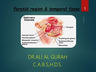
6. Parotid Gland :
Largest of the 3 main salivary glands in the
head & neck
Composed mostly of serous acini.
Enclosed within a tough, unyielding, fascial
capsule, the parotid sheath (capsule), derived
from the investing layer of deep cervical
fascia.
6
7. Surface anatomy of the parotid
gland :
4points:
A-point on the tragus of the ear.
B-point at center of the mastoid process.
C-point 2cm below and behind
angle of the mandible.
D-point at the centre of
the mandibular notch.
7
8. Surface anatomy of the parotid gland
Borders :
Upper end : extend a curved line around the
ext.auditory meatus from tragus to centre of mastiod
process.
The posterior border : astraight line along the ant.
Border of stern.mastiod muscle.
The anterior border : is by 2 lines aline from the piont
below the angle of mandibleto the centre of mandibular
notch and Second line from the last point to the tragus
of the ear.
8
9. Parotid Gland
The apex is posterior to the angle of the mandible.
The base is related to the Zygomatic arch.
9
10. Parotid Gland
Relations
Extends down to the lower border of the mandible and up to
the zygomatic arch.
Posteriorly it covers the anterior part of the
sternocleidomastoid muscle and continues anteriorly to
halfway across the masseter muscle.
The stylomandibular ligament separates the parotid gland
from the submandibular gland .
10
11. Embedded within the substance of the parotid
gland,
from superficial to deep:
1. Facial nerve (CN VII) and its branches.
2. Retromandibular vein.
3. External carotid artery.
The facial nerve divides the gland into superficial
and deep lobes.
11
Parotid Gland
13. Passes horizontally from the anterior edge of the gland.
It runshorizontal at the anterior surface of the
masseter, the duct turns medially, sharply
13
Parotid duct
14. Parotid duct
pierces the following structures
The Buccal pad of fat.
the Buccopharyngeal fascia .
the buccinator muscle(oppesite the
upper 2nd upper molar tooth.
Buccal mucosa and enters the oral
cavity through a small orifice
(opposite the 2nd upper molar tooth.)
14
15. Parotid duct
Surface anatomy:
The duct is represented by the middle 1/3 of a line
extending from the tragus of the auricle to a point
midway between the ala of nose & angle of the
mouth.
The oblique passage of the duct in the buccinator
muscle acts as a valve-like mechanism & prevents
inflation of the duct during blowing
15
17. N.S. of parotid gland
A Sensory
The auriculotemporal nerve supply parenchyma
The great auricular nerve; innervates the parotid capsule
as well as the overlying skin.
17
18. N.S. of parotid gland
B autonomic
1- The parasympathetic (secretomotor):
18
It’s a component of the IX CN supplies presynpatic
secretory fibers to the otic ganglion.
The postsynaptic parasympathetic
fibers are conveyed from the ganglion to the gland
and joining with the auriculo-temporal nerve.
19. N.S. of parotid gland
The nerves reach the gland via the tympanic
branch, the lesser petrosal nerve, the otic
ganglion, and the auriculotemporal nerve.
Stimulation of the parasympathetic fibers
produces a thin, watery saliva.
19
21. B Autonomic
2- Sympathetic
Fibers are derived from the cervical ganglia through the external
carotid nerve plexus on the external carotid artery.
The vasomotor activity of these fibers may reduce secretion from the
gland.
21
N.S. of parotid gland
22. Parotid Gland
Arterial supply: External carotid artery & its terminal branches
Venous drainage: Into the retro-mandibular vein
Lymph Drainage: Into the parotid & then into the deep cervical
lymph nodes
22
23. Relations of parotid gland
A-The postero-med surface:
1-mastiod process,the 2muscles attached to it
-Sterno-mastoid
-Post belly of diagastric
2-styloid process .
3-the carotid sheath and contents.
Internal carotid artery
Internal jugular vein
Last 4 craniaL nerves
23
24. Relations of parotid gland
B-The antero medial surface:
Master m on outer surface
of ramus of mandible
Medial pterygoid m
Maxillary artery
Post. Border of the ramus of Mandible.
24
25. Relations of the parotid gland
(C)The superficial (lateral )surface;
Related to skin.
Superficialfascia
(platysma,great auricularnerve,
preauricular L.N.S).
25
26. D)Relations of the upper end:
Posterioly related to cartilagenous
part of the ext.auditory meatus.
Anteriorly it gives exit to:
a)Temporal br. of facial nerve
b)superficial temporal vessels
c)auriculo-temporal N.
26
Relations of the parotid gland
27. Relations of the parotid gland
Relations lower end
1-it lies on post belly of digastric m.
2-Structures appear under cover
of itExt. carotid a.
3-branches of retromand
.v.(descend)
4-cervicalbr.of facial n.
27
28. Relations of the parotid gland
Relations of the anterior border:
It is overlaps master m and gives exit to the following :
1.zygomatic branches of facial n.
2.transverse facial artery
3-parotid duct
4.buccal br. Of facial n
5.marginal mandibular br of
facial n.
28
30. Temporal fossa
Is the region on the side of the head (the space on
side of calvaria) above the external ear canal, which
is covered by the temporalis muscle.
30
31. Temporal fossa
Temporalis fascia
Tough fascia
Covers the temporalis muscle
Attaching :
1. superiorly: superior temporal line.
2. Inferiorly: the fascia splits into two layers, which attach to the
lateral and medial surfaces of the zygomatic arch.
3. The temporalis fascia also tethers the zygomatic arch
Superiorly.
When the powerful masseter muscle, which is attached to the
inferior border of the zygomatic arch, contracts and exerts a
strong downward pull on the zygomatic arch, the
temporalis fascia provides resistance.
31
32. Temporal fossa
Boundaries:
superiorly: superior temporal line .
Inferiorly: infra temporal crest medially, & zygomatic arch Laterally
Anteriorly: frontal process of zygomatic bone & the zygomatic process of
the frontal bone.
Floor: formed by 4 bones: frontal, parietal, temporal, and sphenoid
forming pterion.
Laterally; it is limited by the temporalis fascia.
32
34. The temporal fossa
Contents:
1. Temporalis muscle.
2. Deep temporal nerves and vessels.
3. Zycomaticotemporal nerve.
4. Auriculotemporal nerve.
5. Superficial temporal vessels.
34
35. Temporalis muscle
Is a large fan-shaped muscle.
Origin : superiorly; the inferior temporal line and laterally temporal
fascia.
Insertion : on the anterior surface of the coronoid process and
along the related margin of the ramus of the mandible, almost to the last
molar tooth.
35
36. Temporalis muscle
Action
1. Powerful elevator of the mandible.
2. Retracts the mandible or pulls it posteriorly.
3. Participates in side-to-side movements of the
mandible.
36
37. Temporalis muscle
NS:
Deep temporal nerves mandibular nerve
[V3] in the infratemporal fossa and then pass
into the temporal fossa.
Blood supply:
Deep temporal arteries, which travel with the
nerves, and the middle temporal artery, which
penetrates the temporal fascia at the root of
the zygomatic arch.
37
39. Deep temporal nerves
Two in number
Originate from the anterior trunk of the mandibular nerve
[V3] in the infratemporal fossa.
They pass superiorly and around the infratemporal crest of
the greater wing of the sphenoid to enter the temporal
fossa deep to the temporalis muscle, and supply it.
39
40. Zygomaticotemporal nerve
Zygomaticotemporal zygomatic nerve
maxillary nerve [V2], which originates in the
pterygopalatine fossa.
Branches of the zygomaticotemporal nerve pass
superiorly between the bone and the temporalis
muscle to penetrate the temporal fascia and
supply the skin of the temple.
40
42. Deep temporal arteries
Two in number
originate from the maxillary artery in the infratemporal
fossa and travel with the deep temporal nerves around
the infratemporal crest of the greater wing of the
sphenoid to supply the temporalis muscle.
They anastomose with branches of the middle
temporal artery.
42
44. Middle temporal artery
Originates from the superficial temporal artery just
superior to the root of the zygomatic arch between
this structure and the external ear.
It penetrates the temporalis fascia and travels
superiorly on the deep surface of the temporalis
muscle.
The middle temporal artery supplies temporalis muscle
and anastomoses with branches of the deep temporal
arteries.
44