
Recommandé
Contenu connexe
Similaire à Bacterial Cell Morphology
Similaire à Bacterial Cell Morphology (20)
Dernier
Dernier (20)
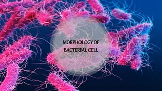
Bacterial Cell Morphology
- 2. Introduction to Bacteria Bacteria constitute a large domain of prokaryotic microorganisms. They are unicellular They are prokaryotic cells because they do not have well developed nucleus. Despite their simplicity they contain a well developed cell structure which is responsible for many of their unique biological properties. The most elemental structural property of bacteria is its cell morphology.
- 3. What is ‘Morphology’ of Bacteria? The morphology of bacteria describes the external appearance of bacterial cells including their shape, arrangement and size.
- 4. In the year 1872 scientist Cohn classified bacteria to 4 major types depending on their shapes as follows – A. COCCI – They are unicellular and spherical or elliptical in shape. They may either remain as a single cell or may aggregate together for various configurations as follows - Monococcus –They are also called Micrococcus and are represented by single, discrete, round cells. Example – Micrococcus flavus Diplococcus – The cells of Diplococcus divides once in a particular plane and after division the cells remain attched to each other. Example – Diplococcus pneumonia Classification of Bacteria on the basis of morphology
- 5. Streptococcus – Here the cells divide repeatedly in one plane to form a chain of cells. Example – Streptococcus pyogenes Tetrad – This consists of four round cells, which defied in two planes at right angles to one another. Example – Gaffkya tetragena. Staphylococcus – Here the cells are divided into three planes forming a structure like bunches of grapes giving an irregular configuration. Example – Staphylococcus aureus Sarcina – In this case the cells divide in three planes but they form a cube like configuration consisting of eight or sixteen cells but they have a regular shape. Example – Sarcina lutea
- 6. B) BACILLI– They are rod shaped or cylindrical which either remain singly or in pairs. Example – Bacillus cereus C) VIBRIO– They are curved, comma shaped and are represented by a single genus. Example – Vibrio cholerae D) SPIRILLA - They are spiral or spring like with multiple curvature and terminal flagella.. Example – Spirillum voluntans
- 7. Overview of Various structures of Bacterial cell
- 8. Bacterial cell wall The bacterial cell wall is uniquely characterized by the presence of peptidoglycan made up of a polysaccharide backbone of alternating N-Acetylmuramic acid (NAM) and N-acetylglucosamine (NAG) residues. It is located immediately outside of the cell membrane and is responsible for the rigidity ,determination of cell shape and transport of substances due to porous nature.
- 9. Bacterial cell membrane Composed of a phospholipid bilayer and thus has all of the general functions of a cell membrane such as acting as a permeability barrier for most molecules. Unlike eukaryotes, bacterial membranes (with some exceptions e.g. Mycoplasma and methanotrophs) generally do not contain sterols. However, many microbes do contain structurally related compounds called hopanoids which likely fulfill the same function. Since the bacteria lack membrane bound organelles all the membrane associated functions are performed in the plasma membrane.
- 10. Gram positive & Gram negative bacteria Apart from the general view, these two forms of bacteria differ greatly in structure of cell wall and cell membrane: Gram-positive cell walls are thick and the peptidoglycan (also known as murein) layer constitutes almost 95% of the cell wall in some gram-positive bacteria and as little as 5-10% of the cell wall in gram-negative bacteria. The presence of teichoic acid is also an exclusive feature of cell wall of gram positive bacteria.
- 11. In addition to the cell membrane and the cell wall, the cellular envelope of Gram negative bacteria has an additional outer membrane located outside the peptidoglycan cell wall. The outer membrane of gram negative bacteria is not a phospholipid bilayer, as the phospholipid is confined to it's inner leaflet. The outer leaflet consists of glycolipids, principally lipopolysaccharide (LPS) which can sensitize the human innate immune system. Gram positive & Gram negative bacteria
- 12. Gram positive & Gram negative bacteria Some of the proteins of outer membrane act as porins for passive diffusion of small molecules such as mono- and disaccharides and amino acids across it. The outer and inner membrane delimit an aqueous cellular compartment called the periplasm which is densely packed with proteins and it is more viscous than the cytoplasm.
- 13. GRAM STAINING The primary stain (crystal violet) binds to peptidoglycan present in both gram-positive and gram- negative cell walls, so initially, all the bacterial cells stain violet. Gram's iodine (iodine and potassium iodide) is applied as a mordant or fixative. Alcohol or acetone is then used to decolorize the cells. Gram-negative bacteria have much less peptidoglycan in their cell walls, so this step essentially renders them colorless, while only some of the color is removed from gram-positive cells. A counterstain safranin is applied. Both gram-positive and gram-negative bacteria pick up the pink stain, but it is not visible over the darker purple of the gram-positive bacteria, which remain purple, while gram-negative bacteria shows pink colouration.
- 14. Cytoplasmic matrix Cytoplasmic matrix is a crystallo-colloid complex. It is composed of 80 %water & 20% solid substances Solid substances are carbohydrate, enzyme, lipid, protein, inorganic ions and low molecular weight components. Membrane bound cell organelles (mitochondria, endoplasmic reticulum etc.) are absent. It is rich in ribosomes. Cytoplasmic streaming is absent.
- 15. Cytoskeleton System Bacteria lack a true cytoskeleton system. But they do have some Protein which functions like the eukaryotic cytoskeletal protein: Tubulin Homologues:- Two types of tubulin homologues FtsZ and BtubA/B, have been identified. FtsZ I essential for bacterial cytokinesis, assembles at the cell division site into circumferential ring, the Z-ring, located at the inner surface of plasma membrane.
- 16. Actin Homologues:- Three best studied actin like cytoskeletal proteins are MreB, ParM & MamK. The extended coil structure of MreB proteins are located on the undersurface of plasma membrane and are important in regulation of cell shape, chromosome segregation and cell polarity by localizing the proteins. ParM function as a partitioning protein in plasmid segregation. Intermediate filament homologues :- Crescentin is the only cytoskeletal Intermediate filament thus far identified in bacterial cell. It is reponsible for the shape of the comma shaped organism C. crescentus.
- 17. Bacterial Cytoskeletal Proteins Other than the homologues proteins, there are some unique bacterial cytoskeletal proteins present in the bacterial cytoplasmic matrix, which are: MinD :- It prevents polymerization of FtsZ at cell poles (specially in many rod shaped bacteria). ParA:- It segregates chromosome and plasmid (observed in many species including vibrio cholerae).
- 18. Internal membrane system Internal membrane system are basically circular to villiform specilization of plasma membrane that develops as an ingrowth from the cell membrane. This structure is called mesosome. It has the shape of vesicle, tubule and lamelle. It is abundant in gram positive bacteria than gram negative bacteria. The recent study confirmed that hydrogen peroxide is involved in meosome formation and excess H2O2 accumulation is associated with mesosome size. May form in bacteria as a defensive mechanism of enviornmental harsh condition. Depending on location and function, mesosome can be divided in two types. • Septal or central mesosome. • Lateral or peripheral mesosome.
- 19. Septal mesosome Connects nucleoid with plasma membrane (long invagination). It takes part in replication of nucleoid by providing point of attachment of the replicated one. It helps in cell wall formation by secreting enzymes such as dehydrogenase. At the time of cell division, plasma membrane grow in the region where the septal mesosome is present. So that it provides membrane for rapid elongation
- 20. Lateral mesosome Lateral mesosome shows a shallow penetration into the cytoplasm and thus is not connected with nucleoid. It contains respiratory enzymes and therefore is called “chondriod” and believed to be equivalent to the mitochondria of eukaryote. It plays a role in electron transport chain, phosphorylation and oxidation-reduction reactions.
- 21. Internal membrane system Many bacteria have internal membrane system quite different from mesosome. They may be spherical vesicle, flattened vesicle or tubular membrane. Plasma membrane infoldings can become extensive and complex in photosynthetic bacteria such as the cyanobacteria and purple bacteria or in bacteria with very high respiratory activity. Its function may be to provide larger membrane surface for greater metabolic activity.
- 22. Inclusion Bodies May occur freely inside the Cytoplasm (e.g Cyanophycean Granules, Volutin Or Phosphate Granules) or covered by 2-4nm thick non-lipid, non-unit protein membrane (e.g Gas Vacuoles, Sulphur Granule and Carboxysome). On the basis of their nature, Inclusion Bodies are of 3 types ~ Gas Vacuoles. ~ Inorganic Inclusion. ~ Food Reserve
- 23. Gas Vacuoles These are gas storing vacuoles,found in Cyanobacteria,Purple and Green Bacteria, which helps to protect the bacteria from harmful radiation. Gas Vacuole consists of variable number of hexagonal,hollow and cylindrical Gas Vesicles.Each Gas Vesicle is surrounded by a single non-unit, non-lipid protein membrane having ribs or folds. The membrane is impermeable to water but permeable to atmospheric gases.
- 24. Fig: Sulphur granules Fig: Polyphosphate granules Inorganic Inclusion Because of the ability to pick up different colour with basic dyes, they are called Metachromatic Granules. Two common type of Inorganic inclusion are – Volutin Granules are Polymetaphosphates which function as storage reserve of Phosphate. Sulphur Granules occur in bacteria living in sulphur rich medium which pick up Hydrogen Sulphide for obtaining reducing power in photosynthesis. Aquaspirillum magnetotacticum contains Magnetosomes which are vesicles having magnetite. The cytoskeletal protein MamK helps to form magnetosome chain. Magnetosome helps the bacteria to orientate themselves along geomagnetic lines.
- 25. Storage Inclusion / Food Reserve Storage of nutrients, metabolic end product, energy, building blocks. Glycogen Storage. Carbon Storage - Poly-beta- hydroxy butyrate(PHB). Phosphate - Polyphosphate( Volutin). Amino Acids - Cyanophycin Granules. Carboxysome - occur in Photosynthetic forms.
- 26. Ribosome Small membrane less, submicroscopic ribonucleoprotein entities having a size of 20nm × 14- 15nm. Ribosomes generally occur in helical groups called Polysomes Or Polyribosomes. Bacterial ribosome is 70S type where the eukaryotic ribosome is 80S type. Subunits of 70S ribosome are- 30S ( one molecule of 16S rRNA) & 50S ( two molecules of 23S & 5S rRNA) It is a mechanism to synthesize several copies of same protein.
- 27. Types of ribosome Free Ribosomes Occur free in the Cytoplasmic Matrix. Synthesize protein for Intracellular use. Fixed Ribosomes Attached to the Plasma Membrane. Synthesize protein for Transport to outside.
- 28. The nucleoid is an irregularly shaped prokaryotic genetic material, composed of about 60% DNA, 30 %RNA, 10% protein by weight. For most bacteria, the nucleoid is simply a region in the cytoplasm; it is not separated from other components of the cytoplasm by a membrane. Prokaryotes contain single, circle of double stranded DNA Unlike the eukaryotes, prokaryotes use NAPs or nucleoid associated proteins for chromosomal supercoiling. Nucleoid division during cell division Chromosome released from a gently lysed E. coli cell. Note how tightly packaged the DNA must be inside the cell. Nucleoid
- 29. Plasmid Extra-chromosomal, small double stranded DNA molecule, usually circular in appearance. Relatively have fewer genes, generally less than 30. Replication of plasmid is independent from chromosomal replication and have no link to any particular stage of cell cycle. Genetic information of plasmid is not essential to the bacterium. However, many plasmids carry genes that confer a selective advantage to the bacterium in certain environments.
- 30. Types of plasmid Type Function Example Size (kbp) Fertility plasmid Carry tra-gene for conjugation F-factor 95-100 Resistance plasmid Carry antibiotics resistance gene RP4 54 Col plasmid Carry genes that encode for antibacterial polypeptide called bacteriocins, a protein that destroy other strain of bacteria. ColE1 9 Virulence plasmid Carry virulence gene Ti 200 Metabolic plasmid Carry genes for enzyme CAM 230
- 31. Depending on the ability to transfer to other bacteria, plasmids may be Conjugative plasmid: Help in initiating conjugation Non-conjugative plasmid: Incapable of initiating conjugation and can only be transferred with the assistance of conjugative plasmid. Episomes: Plasmid of a bacteria or viral DNA that can integrate itself into chromosomal DNA of host organism and replicated as part of the chromosome. Plasmid are inherited during cell division and some time are lost, called curing.
- 32. Plasmid Components of plasmid: An origin of replication Multiple cloning sites An selectable marker (an antibiotic resistance gene) Promoter site Use of plasmid: 1. Genetically engineered plasmid (pBR322,pUC18) are used as cloning vector to make copies of particular genes. 2. Plasmid are used to make a large amount of desired protein by growing bacteria containing plasmid harboring the gene of interest. e.g. human insulin production using E.coli.
- 33. Pilli • Fine, hairlike appendages that are thinner and typically shorter than flagella. • Slender tubes composed of helically arranged protein subunits (pilin) and are about 3 to 10 nm in diameter and up to several micrometers long. Pili grow by adding protein subunits to the base of the pilus.
- 34. Types of Pilli The chaperone-usher pilus system: liner non covalent multi-subunit polymer that are prime virulence factor of proteobacteria and contribute to the establishment and persistence of the infection by mediating host and tissue specific interaction. Type V pilus: Play a roll in bacterial addition, co-aggregation and biofilm formation. Type IV pilus: Multifunctional organelles (help in addition to host cell, biofilm formation etc.) with a distinguishing ability to extend and retract by reversable polymerization and depolymerization. Powers a flagella independent type of bacterial movement known as twitching motility. Type IV secretion pilus: Help in 1. conjugation, 2. DNA uptake (competence).
- 35. Flagella Bacterial flagellum is a helical filamentous appendages extended outwards from the plasma membrane and cell wall, responsible for motility. Rotational movement of flagella is responsible for run and tumble movement of bacteria: counter clock wise movement of flagella: run movement (forward movement) clock wise movement of flagella: tumble movement (reorientation of the cell)
- 36. Monotrichous: have one flagella if it is located in one end then it called polar flagella Amphitrichous: have single flagella in both pole Lophotrichous: have a cluster of flagella at one or both ends Peritrichous: Flagella are spread evenly over the whole surface Classification of bacteria according to flagellal distribution
- 37. Structure of Flagella Flagella composed of mainly two parts 1. Basal body MS rings: located in cell membrane Composed of transmembrane protein FliF C ring: Attach to the cytoplasmic face of MS rings Consist of 3 cytoplasmic proteins FliG, FliM, FliN. P ring: Embedded in the peptidoglycan layer. Made up of periplasmic protein FlgI L ring: Embedded in the outer membrane Made up of lipoprotein FlgH Stator unit: H+ ion coupled MotAB complex composed of MotA and MotB protein. 2.Rod: Proximal rod composed of FliE, FlgB, FlgC,and FlgF Distal rod composed of FlgG. Rod act as drive shaft. 3.Hook: Links filament to its basal body. Composed of about 120 copies of hook protein FlgE. 4.Filament: Made up of flagellin protein FliC. Undergoes polymorphic. transformation for changing the direction.
- 38. Assembly of bacterial flagella Starts from basal body rings. Components of Axial structure are transported by type III protein export apparatus Type III protein export apparatus: • Export gate complex: Composed of FlhA,FlhB, FliP, FliQ and FliR • Cytoplasmic ATPase ring complex: Made up of FliH, FliI and FliJ.
- 39. Chemotaxis: The movement of bacteria toward or away from chemo attractants or chemo repellents is called chemotaxis. Stator unit ( Mot AB complex) use proton motive force as a fuel of energy fot rotational movement. Influx of proton allow the Mot A to associate with and dissociate from FliG to drive the flagellar motor rotation. Rod transmits that torque to filament through hook Flagellar Movement
- 40. Endospores Endospores is a heat resistant & dehydrated structure. It is capable of surviving for long periods in an unfavourable environment ( as long as 20 hrs) & potent to resist environmental stresses such as ultraviolet radiation, gamma radiation, chemical disinfectants and desiccation. Only found in Gram (+) Bacteria – • Bacillus cereus • Bacillus anthracisw • Clostridium tetani • Clostridium botulinum• Clostridium perfringens
- 41. Variations in endospores morphology FIG: (1, 4) central endospore; (2, 3, 5) terminal endospore; (6) lateral endospore.
- 42. Structure of endospores The arrangement of spore layers is as follows: Exosporium : It is a thin delicate protein covering. Spore coat ( acts like a sieve) ; It lies beneath the exosporium composed of several protein layers and may be fairly thick, impermeable & responsible for the spore’s resistance to chemicals.
- 43. Structure of endospores Spore cortex : It is present beneath the spore coat, made up of peptidoglycan. Core wall or spore cell wall : It is present beneath the cortex and surrounds the protoplast or spore core. The spore core has the normal cell structures such as ribosomes and a nucleoid , but is metabolically inactive. The core contains the spore chromosomal DNA which is encased in chromatin-like proteins known as SASPs (small acid-soluble spore proteins), that protect the spore DNA from UV radiation and heat.
- 44. Contents of endospores Up to 20% of the dry weight of the endospore consists of calcium dipicolinate within the core, which is thought to stabilize the DNA. Dipicolinic acid could be responsible for the heat resistance of the spore, and calcium may aid in resistance to heat and oxidizing agents. Small acid-soluble proteins (SASPs) are found in endospores. These proteins tightly bind and condense the DNA, and are in part responsible for resistance to UV light and DNA-damaging chemicals.
- 45. Staining procedures of endospores Staining is used to determine the morphology and the location of endospores. A special stain technique called a Moeller stain is used. That allows the endospore to show up as red, while the rest of the cell stains blue. Another type of staining of endospores through Schaeffer-Fulton procedure first involves heating of bacteria with malachite green that penetrates endospores and gives greenish colour. After the malachite treatment cell is washed with water & is counterstained with safranin.
- 47. Comparison of prokaryotes & eukaryotes
- 48. Comparison of prokaryotes & eukaryotes