2. INTRODUCTION
• Zygomatic fractures are common facial injuries, representing the most
common facial fracture or the second in frequency after nasal fracture.
• The high incidence of these fracture - the zygoma’s prominent position.
• Male over female ratio - 4 : 1.
• peak incidence - second and third decades of life. 80% - motor vehicle
accident.
• 20%- interpersonal violence and falls.
• The left zygoma is most commonly affected.
• Disruption of zygomatic position also has great functional significance
because it causes impairment of ocular and mandibular function.
Therefore, for cosmetic and functional reasons, it is imperative that
zygomatic injuries be properly and fully diagnosed and adequately treated.
3. SURGICAL ANATOMY
The zygoma is a four sided pyramid.
• This tetrapod configuration lends itself to
complex injuries, as fractures here rarely occur
in isolation.
1. Frontal process
2. Zygomaticomaxillary
buttress
3. Infraorbital rim
4. Zygomatic arch
5. Lateral orbital wall
5. • The malar bone represents a strong bone on fragile supports, and it is for this
reason that, though the body of the bone is rarely broken, the four processes-
frontal, orbital, maxillary, and zygomatic – are frequent sites of fracture.
• H.D.Gillies,T.P.Kilner, and D.Stone,1927
6. BIOMECHANICS OF FRACTURE
• The fracture pattern of any bone depends mainly on the
direction and magnitude of the force.
• The fracture lines pass through the areas of greatest weakness of
a bone or between bones.
• The inferior orbital fissure is the key to remember the usual
lines of ZMC fractures.
• Three fracture lines extend from the inferior orbital fissure in an
anteromedial, a superolateral, and an inferior direction.
7. Fracture patterns in zmc injury
FRONTAL VIEW
1)Anteromedially – orbital floor – orbital process towards infraorbital rim
2)Inferiorly – posterior aspect (infratemporal) of maxilla
3)Superiorly – lateral orbital wall – posterior to rim – seperates ZS suture
Anteriorly – seperates FZ suture.
8. • However, the fracture through the lateral orbital
rim is occasionally superior or inferior to the FZ
suture. A ZMC fracture that follows this pattern
usually has one additional fracture line through the
zygomatic arch. Because the point of least
resistance to fracture is not at the ZT suture, but
approximately 1.5 cm more posteriorly, the point of
fracture when a single fracture exists is usually in
the approximate middle of the zygomatic arch, in
the zygomatic process of the temporal bone.
• The variability of these fractures is great, owing to
the difference in magnitude and direction of force,
the amount of soft tissue covering the zygoma, and
the density of the adjacent bones.
•
9. CLASSIFICATIONS OF ZMC FRACTURES
• Knight and North (1961)
• Rowe and Killey (1968)
• Yanagisawa ( 1973)
• Larsen and Thomson (1978)
• Fuji and Yamashiro (1983)
• Rowe and Williams (1985)
• Poswillo (1988)
• Zingg et al Classification (1992)
• Ozyazgen et al classification(2007)
10. Knight and North Classification (1961)
Based on the direction of the displacement based on a
water’s view:
Group 1: Undisplaced fractures.
Group 2: Isolated displaced fractures.
Group 3: Displaced Body fractures (Unrotated)
Group 4: Medially rotated
4a: Outward at malar buttress
4b: Inward at the FZ suture.
Group 5: Laterally rotated
5a: Upward at the infraorbital margin
5b: Outward at the FZ suture
Group 6: Any additional fracture lines across the main
fragment.
11. Rowe has suggested that the displacement of the zygomatic bone can be
best understood by reference to rotation about different axes.
12.
13. • Row and Killey classification(1968)
– Type I – No significant displacement
– Type II – Fracture of zygomatic arch
– Type III – Rotation around vertical axis (inward or outward displacement of
orbital rim)
– Type IV – Rotation around longitudinal axis (medial or lateral displacement
of frontal process)
– Type V – Displacement of complex enblock
– Type VI – Displacement of orbitoantral partition – superiorly or inferiorly
– Type VII – Displacement of orbital rim segment
– Type VIII - complex comminuted fractures
14. Rowe & William’s Classification (1985):
Fractures stable after elevation:
• Arch Only
• Rotation around a vertical axis: Medially
Laterally
Fractures unstable after elevation:
• Arch only (inferiorly displaced)
• Rotation around a horizontal axis: Medially
Laterally
• Dislocations en bloc: Inferiorly
Medially
Laterally
• Comminuted fractures of the Zygomatic complex
15. LARSEN AND THOMSEN CLASSIFICATION
• Group A: Stable fracture—showing minimal or no
displacement and requires no intervention.
• Group B: Unstable fracture—with great displacement and
disruption at the frontozygomatic suture and comminuted
fractures. Requires reduction as well as fixation.
• Group C: Stable fracture—other types of zygomatic
fractures, which require reduction, but no fixation.
16. ZINGG etal classification (1992)
Type A: Incomplete zygomatic fracture. Isolated fractures
involving only one zygomatic pillar:
• Type A1: Isolated ZA fracture
• Type A2: Lateral orbital wall fracture
• Type A3: Infra orbital Rim fracture
17. • Type B: Complete monofragment zygomatic fracture
(tetrapod fracture). All 4 pillars of the zygomatic bone are
fractured
18. • Type C: Multifragment zygomatic fracture. Same as type
B, but with fragmentation, including the body of the
zygoma
19. DIAGNOSIS OF ZMC FRACTURE
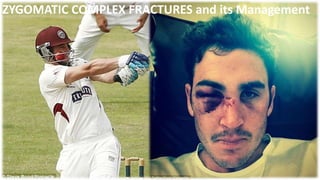
• In a typical case, diagnosis may be made at sight once the
characteristic appearance has been fully recognized. A peculiar facies
is present, due chiefly to a certain flatness of contour and an absence
of expression on the affected side.
• H.D. Gillies, T.P. Kilner, and D. Stone, 1927
20. CLINICAL EXAMINATION
• After accessing neurological status , the first priority - visual status
of the involved eye.
• One third of all pateints with comminuted ZMC fractures suffer
severe ocular disorders.
• Examination of zygoma- Inspection and palpation
• INSPECTION – Frontal, lateral ,superior and inferior views. Note –
symmetry , pupillary levels, presence of orbital edema,
subconjunctival ecchymosis, and anterior and lateral projection of
the zygomatic bodies.
• Superior view – most useful method of evaluating the position of
21. Method of assessing posterior displacement of
the ZMC from behind the patient. The clinician
should firmly depress the fingers into the
edematous soft tissue while palpating along
infraorbital areas.
BIRD’S EYE VEIW
22. • Clinical Features:
a) Skeletal Deformities:
• Asymmetry of the midface
• Depression or flattening of Malar Prominence
• Flattening, Hollowing or Broadening over the Zygomatic Arch
• Step Deformity of orbital margins
– b) Ocular/Ophthalmic Symptoms:
• Periorbital edema
• Enopthalmos
• Increased visibility of sclera
• Downward slant of palpebral fissure
• Malposition of the lateral canthus
• Vertical shortening of the lower eye lid
23. – Subconjunctival ecchymosis
– Hypoglobus
– Proptosis bulbi
– Enophthalmos , unequal pupillary level
– Subcutaneous periorbital air emphysema
– displacement of the palpebral fissure
– Diplopia
24. Inferior displacement of the zygoma results in depression of the
lateral canthus and pupil because of depression of the
suspensory ligaments that attach to the lateral orbital (Whitnall’s)
tubercle.
25. • The diagnosis of diplopia can be difficult in the early stages
of an injury, when severe edema of the orbit and eyelids is
present.
• Diplopia of edema or hemorrhagic origin should resolve in
a few days, whereas diplopia caused by entrapment of
orbital tissue does not.
• FORCED DUCTION TEST – determines whether there is
physical impediment to ocular motility.
26. • Finger gaze test -
Testing the motions of the eyes in all nine positions of
gazes (Arrows). At the same time diplopia and ocular level
is also checked
Hess test: . The test is based upon the projection of
dissimilar images from each eye. The patient sits in
front, at a distance of one meter from a screen
wearing a red/green goggle. The examiner holds a
red test object against the screen and the patient
tries to indicate the position of the object by
touching it with a green tipped wand. The result of
his efforts is charted when his head is held still and
he moves his eyes from the primary position to the
horizontal right and left extremes of movements.
This is repeated when looking above, to the right
above and to left above. The equivalent lower
positions are also charted.
27. – Superior Orbital Fissure Syndrome
Superior orbital fissure syndrome, also known as Rochon-Duvigneaud's syndrome, is
a neurological disorder that results if the superior orbital fissure is fractured.
Involvement of the cranial nerves that pass through the superior orbital fissure may
lead to diplopia, paralysis of extraocular muscles, exophthalmos, and ptosis.
Blindness or loss of vision indicates involvement of the orbital apex, which is more
serious, requiring urgent surgical intervention.
Typically, if blindness is present with superior orbital syndrome, it is called orbital apex
syndrome
28. • ) Neurological Symptoms :
• Paresthesia of infraorbital nerve -50- 90% of zmc injuries.
• Paresthesia of supraorbital and supratrochlear nerve
• Paresthesia of zygomatico temporal and zygomatico facial nerve
• Paresthesia of facial nerve
• Paresthesia of extraocular muscles
29. • Oral Symptoms
• Ecchymosis in the buccal sulcus of maxillary arch
• Deformity of zygomatic buttress of maxilla
• Trismus – high incidence in isolated fractures of zyg arch (45%)
• Pain
• Impacted / flattened zygomatic arch
e) Nasal Symptoms:
• Ipsilateral epistaxis – 30 -50 % of zmc injuries.
30. Depressed zygomatic arch impinging on the
temporal muscle and/or coronoid process,
limiting mandibular excursions.
31. • The preferred radiologic modality in zygoma injuries is the
computed tomography (CT) scan, for it provides greater
detail, especially if a floor or wall disruption has occurred.
• CT scans using axial and coronal cuts provide the best
evaluation of the orbit, with axial cuts preferred for
assessing damage to the lateral and medial walls, and
coronal cuts preferred for floor disruption.
• Hammerschlag and coworkers reported 100% accuracy in
the diagnosis of floor blow-out fractures when using 2-
mm-thiek sagittal section CT scans as well as lateral
tomograms.
• A sagittal scan is useful in determining the posterior extent
32. MANAGEMENT OF ZMC
Historical review:
• Various authors have given various treatment modalities and
techniques for the management of ZMC fractures.
• Dating back to 1751, when Duverney stressed the role of contraction of
temporal muscle in realigning the medial displacement of the zygomatic
arch.
• Ferrier in 1825, attempted to reduce fracture of zygomatic arch through
an incision above the arch.
Dupuytren in 1847, discovered the important relationship of the
temporal fascia and the muscle as a pathway to the zygomatic arch.
33. • Gillies in 1927 emphasised the cosmetic value of placing the
incision within the hair line.
• Stroymeyer in 1844 described the percuteneous hook technique.
• Cheyne and Burghard in 1901 discussed the intraoral digital
manipulation technique.
• Smith and Yanagisawa in 1961 stressed the importance of
cosmetic aspects of the treatment.
34. • GENERAL PRINCIPLES OF TREATMENT
• No treatment
• Indirect reduction with,
• a. No fixation
• b. Temporary support
• c. Direct fixation
• d. Indirect fixation
• Direct reduction and fixation
35. NO TREATMENT
• Cases with a minimal degree of displacement, which
following union, are considered unlikely to result any
cosmetic deformity, disturbance of vision, persistent
paraesthesia or impairment of mandibular movement.
36. INDIRECT REDUCTION
NO FIXATION:
• Includes procedures which do not involve exposure of
the fracture sites.
• The principle is to disimpact and reduce the fracture by
direct application of an instrument, through an indirect
approach remote from the fracture line.
37. • The techniques which have been developed for this operative
approach, are based upon the introduction of an instrument
through,
• a. the temporal fossa,
• b. the upper buccal sulcus (intraoral),
• c. the cheek (percutaneous),
• d. the nose (transantral)
• e. the eyebrow (lateral brow)
38. • Temporal fossa approach:
• This method was introduced by Gillies et al (1927) for elevation
of the zygomatic arch.
• Incision of (2 cm in length), made 2.5 cm superior and anterior
to the helix, within the hairline made above and parallel to the
anterior branch of the temporal artery and dissection is carried
down to the temporal fascia. This fascia is then incised to
expose the temporalis muscle. An instrument is inserted deep
to the temporalis fascia and superficial to the temporalis
muscle
• Using a Back- and- Forth Motion the instrument is advanced
until it is medial to the depressed zygomatic arch.
39. Firm Upward and outward force to the lifting
handle
Use of Rowe’s zygomatic elevator (1966)
40. • Elevation from eye brow approach: (Dingman & Natwig 1964)
• The advantage of this technique is that the fracture at the
orbital rim is visualized directly.
• The frontozygomatic area of the lateral orbital rim is
exposed by the eyebrow incision.
• The instrument is inserted to lift the zygoma anteriorly,
laterally and superiorly.
• Useful instruments for this purpose are – Dingman
zygomatic elevator , urethral sound, or even large Kelly
hemostat.
41. Dingman zygomatic elevator is placed along the temporal surface of zygoma for
anterior , lateral and superior elevation
42. Zygomatic Arch Fracture
Todd G. Carter, DMD,* Shahrokh Bagheri, DMD,
MD,†
and Eric J. Dierks, DMD
J Oral Maxillofac Surg
63:1244-1246, 2005
43.
44. Upper buccal sulcus: (keen’s approach)
• The advantages of this technique have been discussed by
Balasubramaniam (1967) who considers that “ less force
is required by the intraoral approach than by the
extraoral, because the force is exerted where it should
be, i.e., more at the centre of the fractured fragment”.
• Access is gained by an incision of about 1cm in length at
the reflection of the upper buccal sulcus immediately
behind the zygomatic buttress, so that a pointed curved
elevator can be passed upwards supraperiosteally to
contact the infratemporal surface of the zygomatic bone.
• This enables upward ,forward and outward pressure to
45. • The elevator by Monks is suitable
for this purpose. (Taylor monk’s pattern)
A right angle retractor,bone hook, large Kelly
Hemostat, simple dental extraction forceps and a
Flat instrument –Seldin retractor to follow medial
Surface of zyg arch and elevate it laterally.
Quinn in 1977 described a modification which is of value for
medially displaced fractures of zygomatic arch.
• This employs a lateral coronoid approach through an
incision situated over the anterior border of ramus.
47. • Percutaneous approach: (Stroymeyer 1844)
• Simplest of all because no soft tissue dissection is necessary.
• Several instruments – bone hook , carroll –Girard screw(
large bone screw ) for elevating zygomas.
• This method consists of inserting a hook through the soft
tissue of the malar area at a point just inferior and posterior
to the prominence of the zygoma so that it engages the
infratemporal aspect.
• Poswillo advises that the exact location of the initial stab
wound for insertion is found at the intersection of a
perpendicular line dropped from the outer canthus of the
eye and a horizontal line extended posteriorly from the alar
margin of the nostril.
48.
49. Anterior and lateral traction with bone
hook
Carroll-Girard screw – elongated cork screw
with a T bar handle and threads on its working
end. This screw can be threaded into the body
of zygoma following placement of hole
Adv – control ZMC position in all 3 planes of
space.
50. • Intranasal transantral approach: (Lothrop’s approach 1906)
• Not common in use.
• An opening is made into the antrum below the inferior meatus,
and a curved urethral sound introduced and manipulated so that
its tip lies on the antral aspect of the zygomatic bone. Firm
outward and upward pressure is applied to reposition the bone.
51. • ASSESSMENT OF REDUCTION
• The success or failure of reduction will be obvious for those
who have opened the fracture at three sites. If exposure at
three sites has not been performed, the orbital margins are
the areas that should be palpated first to determine
reduction.
• If reduction has been satisfactory, these margins will be
smooth and continuous. This finding by itself, however, is
inadequate verification . Although the zygomaticofrontal
suture area provides the strongest pillar of the zygoma, it is
one of the worst indicators of proper reduction of the entire
complex, even when surgically exposed and evaluated
directly.
52. • One should also palpate in the maxillary vestibule. If there is
any flatness still visible, then zygoma has not been properly
elevated. If there is any doubt about proper reduction,
exposure is mandatory. In this case, an incision in the maxillary
vestibule offers excellent exposure of the zygomaticomaxillary
buttress and the infraorbital rim.
53. TEMPORARY SUPPORT
This may be indicated, as a supplementary measure,
under the following circumstances:
• When the zygomatic complex is unstable following
reduction,
• When there is gross comminution of the zygomatic bone.
• When there is comminution without bone loss of the
orbital floor.
Instability following adequate reduction
could be due to:
• Rupture of the enveloping periosteum or attached
temporal fascia.
54. • Temporary support is a concept which is primarily based upon the
introduction of a pack or other material into the antrum so as to
exert counter-pressure against those forces which tend to bring
about a relapse of the position achieved by indirect reduction.
• Since the pack will bring about repositioning of fragments by
pressure from their antral aspect, it will be evident that this selective
effect can only take place if there is absolute stability of the
remainder of the antrum and the other elements of the zygomatic
complex.
55. DIRECT FIXATION
• Indirect reduction, combined with direct fixation following
exposure of the fracture site, provides an excellent method
of treatment.
• Need - when the fractures remain unstable after indirect
reduction
• Transosseous wiring or osteosynthesis :
• Separation at the frontozygomatic suture Line with
displacement in excess of 2-3 mm, is likely to cause
detachment of the periosteal and fascial attachments, and
hence the direct fixation becomes necessary.
56. • Elevation of the zygomatic bone and transosseous
wiring or boneplating at the frontozygomatic suture will
achieve a stable realignment of the orbital rim
• A more severe displacement, a dislocation of the
zygomatic complex en bloc, will result in separation of
the fracture ends at the inferior orbital margin, with the
fracture passing into the orbital floor.
• In this type of unstable fracture it is essential to carry
out an osteosynthesis (transosseous wiring) or
microplating technique.
57. Comminuted fracture of the
inferior orbital margin and frontal
process of the left maxilla .
The fractures are maintained in
position by a direct and a figure of
eight wire suture.
58. INDIRECT FIXATION
• Indirect fixation implies that the zygomatic bone will
be rigidly secured to some point elsewhere on the
facial skeleton until union occurs , after which
connecting apparatus may be removed.
• The required degree of firmness can only be achieved
by means of internal (intramedullary) pins or wires or
external pins and rods which are linked together.
• Indirect fixation has limited application at the present
time in view of greater efficiency and comfort by
internal fixation techniques.
• However , it provides means of fixation when there has
been gross loss of bone in the region of
59. • The indirect fixation can be achieved by the following
methods:
• Zygomatico-zygomatic (Trans-maxillary)
• Naso-zygomatic
• Zygomatico-palatal
• Maxillo-zygomatic
• Fronto-zygomatic
• Cranio-zygomatic
60. 1)Maxillo-zygomatic
In this type of fixation a coarse threaded pin is
inserted into fractured zygoma and it is attached via
universal joints and rods to a dental splint fitted to maxilla.
2) Cranio-zygomatic
Here zygoma is fixed to the cranium by a plaster of
paris (headcap). This is the particular value when Lefort
fracture exists.
3) Fronto-zygomatic
This technique employs the fixation of one pin in
zygomatic bone body and another in zygomatic process in
frontal bone
61. 4) Zygomatico-zygomatic
In this technique Krichner wire is passed from body of
normal zygoma via nasal cavity into the fracture site in the
wire projects on to the cheek
5) Naso-zygomatic
Brown and Bernard recommended in the use of
transnasal Krichner wire which is inserted with the hand drill
passed from frontal process of maxilla on the controlateral
aspect of the nose to engage the antral surface of the
zygomatic bone.
6) Zygomatico-palatal –
Matsunga in 1977 advocated a technique which is
62. Direct reduction and fixation
Objective- An exposure of the fractured site by an
incision placed as close as possible to the bone ends , so that
these may be directly manipulated and firmly secured either
by transosseous wires or bone plates.
63. Fixation techniques
• The application of plate and screw fixation techniques to
ZMC fractures has replaced all the older techniques of
fixation.
• There is no better method of providing stable fixation to
an unstable ZMC fracture than to secure it rigidly
internally with bone plates and screws.
• The obvious advantage to bone plates is that stabilization
in three planes of space can be provided, even across
areas of comminution or bone loss.
64. • When plate and screw fixation is used, there are general
principles of its application for ZMC fractures. They are:
1. Use self-threading bone screws. The thin bones of the
midface lend themselves to the application of self-threading
screws. selfthreading screws - more holding power in thin bones
than when the holes are tapped.
2. Use hardware that will not scatter postoperative CT scans.
Titanium plates and screws have the advantage of not causing
scatter in CT scans. Vitallium causes more scatter, so if it is
selected, smaller plates and screws should be used to minimize
CT artifacts.
3 .At least two screws are necessary for stabilizing a bone plate
65. • 4. Avoid important anatomic structures, such as the
tooth roots and infraorbital nerve. If the fracture
through the zygomaticomaxillary buttress is low, one
should select an L-, a T-, or Y-shaped bone plate so that
both of the lower screws are positioned horizontally in
the alveolar process.
66. • 5. Use as thin plate as possible in the periorbital areas.
• 6. Place as many bone plates in as many locations as
necessary to ensure stability. Many fractures can be
adequately stabilized with a single bone plate applied at the
frontozygomatic area* or at the zygomaticomaxillary
buttress. However, when the articulations between the ZMC
and adjacent bones are comminuted -- additional bone
plates in additional areas.
• 7. In areas of comminution or bone loss, span the gap with
the bone plate. Comminution of fractures through the
zygomaticomaxillary buttress and infraorbital rim is
common. If small bone fragments are missing, it is
67. Fixation:
• 1 Point Fixation
• 2 Point fixation
• 3 point fixation
• 4 point fixation
68. • One point fixation:
• Indication:
• Undisplaced fracture.
• Simple non comminuted zygomatic complex fracture
• Approach :
• • Frontozygomatic suture approached through
supraorbital eyebrow
• approach.
• • Zygomaticomaxillary buttress approached through
maxillary
• vestibular approach.
69. • Two point fixation:
• Indication:
• • Displaced fracture unstable after reduction
• • Fracture at Frontozygomatic suture, Infraorbital rim and
• buttress.
• Approach:
• • Exposure of frontozygomatic suture
• • A 2 point fixation using low profile plate at
• zygomaticomaxillary buttress or at the infra
• orbital rim
70. • Three point fixation
Fixation is done at Frontozygomatic suture, Zygomaticomaxillary
• buttress and the Infraorbital rim.
Good reduction of these 3 sites mostly reduces the arch fracture
• which is not fixed
71. Four point fixation:
Unique from 3 point technique in that the surgeon
visualizes
the Zygomatic arch.
The order of placement of the plates will be dependant
on
the least damaged landmarks.
The zygomatic arch is an excellent reference to restore
72. Other approaches to zmc
• Supraorbital eyebrow approach
• Upper eye lid approach
• Lower eye lid approaches- sub tarsal , subciliary,
transconjunctival
• Coronal approach
73. • Supra orbital eyebrow approach
• popular approach used to gain access to the lateral orbital rim.
• No important neurovascular structures of any significance are
at risk.
• simple and rapid access to the frontozygomatic area.
• Because the incision is made almost entirely within the
confines of the eyebrow, the scar is usually imperceptible.
74. • Upper eye lid approach- upper blepharoplasty
• One of the best approaches to the region of superolateral
orbital complex.
• a natural skin crease in the upper eyelid is used to make
the incision.
• The advantage - inconspicuous scar.
• Incision -10mm superior to the upper lid margin and 6mm
above lateral canthus as it extends laterally.
75. • Coronal approach
• Coronal or bifrontal flap modified to include some
advantages of mod. Preauricular flap of alkayat bramley
incision.
• Although it may initially appear as a radical approach to
the management of zygomatic fractures, it provides
excellent access to the orbits, zygomatic bodies, and
zygomatic arches, with almost no complications.
• It is an extremely useful incision if there is comminution
of the supraorbital and lateral orbital rims, and
zygomatic body and arch.
• The scar produced is hidden within the hairline and is
therefore invisible.
76. How to protect the reduced zygomatic arch fractures ?
• Isolated zygomatic arch fractures constitute fewer than 10% of
zygomatic injuries.
• Ellis et al have found that 10 of 126 (7.3%) isolated zygomatic arch
fractures treated in their study required fixation. Others have reported
that almost every zygomatic arch fracture is stable, once elevated.
• Stabilization of depressed zygomatic arch fractures has been achieved
with the use of percutaneous circumferential wires or heavy sutures
passed around the arch with an aneurysm needle or Mayo trocar and
tied to an external object has served this purpose well. Plastic oral
airways, metal eye shields, short pieces of endotracheal tubing, and
orthopedic finger splints all have been used as the external devices.
77. • Following reduction of zygomatic arch fractures, one must protect
the side of the head from injury.
• The force of the weight of the head resting on a pillow is
sufficient to displace even a properly reduced fracture.
• Commonly used and readily available materials are paper cups,
metal eye patches, aluminum finger splints bent in a staple
configuration. Ideally, they should be left in place for 2 to 3
weeks.
78. ORAL AIRWAY FOR PROTECTION OF
REDUCED ZYGOMATIC ARCH
ALUMINIUM FINGER SPLINT
79. complications
• Infraorbital nerve disorders
• Persistent diplopia
• Enopthalmos
• Blindness
• Retrobulbar and intraorbital haemorrhage
• Malunion of the zygoma
80. Infraorbital nerve disorders
• De Man and Bax, in a study of 273 isolated ZMC
fractures, found that 80% suffered from dysesthesia on
admission.
• Nordgaard found sensory disturbance in 96% of 100
patients immediately after fracture.
• Jungell and Lindqvist - 81% of patients with ZMC
fractures had paresthesia of the infraorbital nerve. The
figure was even higher (94%) in those who required
surgical treatment. Most patients had regeneration, but
42% of patients had some degree of persisting sensory
disturbance.
81. • They maintain that fixation of the fracture line by a
miniplate, mainly in the frontozygomatic area, achieves
the fastest recovery rate of neurosensory dysfunction.
• The proposed method whereby recovery improves is
that improved stability prevents continued compression
on the nerve after reduction
• In cases of persistent dysesthesia, anesthetization of the
superior alveolar nerves by local infiltration should be
attempted. If symptoms are not alleviated, the clinician
should suspect a disruption of the infraorbital nerve
within its canal where the middle and anterior superior
alveolar nerves take origin, with possible neuroma
82. • Persistent diplopia
Binocular diplopia present initially after zygomatic fracture is
generally a result of edema or hematoma of one or more
extraocular muscles or their nerves and intraorbital edema or
hematoma.
In these cases, resolution of diplopia following fracture treatment
(if necessary) usually occurs spontaneously within 5 to 7 days.
Neural injuries from the trauma or from surgery may also produce
persistent diplopia.
83. Enopthalmos
causes - by a decrease in volume of the orbital contents,
increase in volume of the bony orbit,
loss of ligament support,
scar contracture, or combination of these.
The most popular theories of the mechanism of enophthalmos have been
bony orbit enlargement and fat atrophy.
• post-traumatic enophthalmos is usually caused by an increase in bony
orbital volume . Even after restoration of the orbital rims and floor at
the time of surgery, defects located posteriorly along the medial and/or
lateral walls are common and frequently overlooked, and are probably
the main reason for postoperative enophthalmos.
84. • The signs and symptoms of retrobulbar hematoma
include a tense proptosis (exophthalmos), periorbital
swelling that may be in the process of increasing in size,
retroorbital pain, dilation of the pupil, and
ophthalmoplegia.
• Hueston and Heinze have stated that “retrobulbar
hemorrhage is not an emergency, total blindness is.”
• Most ophthalmologists do not treat retrobulbar
hemorrhages or treat them conservatively with the
application of ice, sedatives, bed rest, and/or diuretics.
• Gradual absorption of the hemorrhage occurs and full
range of motion usually returns in several weeks but
85. • When the point is reached that the optic nerve or
retinal artery becomes involved - the pupil becomes
fixed and nonreactive to light.
• These findings, associated with visual loss, constitute
and should be considered a medical emergency because
permanent loss of vision will occur in several minutes if
the orbit is not immediately decompressed.
• Hueston and Heinze have claimed that survival of the
optic nerve head is at stake in this situation and 60
minutes of ischemia appears to be the limit for survival
and recovery. Row – 15- 20min.
86. • MALUNION OF ZYGOMA
• SIGNS & SYMPTOMS
• Cosmetic – Loss of contour/prominence of cheek.
• Neurological – Paresthesia / Anaesthesia.
• Antral - persistent sinusitis due to presence of loose
necrotic bone pieces / foreign body.
• Masticatory – Depressed zygomatic arch fracture,
impinges on the coronoid process.
• Opthalmic – Change of ocular level, diplopia and
enopthalmos.
87. CONCLUSION
• Face is the most prominent and expressive part of
human body and adds to well being of a personality.
• Maxillofacial region is vulnerable for various injuries due
to variety of external causative factors.
• Zygomatic complex forms key to structure of
anterolateral surface of face.
• The importance of zygomatic complex in facial skeleton
lies in protecting globe of eye and absorbing and
redistributing masticatory and external load.
88. REFERENCES
• Oral & Maxillofacial Trauma – Raymond J. Fonseca
• Rowe and Williams – Maxillofacial injuries.
• Oral and Maxillofacial surgery, – Peterson.
• Journal of oral and maxillofacial surgery
Malar surface – z.major,minor lv labii superioris. Masetter – along the body and temporal process.
Temporal fascia – along the arch and post lateral edge of temporal process. 2 foramens –zf and zt foramen
One fracture extends from the inferior orbital fissure anteromedially along the orbital floor mostly through the orbital process of the maxilla towards infraorbital rim.
2nd line from from the inferior orbital fissure runs inferiorly through the posterior (infratemporal) aspect of maxilla and joins the fracture from the anterior aspect of maxilla, under the zygomatico maxillary buttress. 3rd line runs superiorly from inf orbit fissure along the lat orbital wall posterior to the rim, separating Zsphenoid suture and extending anteriorly it seperates FZ suture at lateral orbital rim.
, however, three fracture lines exist through the arch, producing two free segments when the fractures are complete.
Vertical axis- Fzsuture downward to center of body to the zyg buttress.
If blow – infront of the v axis, rotation of H plane, there wil b outward mov of center of the arch. Behind the axis then, outward mov of orbital floor and rim.
line at the level of the infraorbital foramen- from in front horizontally backward through the center of the body of the bone and the zygomatic arch.
if An impact above h axis- rotation of vertical plane with medial mov of frontal process and outward mov at the zyg buttress
Impact – at level enbloc displacement. Below the level- lat mov of frontal process and medial mov of buttress into antral cavity.
The patient can be placed in recumbent or recline position. Inspect from superior position evaluating how the zygomatic bodies project anteriorly and laterally to the supraorbital rims, comparing one side with the other. Don’t forget to intraoral exam, ecchymosis in superior buccal sulcus and maxillary dentoalv fractures.
Enopthalmos – means decrease in orbital softtissue volume by herniation of orbital softtissue. Enopthalmos -narrowing of palpebral fissure causing pseudoptosis of upper eyelid.
Small forceps grasp the tendon of inferior rectus through conjunctiva of inferior fornix and globe is manipulated thru its entire range of motion. Inability to rotate the eye superiorly signifies entrapment of muscles in orbital floor. This test shoulb be performed routinely in those who cannot rotate the globe in an upward gaze.
Epistaxis – sinus mucosa disrupted –hemarhage into sinus –as sinus is connected to nasal cavity thru middle meatus unilateral hemrge frm nose
A spcl elevator designed by row in 1966 has (lifting handle /external handle) and internal handle with /elevating blade with same length and so extent of insertion may be assessed accurately. Lifting handle is attached by strong hinge with a positive stop near origin of other handle. Audible click
Disadvantage – difficult to generate large amount of force especially in superior direction.
Poswillows lines of insertion for bone hook. Line 4m outer canthus vertically .horizn 4m alar margin of nostril.
The most important step in the management of ZMC fractures is to determine at the table whether the fracture has been properly reduced
At one time use of antral pack was popular but with introduction of improved techniques for direction and fixation of fractures , the indications for this concept frequently reduced.
The use of straight plate in this case might cause the lower screw to impale a tooth root