1. 2.207. Extracellular Matrix: Inspired Biomaterials
H M Waldeck and W J Kao, University of Wisconsin-Madison, Madison, WI, USA
ã 2011 Elsevier Ltd. All rights reserved.
2.207.1. Introduction 114
2.207.2. Overview of ECM Structure and Function 115
2.207.3. Types of ECM Mimicry 116
2.207.3.1. Mimicking ECM Functions by Using ECM Components 116
2.207.3.1.1. Full/direct copy 116
2.207.3.1.2. Partial copy 119
2.207.3.2. Mimicking ECM Function Through ECM Architecture and Topography 121
2.207.3.2.1. Hierarchical microstructure and porosity 121
2.207.3.2.2. Topographical features and patterning 122
2.207.3.3. Mimicking ECM Protein Design and Assembly 125
2.207.4. Future Directions 126
References 126
Glossary
Affinity (protein-ligand) Characteristic binding strength
between a single protein and ligand; commonly described
by the dissociation constant.
Allogeneic immunologic response Immune response to
cells or other materials derived from a genetically
nonidentical donor of the same species (allograft).
Angiogenesis The process by which new blood vessels are
grown from preexisting vessels.
Anisotropy A property of a material having directional
dependence.
Avidity (protein-ligand) Strength of binding derived from
multiple bond interactions.
Basement lamina Characteristic extracellular matrix
found under epithelial or endothelial cells consisting of the
fusion of two distinct layers: the basal lamina and the
reticular lamina.
Bioderivation Extracting certain components/information
from the biological system and then applying that to a
different area of use.
Bioinspiration Deriving strategies different from that
employed by nature to achieve the same function and
properties.
Biomimicry Learning the principles governing the function
of a biological system and then using that same strategy to
create a synthetic system that functions with a similar
precision.
Bioresorption A form of resorption in which materials
degrade when they come into contact with one or more
specific biological molecules.
Cell differentiation Process by which a cell becomes more
specialized through modifications in gene expression that
can lead to alteration in morphology and activity.
Collagen Primary structural ECM protein family. All
collagen subunits initially organize into triple-helical fibers
and then these fibers associate further to form fibrillar,
sheet-like, or fibril-associated structures.
Cytokine Small cell-signaling proteins, peptides, or
glycoproteins which are released into the extracellular
environment by various cell types for intercellular
communication.
Cytoskeleton Intracellular protein scaffolding which plays
important roles in cell morphology, cellular mobility,
intracellular transport, and cellular division.
Denaturation The process through which proteins or
nucleic acids lose either tertiary or secondary structure
due to the application of an external stress. A loss of
function is typically seen in conjunction with this loss
of structure.
Electrospinning Process by which continuous nano- to
microscale fibers are formed by using electric forces to
overcome surface tension and thereby elongate droplets of
polymer melt or solution into a stream.
Excitation threshold Minimum amount of stimulus
necessary to create an action potential in a nerve cell.
Extracellular matrix (ECM) The native cell
microenvironment composed of specific proteins,
proteoglycans, small molecules, water, etc. depending on
the cell type and resident tissue. Through adhesion to the
ECM, cells are exposed to physical, mechanical, and
biochemical cues capable of altering cell differentiation,
activation, migration, and adhesion strength.
Fibrillogenesis Creation of thin fibril structures present in
the collagen fiber architecture.
Filopedia Nanoscale cellular extensions capable of
interacting with nanoscale physical cues.
Glycoprotein Proteins which contain covalently attached
oligosaccharide chains.
Glycosaminoglycan (GAG) Long, unbranched
polysaccharides consisting of repeating disaccharide units.
Hydrogel Water-swollen, polymeric structures containing
either covalent or physical cross-links. Hydrogels are highly
varied in composition, construction method, and the
resulting chemical and physical characteristics.
113
2. Hydrolysis A chemical process in which a molecule, such as
a polymer, is split into two parts through the addition of a
molecule of water.
Hydrophobicity/hydrophilicity A physical property which
characterizes the ability of a molecule to repel/attract water
molecules.
Hydroxyapatite The naturally occurring mineral form of
calcium apatite. A modified form of this substance is a
primary component of bone tissue.
Integrins A large family of heterodimeric transmembrane
proteins which serve as the primary receptors between cells
and ECM proteins.
Isotropy A property of a material being homogeneous in all
directions.
Ligand Substance capable of forming a complex through
binding to another molecule or binding site. Typically in
biology, ligands are small molecules or functional groups
which are able to bind to specific sites on proteins.
Mechanotransduction Mechanism though which cells
convert mechanical stimuli into chemical activity.
Microablation Microscale removal of material from a
surface.
Microstructure Structural features of a material which can
be visualized using a microscope at 25Â magnification.
Morphogenesis The process by which an organism, tissue,
or cell develops its shape and/or structure.
Morphology Form or structure of an organism including
shape, size, and structural features.
Motif Characteristic sequence or structure found in a
protein which performs a biological function or results in
specific higher-order structures, respectively.
Nanocomposite A solid multiphase material in which the
size of the phases, the distance between the phases, or the
structural repeats have nanoscale dimensions.
Nanopatterning Fabrication of a nanoscale pattern on a
surface.
Nanostructure Structural features which range in size
between molecular and micrometer dimensions.
Osteogenesis The process by which new bone tissue is
developed through the laying down of new bone material by
osteoblasts.
Phenotype Observable physical, biochemical, or behavioral
characteristics of an organism.
Porosity A measure of the void spaces within a material.
Protease An enzyme which can catalyze the breakdown of
peptide bonds within the primary protein structure.
Proteoglycans A diverse family of molecules whose
structure consists of a core protein covalently attached to
multiple glycosaminoglycan chains.
Recombinant DNA/recombinant protein Engineered DNA
which is developed by combining sequences which would
not normally occur in nature. Often this process is
accomplished by introducing foreign DNA into an existing
organismal DNA, such as the plasmids of bacteria.
Recombinant DNA technology is used to manufacture
recombinant proteins.
Tangent modulus The slope of the compression
stress–strain curve as a specified stress or strain. Typically,
the tangent modulus is used to describe the behavior of
materials beyond their elastic regions.
Topography The composition and configuration of
physical features on a surface.
Ultimate strength Maximum load or stress a material can
withstand before necking of the material occurs.
Ultrastructure Detailed biological structure of a specimen
not able to be visualized using a light microscope.
Viscoelasticity A property of materials that demonstrate
both viscous and elastic characteristics when undergoing
deformation.
Xenogeneic immunologic response Immune response to
cells or other material derived from a donor of a different
species (xenograft).
Abbreviations
3D Three-dimensional
DNA Deoxyribonucleic acid
DRG Dorsal root ganglia
ECM Extracellular matrix
FAK Focal adhesion kinase
GAG Glycosaminoglycan
HAp/
Col
Hydroxyapitite/collagen nanocomposite
hMSC Human mesenchymal stem cell
MCL Medial collateral ligament
MMP Matrix metalloproteinase
PCL Poly(e-caprolactone)
PEG Poly(ethylene glycol)
PGS Poly(glycerol sebacte)
PLGA Poly(DL-lactic-co-glycolide)
rhBMP Recombinant human bone morphogenetic
protein
RT-PCR Reverse transcriptase polymerase chain
reaction
SIS Small intestinal submucosa
SLRP Small leucine-rich proteoglycan
2.207.1. Introduction
Of all the advances in science, technology, and engineering in
the past few decades, the deeper understanding of biological
systems has led to an ever more intimate contact between
individuals and technology. This personal impact of biology-
based technologies has been demonstrated to be a very power-
ful force in shaping the scientific community. The creation
of biology-centered enablers is the result of the pursuit of
three general research paradigms: biomimicry, bioinspiration,
114 Biologically Inspired and Biomolecular Materials and Interfaces
3. and bioderivation. Biomimicry is defined as “learning the
principles governing the function of a biological system and
then using that same strategy to create a synthetic system
that functions with similar precisions.” Examples of this are
protein/cell-based biosensors for detecting (bio)hazards and
self-assembled materials with unprecedented responsiveness,
complexity, and ability to interact and evolve. Bioinspiration is
defined as “devising strategies different from that employed
by nature to achieve the same function and properties.” Exam-
ples of this are novel light harvesting methods based on nano-
technology and microorganism-based air/water filtration
systems to control pollution including greenhouse gases.
Bioderivation is viewed as extracting certain components/
information from the biological system and then applying
that to a different area of use. Examples of this are also plenti-
ful: alternative fuel sources, incorporation of biologically
derived molecules for targeted drug delivery, and biofunctio-
nalized materials for advanced cellular and molecular medi-
cine such as stem cell and gene therapies.
Earlier biomaterial research has mainly focused on biocom-
patibility and application-specific macroscale requirements such
as mechanical, adhesive, or optical properties. The design con-
straints were therefore primarily concerned with preventing
adverse effects on the body caused by a foreign material.
Although these subjects remain to be critical issues in biomedi-
cal research with incomplete mechanistic insights, the advance-
ment in biological sciences have influenced and expanded
current biomaterial research and design. By applying biological
principles through biomimicry, bioinspiration, or bioderivation
to material design, the overall rationale and motivation is to
expand the property and to improve the biological interaction of
these materials. This chapter focuses on how the structural and
functional principles derived from the native cell microenviron-
ment, the extracellular matrix (ECM), have been applied to
biomaterial design and construction. By mimicking the ECM,
researchers are able to take inspiration from defined cell–matrix
interfaces to subsequently control cell–biomaterial interactions.
Furthermore, the hierarchical structure of ECM architecture is a
desirable characteristic to incorporate into material design. Sev-
eral methodologies are covered ranging from directly incorpor-
ating ECM proteins to utilizing self-assembly principles to
construct materials. An overview of basic ECM structural and
functional features is given followed by selected examples
throughout the chapter that illustrate how these bioactive attri-
butes are incorporated into biomaterials.
2.207.2. Overview of ECM Structure and Function
The interplay between cells and the surrounding ECM estab-
lishes a dynamic tissue microenvironment capable of
performing the varied functions seen in biological systems.
Cells secrete ECM molecules and maintain the matrix through
continuous remodeling of the structure. The ECM, in turn, sup-
ports and maintains the cells by providing nutrition and indi-
rectly mediating cell–cell communication. Furthermore, in
some tissues, the ECM also acts as the structure responsible for
carrying out the central function of a tissue. The organization
and composition operate cooperatively to balance strength,
flexibility, and complexity to establish specific tissue properties.
ECM-inspired biomaterials attempt to mimic the bioactive and
bioresponsive relationship between cells and ECM as well as the
ECM’s hierarchical structure through the incorporation of ECM-
derived biochemical and structural components. This section
focuses on the basis for such mimicry by providing an overview
of how the main constituents and structural characteristics of
native ECM contribute to overall tissue function (for a more in-
depth examination of the ECM structure–function relationship,
see Plopper1
).
The tissue-specific ECM is composed of a unique combina-
tion of water, glycoproteins, proteoglycans, and sequestered
signaling molecules integrated into a highly complex three-
dimensional (3D) cell scaffold. The individual properties and
subsequent configuration of these components play multiple
roles in the tissue hierarchy. Proteoglycans are a diverse family
of molecules whose structure consists of a core protein cova-
lently attached to one or more glycosaminoglycan (GAG)
chains. The anionic nature of the GAG leads to electrically
driven association with cations and these, in turn, attract
water molecules. The association of a large amount of water
with the GAG chains causes overall structural rigidity as well as
hydration. As such, large interstitial proteoglycans, for exam-
ple, aggrecan, are able to maintain tissue hydration and estab-
lish tissue structure. Proteoglycan-mediated hydration allows
diffusion of small molecules and increases the tissue’s ability to
resist compression forces. The relatively rigid and large struc-
ture helps define ECM architecture and can also act to prevent
bacterial infiltration. Additionally, the ability of proteoglycans
to bind other components facilitates organization of the ECM
into functional structures. For example, binding of small
leucine-rich proteoglycans (SLRPs) to collagen helps stabilize
and align fibers leading to mechanical anisotropy. Soluble
factors, such as cytokines or proteases, are sequestered through
interactions with proteoglycans creating depots or gradients of
these regulatory molecules throughout the ECM. Subsequent
cell-mediated degradation of the matrix results in liberation
and/or activation of these soluble proteins, enabling biore-
sponsive cell signaling.
The principal structural ECM proteins, collagens, are
organized into a variety of functionally relevant configurations.
All collagen subunits initially organize into approximately
1.5-nm triple-helical fibers and then these fibers associate further
to form fibrillar, sheet-like, or fibril-associated structures. Fibril-
lar collagens are formed through bundling of progressively larger
fibers along a single axis till eventually fibers with diameters up
to approximately 3 mm are achieved. The final structure displays
directionally dependent mechanical properties, for example,
increased strength in the direction of alignment. Sheet-like col-
lagens, on the other hand, are organized into defined planar
networks and are better able to withstand forces in multiple
directions. Collagen is often paired with elastin in tissues requir-
ing flexibility, because of collagen’s strength and elastin’s ability
to resist structural deformation. Alternating regions of hydro-
phobic and hydrophilic peptide sequences in the structure of
elastin’s subunit allow it to return to its original coil shape
without any loss of energy. This property is advantageous for
tissues undergoing constant deformation, such as blood vessels
and skin because the tissue superstructure is maintained without
excessive expenditure of energy. Similar association of collagens
with other cell-adhesive ECM proteins, such as the bifunctional
Extracellular Matrix: Inspired Biomaterials 115
4. glycoproteins, fibronectin, and laminin, may also influence
mechanical properties of tissues by defining the arrangement of
mechanically active cells.
Through adhesion to the ECM, cells are exposed to physical,
mechanical, and biochemical cues capable of altering cell dif-
ferentiation, activation, migration, and adhesion strength. Col-
lagen, fibronectin, laminin, and other ECM proteins contain
adhesive motifs within their structure to which cells can bind
through transmembrane integrin receptors (Figure 1). Integrin
activation through binding can in itself trigger a variety of
downstream effects through intracellular signaling pathways.
Increased understanding of cell–ECM interactions, however,
has revealed a more complex relationship with spatial, tempo-
ral, and multisignal components. The spatial arrangement of
the ECM components leads to cell interaction with differential
densities and types of adhesive motifs, 3D constructs, and
micro- and nanoscale topographical features. Furthermore,
mechanical characteristics of the ECM are able to alter cell
behavior to a similar degree as biochemical signals.2
This con-
cept is true not only for the overall mechanics of the tissue
but also for the mechanical differences established by cellular
and subcellular scale ECM structural features such as fibers
or pores. All of these different types of signals coalesce to create
a niche with the ability to control the adhesion strength,
rate of proliferation, differentiation state, migration, and mor-
phology of cells.
Cells, in turn, are able to manipulate and remodel the ECM
environment. For example, after injury, through complex, yet
coordinated, phases of healing, a temporary fibrin scaffold must
be established and then quickly degraded to be replaced by
fibrous tissue consisting mainly of loose bundles of type III colla-
gen as well as new vascularization. To fully regain tissue function,
this fibrous tissue must be remodeled into the native ECM of the
injured tissue. Throughout these phases, a continuous progres-
sion of cell-mediated degradation, synthesis, and organization of
ECM molecules occurs. Degradation is mediated by recognition
of cleavable peptide sequences contained within the ECM mole-
cules by proteases released from or located within the mem-
brane of cells. Assembly of ECM structures is primarily driven
through extracellular protein–protein interactions; however,
cells participate by drawing together and orienting higher-
order ECM structures through application of traction forces to
the networks to which they are bound. For example, mechani-
cal loading of collagen scaffolds stimulates cells to align colla-
gen fibers in the direction of tension. The ECM’s ability to
influence and respond to the cellular environment makes it a
crucial factor in important biological processes such as tissue
development, blood clotting, wound healing, and cancer
metastasis, all of which are targets for biomaterial applications.
2.207.3. Types of ECM Mimicry
In order to take advantage of both the structural and biological
functions of ECM, material design has drawn inspiration from
the structure–function principles of ECM. There are various
approaches in which both natural and synthetic materials can
be formulated to mimic either the function and/or structure
of the ECM (Figure 2). In this section, an overview of main
methods of mimicry is given and selected case studies are
presented to demonstrate different methods of incorporating
ECM-derived biological principles (for an excellent review
of tissue engineering focused ECM mimicry, see Place et al.3
).
2.207.3.1. Mimicking ECM Functions by Using ECM
Components
2.207.3.1.1. Full/direct copy
The most straightforward methods to achieve ECM mimicry in-
volve incorporating native ECM components into biomaterials
as structural and/or functional factors. The vast array of both
Alterations in cell behavior
Interaction modulators
Adhesion strength
Differentiation
Proliferation
Motility
Morphology
Molecule identity
β
βα
α
Spatial arrangement
Mechanical forces
Activation
Integrin
clustering
Extracellular
space
Cytosol
ECM component
Figure 1 Integrin binding to the extracellular matrix (ECM) and
subsequent extra- and intracellular clustering can lead to a variety of
downstream effects causing changes in cell behavior. The ECM
properties participate in the determination of cellular responses.
ECM components
Self-assembly
Topographical features
Spatial patterning
Overall scaffold structure
Three-dimensional
Mass transport/mechanics
Full molecules/functional motifs
Cell−substrate interactions
Figure 2 Extracellular matrix properties targeted for mimicry.
116 Biologically Inspired and Biomolecular Materials and Interfaces
5. the types of ECM components and methods of incorporation
provides a flexible and relatively controllable platform to
incorporate bioactivity into material design. For example,
structural proteins, mainly collagen type I or III, may be
manipulated to form 3D matrices or combined with synthetic
materials as a bioactive component. Soluble factors, such as
growth factors, are often sequestered into a matrix, to be deliv-
ered to the surrounding tissue upon implantation or to cells
encapsulated within or adhered to the biomaterial. Full ver-
sions of the molecules have the potential to retain complete
functionality in terms of physical properties and/or the ability
to interact with the cellular environment. By controlling
the presentation of these molecules, the correct cues for
various cell processes, for example, differentiation, may be
accomplished through biomaterial applications (reviewed in
Abraham et al.4
). ECM molecules for use in biomaterial pro-
duction are derived from a variety of harvested tissues, typically
from animal sources, or through chemical synthesis or biosyn-
thesis using recombinant DNA technology. The primary chal-
lenge of this methodology is maintaining the bioactivity of full
copies of ECM components, in particular proteins, during
material construction or processing where denaturation and
loss of higher-order structures are common. Additionally, sim-
ply incorporating individual native ECM components may not
fully represent the complexities of the ECM to the extent
needed for certain cell interactive applications.
The multicomponent, hierarchical structure of the ECM can
be more closely mimicked by decellularized ECM scaffold
material derived from intact mammalian tissue (see Badylak
et al.5
for a more in-depth review). A variety of tissues can be
used as source material including small intestinal submucosa
(SIS), heart valves, blood vessels, ligaments, nerves, and ten-
dons. After removing the cellular material to avoid adverse
allogeneic or xenogeneic immunologic responses, what
remains is a scaffold consisting of a preformed 3D matrix
composed of the necessary ECM molecules and architecture
appropriate for a particular tissue. Despite their xenogeneic
source, decellularized ECM scaffolds stimulate a minimal
inflammatory response6
and demonstrate prominent host
cell infiltration leading to successful engraftment and resolu-
tion of the inflammatory response. Facilitation of tissue recon-
struction in both animal and preclinical models occurs though
the scaffold’s ability to affect cell proliferation, migration, and
differentiation as well as stimulating angiogenesis. These
effects are attributed to the presence of a combination of
different types of collagen, sequestered growth factors, bifunc-
tional glycoproteins, and other soluble and insoluble bioactive
molecules. Additionally, the degradation products of these
scaffolds have also been shown to stimulate cell proliferation
and cell migration. However, the decellularization process can
lead to alterations in the structure, composition, and type of
host response that these materials elicit.7
For example, the loss
of water may lead to changes in the scaffold’s mechanical
properties caused by alterations to collagen fiber morphology
or creation of physical bonds between proteins. Lack of proper
hydration may also lead to reduced bioactivity because of
protein denaturation or the aforementioned creation of physi-
cal bonds. Conversely, maintaining the ECM scaffolds in a
hydrated state could lead to other consequences including
the loss of proteoglycans or other sequestered soluble factors.
A few options exist to manipulate the ECM scaffolds’ mechani-
cal and functional properties including introducing cross-links
between proteins within the matrix, creating multilaminate
constructs from several ECM scaffold sheets, preloading the
tissue to cause collagen fiber alignment, or combining pow-
dered (50–200 mm particles) or gel forms of the ECM scaffold
material with synthetic materials with the appropriate mechan-
ical and/or degradation properties. These methods, however,
may also lead to changes in the bioactivity of the material and
alter the host immunological response.
2.207.3.1.1.1. Case study: hydroxyapatite/collagen
nanocomposite based material for bone regeneration
Skeletal bones consist of a complex hierarchical porous struc-
ture comprised of mainly collagen type I and hydroxyapatite
crystals ranging in size from 30 to 120 nm. Bone defects result-
ing from injury or disease have the ability to self-repair due
to the osteoconductive nature of the native ECM components.
When the defect size is large, however, the tissue framework
is too damaged to effectively support the regrowth of bone
tissue. Tissue engineering strategies to induce bone formation
often incorporate native ECM components in order to take
advantage of their ability to stimulate osteogenesis as well as
for their natural biocompatibility. For example, collagen I
isolated from xenogenic tissues such as skin, bone, tendons,
ligaments, and cornea are able to be employed after undergoing
a purification process to remove a majority of the antigenic
components such as the telopeptide regions. Hydroxyapatite
can also be derived from xenogenic tissues, but is more com-
monly obtained through direct precipitation of calcium and
phosphate ions. Chemical methods, however, may result in
an impure product or nonmimetic crystal sizes. Third, the
delivery of recombinant bone morphogenic proteins (BMPs)
has been shown to stimulate complete bone morphogenesis
and has been approved by the USDA for clinical applications.
The success of these individual native ECM components in
supporting and enhancing repair of large bone defects has
prompted creation of scaffolds containing various combina-
tions and arrangements of these molecules to more closely
mimic native bone ECM.
In a study by Sotome et al.,8
a hydroxyapatite/collagen
nanocomposite (HAp/Col) and alginate hybrid material was
investigated as a bone filler and BMP delivery system using a
rat model. Previous work had demonstrated the bone-like
nanostructure of the HAp/Col composite with hydroxyapatite
crystals of up to 50 nm aligned along the collagen fibers.
However, the density of the blocks of the HAp/Col composite
prohibited tissue invasion and thus, limited its efficacy. By
combining a powder form of the HAp/Col nanocomposite
with alginate, they were able to create an injectable, porous
scaffold. Application of this scaffold to holes in a rat femur
demonstrated enhanced proximal bone formation and tissue
ingrowth over 8 weeks compared to both porous HAp scaf-
fold and alginate gels. Additionally, recombinant human
BMP-2 (rhBMP-2)-induced ectopic bone formation showed
dose dependent amounts of bone growth throughout almost
the entire matrix as compared to the isolated small area of
bone growth seen with rhBMP-2 loaded collagen scaffolds.
Coupled with the scaffold’s ability to maintain its shape in
the compressive environment of bone tissue, the results
Extracellular Matrix: Inspired Biomaterials 117
6. suggest that the HAp/Col-alginate scaffold would be a more
effective BMP delivery vehicle in bone tissue than tradition-
ally employed collagen matrices.
By incorporating multiple native components, the material
more closely mimicked the native structure of bone ECM in
both structural features and the ability to direct cell behavior
in vivo. The results of this study, however, represent preliminary
comparisons of feasibility and do not examine the quality
of the regenerated tissue. While the material may have been
able to stimulate cellular ingrowth and some bone formation,
the ideal outcome of scaffold replacement with functional
bone tissue was not demonstrated within the time frame or
with the materials employed. The dependence of tissue
ingrowth on swelling limits the possible applications of this
material and may lead to an eventual lower rate of healing.
Thus, while the use of ECM molecules provides advantageous
bioactivity through stimulation of osteogenesis, further con-
sideration must be given to mimic the mechanical and cell
permissive properties of native ECM to achieve optimal tissue
regeneration.
2.207.3.1.1.2. Case study: use of ECM scaffolds to repair
ligaments
Ligaments are highly hydrated tissues featuring closely packed
fibers composed of mainly collagen type I as well as collagen
types III, V, and SLRPs. Type I collagen dominates the structure
and is the main component responsible for the tissue’s
mechanical properties. Variations in the relative quantities or
organization of these components can lead to significant dif-
ferences in the mechanical properties of the tissue. Injuries
to ligament tissue in the form of ruptures or tears usually result
in regenerated tissue with significantly inferior mechanical
properties to normal ligaments because of alterations in com-
position and ultrastructure even after years of remodeling.
In particular, increases in the relative concentration of collagen
type V and the amount of proteoglycans as well as a decrease
in fiber diameter are commonly seen in the healed tissue.9
One strategy employed to improve healing efficacy is surgi-
cally implanting a SIS-derived biological scaffold into the site
of ligament injury. In a series of investigations, a single layer
of SIS was sutured into a 6 mm gap medial collateral ligament
(MCL) injury in a rabbit model and compared to nontreated
injuries and a sham control of undermined, but not injured,
ligament.10,11
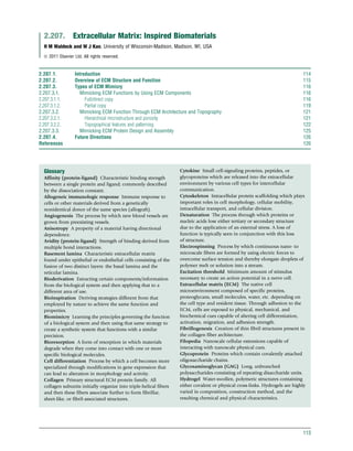
After 12 weeks, SIS-treated MCL had greater
collagen density, cellularity, overall collagen fiber diameter
(Figure 3), and fiber alignment than nontreated controls.
Additionally, RT-PCR investigations of the healed tissues
demonstrated lower relative concentrations of collagen type V
and certain SLRPs in SIS-treated conditions than in nontreated
conditions. These compositional differences corresponded
to at least 50% increases in the stiffness, tangent modulus,
and ultimate strength of the SIS-treated healing tissue com-
pared to nontreated controls. Such effects are attributed to cell
signaling through the presence of growth factors and the
degradable collagenous cell scaffold material as well as to
the SIS scaffold’s ability to maintain hydration within the
wound environment.
Despite the improvements seen in SIS-treated healing tissue
as compared to nontreated tissue, the composition and
mechanical properties of the healed tissue from each condition
were still vastly different from those of the sham controls even
after 26 weeks. One important requirement of scaffolds
(a) (b)
200nm 200 nm
(c)
200nm
Figure 3 Transmission electron micrographs (70 000Â) of cross-sectional collagen fibrils in (a) sham-operated medial collateral ligament (MCL),
(b) small intestinal submucosa-treated MCL, and (c) nontreated MCL at 12weeks post-6-mm gap injury. Adapted from Woo, S. L. Y.; Abramowitch, S. D.;
Kilger, R.; Liang, R. J. Biomech. 2006, 39, 1–20.
118 Biologically Inspired and Biomolecular Materials and Interfaces
7. employed to regenerate tissue is to be able to transmit the
mechanical forces across the injury. This requisite not only
provides the replacement of lost function but also allows for
mechanical conditioning of the regenerating tissue. As ECM
scaffolds, including SIS, reflect the composition and architec-
ture of the source tissue, application of these materials to
different types of allogenic or xenogenic tissue may not fully
replicate the environment necessary to restore functioning tis-
sue. In this case, SIS lacking some crucial characteristics of
ligaments, such as collagen fiber orientation in space, may
have limited its ability to fully stimulate remodeling of the
healing tissue despite containing many of the same structural
molecules. Some current research focuses on mechanically
preconditioning cell-seeded ECM scaffolds that reorient fibers
and increase mechanical properties.12
Such steps may allow
ECM scaffolds from more accessible tissues to be more effective
in mimicking the properties of ECM from different tissues.
2.207.3.1.2. Partial copy
Using full-length copies of proteins is not always feasible or
appropriate for certain biomaterial applications. ECM scaffolds
may not fulfill either the required mechanical or bioactive role
or the rapid degradation may compromise the material effi-
cacy. Individual ECM proteins are often not sufficiently stable
to successfully retain bioactivity after incorporation into the
biomaterial and the amount of source material or the cost of
production to achieve the required bioactive concentration can
be prohibitory. Additionally, full proteins may stimulate unde-
sired cell responses, especially in cases where certain cell differ-
entiation states must be maintained (reviewed in Carson and
Barker13
). As mentioned previously, ECM–cell interactions
and interactions between ECM proteins are mediated by recog-
nition of small peptide sequences contained within ECM pro-
teins. Exploiting this relationship provides an alternative
method to attaining the desired protein–protein interaction
though incorporating a fraction of the native ECM protein in
the form of a functional motif. Through these protein–protein
interactions, changes in cell behavior and ECM structure can be
stimulated through alterations of protein configuration, forma-
tion of membrane-proximinal protein clusters, transmission of
mechanical forces between cells and ECM, and organization
or enzymatic cleavage of ECM proteins. By taking advantage of
the relatively low complexity of these recognition motifs, bio-
activity and bioresponsiveness can be incorporated and con-
trolled in synthetic or natural biomaterials.
The techniques used to modify biomaterials with these
functional peptide motifs generally fall into two categories:
‘surface modification’ and ‘incorporation’ into the scaffold
structure. Surface modification methods, as opposed to bulk
material modification, are typically employed with materials
having a lower surface to volume ratio where cell–material
interactions occur within a limited surface area. This approach
is also advantageous in in vivo settings as it allows the material
to maintain its bulk mechanical properties while adding sur-
face bioactivity. The major methods of immobilizing peptides
to the biomaterial surface include electrostatic interaction
(e.g., adsorption, self-assembled monolayers), ligand–receptor
interactions (e.g., biotin–avidin, antibody–antigen), and cova-
lent attachment (e.g., silanization, polymer tethering; see
Garcia,14
Goddard and Hotchkiss,15
Raynor et al.16
for more
detailed reviews). Limited availability of the ligand due to
surface rearrangement, nonspecific adsorption of proteins,
and changes in ligand conformation is a common challenge
that reduces the efficacy of these techniques. To overcome
some of these limitations, one common strategy is to intro-
duce a spacer group, typically in the form of a hydrophilic
polymer, between the surface and the bioactive molecule
to increase availability, prevent denaturation, and reduce
nonspecific protein adsorption. For highly porous materi-
als, typically hydrogels or other polymeric scaffolds, ECM
mimetic ligands are often incorporated directly into the poly-
meric structure. Peptide sequences can be covalently attached
to polymer chains prior to formation of the scaffold or a
peptide sequence can be incorporated on formation of the
scaffold through the attachment of cross-link-susceptible
chemical groups or interactions with proteins. For example,
SLRP-mediated collagen fibrillogenesis has been mimicked in
collagen scaffolds by incorporating a small peptidoglycan
containing collagen-binding peptide sequences derived from
these SLRPs.17
In this way, functional peptide sequences are
presented throughout the scaffold in a similar manner, as
seen with surface modification, or as an integral, biorespon-
sive segment of the polymeric chains.
2.207.3.1.2.1. Case study: multiple ligand–integrin interactions
alter intracellular signaling
Integrin binding to adhesive motifs that are present in a variety
of ECM proteins may direct cell phenotypes and guide various
cell processes such as adhesion, intra- and intercellular signal-
ing, and cell death. Affinity between the ligand and integrin,
avidity of the ligand, and integrin specificity are all influential
factors in the subsequent downstream cellular effects. One of
the first and most commonly employed peptide motifs in
biomaterial design is the ECM ubiquitous integrin-binding
tripeptide sequence arginine-glycine-aspartic acid (RGD).
RGD was first employed to increase or control adhesion to
materials which normally may not support adhesion, but has
been found to influence a variety of cell behaviors including
cell phenotype. For example, variations in monocyte behavior
seeded onto poly(ethylene glycol) (PEG)-based hydrogels with
or without incorporation of a tethered RGD motif demonstrate
possible implications toward modifying the host response
using functional motifs.18
Both adhesion of and inflammatory
cytokine and protease release from primary monocytes were
shown to be modulated by the presence and density of RGD
within the scaffold. While the ability of simple peptide
sequences, such as RGD, to bind multiple integrin pairs is
advantageous for increasing cell adhesion, in cases where
unambiguous downstream outcomes are desired, integrin-
specific binding is necessary. Moreover, additional complexity
present in the native protein may provide further bioactivity
than is seen with binding to just a simple, small peptide
sequence. For example, presentation of the collagen I derived
adhesive motif, GFOGER, in a triple-helical conformation sim-
ilar to that of native collagen is critical for a2b1 integrin bind-
ing. By incorporating integrin-specific protein fragments into
biomaterials, researchers hope to exploit individual integrin-
mediated bidirectional transfer of biochemical signals.
Both a2b1 and a5b1 integrin pairs have been shown to play
integral roles in mediating the interaction of several cell types,
Extracellular Matrix: Inspired Biomaterials 119
8. for example osteoblasts, fibroblasts, and chondrocytes, with
their native ECM. Most studies, however, deviate from what
is seen in the ECM by incorporating only a single adhesive
motif. In a study by Reyes et al.,19
surfaces combining two
specific integrin-binding motifs were employed to elucidate
possible synergistic effects on fibrosarcoma cell adhesion,
integrin binding, and integrin-mediated signaling responses.
Biotinylated triple-helical GFOGER and a fibronectin fragment
(FNIII7-10) were attached in various ratios to avidin-adsorbed
tissue culture polystyrene surfaces using the well-known high-
affinity interaction between avidin and biotin. The fibronectin
fragment spanned the 7th–10th type III repeats containing
the adhesive motif RGD and its synergistic binding domain
PHSRN. Previous work had demonstrated the increase in a5b1
binding specificity of this fragment as compared to that of
the linear RGDS peptide. The presentation of both GFOGER
and FNIII7-10 demonstrated synergistic enhancement of cell
adhesion, FAK activation (implicated in integrin-mediated
intracellular signaling), and cell proliferation as compared
to single ligand or no ligand surfaces (Figure 4). The use of li-
gand mimics providing specific integrin binding as well as
antiintegrin antibody controls point to specific coordinated
integrin binding of the adhesion motifs leading to membrane-
proximal clustering of the two integrin types and possible fur-
ther downstream interaction between signaling pathways.
The results of these studies demonstrated intracellular con-
vergence of integrin-activated signaling pathways through use
of fibronectin and collagen mimetic ligands presented on a
material surface. Such synergistic effects demonstrate some of
the complexity involved in mimicking cell–ECM adhesive
interactions. While there are prevalent examples of incorpora-
tion of the single ECM mimetic ligands into biomaterial scaf-
folds, cell-responsive benefits may be gained by expanding the
number and types of motifs. However, further material devel-
opment and cell-based studies are needed to determine if
multiligand materials could be employed to improve material
efficacy in an in vivo or in a clinical setting.
2.207.3.1.2.2. Case study: enhancement of chrondrogenic
differentiation by MMP-13 degradable hydrogels
Differentiation of stem cells to dedicated cell types requires
highly coordinated processes integrating multiple types of sig-
nals derived from growth factor–receptor binding to mechan-
otransduction. An additional level of complexity is also
presented in the form of temporal synchronization of these
varied signals. As such, while stem cells hold much promise as
a tool for research and clinical purposes, many challenges
remain concerning both stimulating and inhibiting differenti-
ation. Designing and constructing biomaterials to act as plat-
forms for controlling differentiation states will require a
similar level of complexity as seen in the native ECM environ-
ment which normally mediates stem cell fate. While research
into this field is still in its infancy, several promising bioactive
materials have been developed by incorporating ECM func-
tional motifs.
Temporal principles of chondrogenesis were applied to
enhance human mesenchymal stem cells (hMSC) differentia-
tion seeded on an enzymatically responsive PEG hydrogel.20,21
Differentiation of hMSC into chrondrocytes has been shown
to require an increase in fibronectin, specifically the RGD
adhesive motif, during preliminary phases of chondrogenesis,
likely to stimulate cell–cell interactions. A subsequent decrease
in fibronectin is then seen as differentiation proceeds and cells
adapt a more spherical shape. In fact, the persistent presence of
fibronectin may be inhibitive to chondrocyte function as was
seen in in vitro work from another group using RGD-conjugated
alginate gels. Incorporation of RGD into a PEG hydrogel
was shown to support hMSC viability and initiate chondro-
genic differentiation; however, results also demonstrated that
extended incubation of the hMSC reduced the percent differen-
tiation. On the basis of studies which demonstrated matrix
metalloprotease-13 (MMP-13) upregulation at 7–12 days of
hMSC chondrogenesis, a MMP-13 cleavage site, derived from the
cartilage ECM component aggrecan, was incorporated into the
PEG hydrogel. By integrating a 12-mer peptide containing both
350
300
250
200
150
100
50
0 100
0
200 300 400
GFOGER peptide density (fmolcm−2) GFOGER peptide density (fmolcm−2)
350
(a) (b)
FNIII7-10density(fmolcm−2)
300
250
200
150
100
50
0
0.1
0.2
0.3
0.4
0.5
0.6
0.6
0.8
1.0
1.2
1.4
1.6
2.0
1.8
100 4000 200 300
FNIII7-10density(fmolcm−2)
Figure 4 Contour plots displaying the effect of the GFOGER and fibronectin fragment mixed densities on adhesion ligands on fibrosarcoma;
(a) adhesion and (b) FAK phosphorylation. Results are presented as (a) postcentrifugation calcein-AM signal normalized to the precentrifugation
signal and (b) activated FAK normalized to total FAK detected for those conditions. Adapted from Reyes, C. D.; Petrie, T. A.; Garcia, A. J.
J. Cell Physiol. 2008, 217, 450–458.
120 Biologically Inspired and Biomolecular Materials and Interfaces
9. the cleavage site, PENFF (proline-glutamic acid-asparagine-
phenylalanine-phenylalanine), and RGD into the PEG hydro-
gel, bioresponsive enzymatic cleavage of the RGD sequence
was achieved. While there was a loss of viability of hMSC
encapsulated in hydrogels containing the cleavage site after
11 days, likely because of the loss of adhesion sites, there was
a dramatic increase in glycosaminoglycan deposition, an indi-
cator of chondrogenesis, compared to RGD-only controls.
These studies demonstrated effective temporal and biore-
sponsive presentation of ECM cues to modulate differentiation
of hMSC through incorporation of biological principals to
material design. While these studies address mimicking down-
regulation of signals in the extracellular environment, one
important function of native ECM, maintenance of cell viabil-
ity, was not achieved, and it demonstrates the need for further
material development to fully realize how materials can con-
trol cell fate. Furthermore, additional levels complexity must
be considered for possible future in vivo applications of bior-
esponsive materials. For example, levels of MMP-13 may be
altered in an inflammatory environment as compared to an
in vitro hMSC culture system leading to possible incorrect
timing of RGD cleavage.
2.207.3.2. Mimicking ECM Function Through ECM
Architecture and Topography
While specific ligand–receptor interactions between ECM
components and the cellular environment are the primary
interface responsible for mediating ECM functions, the way in
which these components are organized play a major role
in controlling both the downstream cellular effects and overall
function of the tissue. The hierarchical configurations of
the ECM ultrastructure establish macrolevel mechanical and
mass-transport properties in a tissue. Well-defined nanostruc-
tural topographical and mechanical cues are able to influence
cell–material interaction by promoting cell proliferation,
differentiation, adhesion, and migration. Additionally, nano-
patterning of cell-adhesive motifs provides a secondary level of
cell behavioral control. In this section, an overview of how
these ECM features are replicated and employed in material
design is discussed.
2.207.3.2.1. Hierarchical microstructure and porosity
The core structure of the ECM across tissues consists of a 3D,
highly hydrated, porous matrix. This configuration allows for
water retention, mass transport of nutrients such as glucose
and oxygen, as well as directed cell migration and soluble
factor storage. For example, cell migration efficiency has been
found to be optimal at pore diameters that are the same or
slightly smaller than the diameter of polarized cells (reviewed
in Friedl and Wolf22
). Larger and smaller pore sizes lead to
reduced cell migration rates because of decreased amounts
of cell–ECM contacts and steric hindrance, respectively. Also,
the additional complexity incorporated into the porous
structure of the ECM creates differential physical properties
of a tissue. Simple architectural features, such as fibers (see
Section 2.207.3.2.2), are able to undergo further organization
to form multifunctional lattice structures with specific densi-
ties and spatial arrangements. Developing these hierarchical
arrangements establishes tissue-specific directionally dependent
mechanical properties and cell arrangement (see Isenberg and
Wong23
for further review). For example, helical arrangement
of successive layers of collagen- and elastin-embedded smooth
muscle cells provides enhanced circumferential load-bearing
properties and high torsional stability in arterial walls. Another
widespread example is the organization of the basement mem-
brane in a variety of endothelial tissues: the high density of
structural ECM components forms nearly a 2D platform for
cell attachment and organization through steric- and adhesion-
based inhibition of endothelial cell migration. Mimicking the
structural properties of the ECM in biomaterial design can
range from simply imitating the properties of the core structure
to incorporating mechanically effective higher-order lattice
construction.
A major biomaterial application requirement is to support
cell viability and growth, particularly in tissue engineering and
wound healing applications. Hydrogels composed of both
hydrophilic synthetic polymers (e.g., PEG) and natural macro-
molecules (e.g., collagen) have garnered attention for their
similarity to the ECM core structure in terms of possessing
basic cell-supportive properties including providing hydration,
mass transport, and a 3D environment. This broad class
of materials is highly varied in terms of polymer chemistry,
construction methods, and types of functional modifications
(see Andriola Silva et al.,24
Jia and Kiick,25
and Tibbett and
Anseth26
for relevant reviews) allowing extensive customiza-
tion of hydrogels for different applications. By controlling the
material chemistry and the porosity, specific mechanical prop-
erties and transport characteristics can be achieved. For exam-
ple, hydrogels or similar systems can act as local reservoirs
for soluble proteins where diffusion of soluble factors can
be controlled, in part, by pore size and interactions with the
polymer backbone similar to what is seen in native ECM.
Additionally, controlling the density of physical or chemical
cross-links will alter mechanical properties of the hydrogel.
However, highly porous materials are traditionally limited in
both the strength and complexity of the mechanical char-
acteristics they can achieve. Additionally, pore size is typically
heterogeneous and not able to be precisely controlled using
simpler scaffold construction techniques. Therefore, while
the core structural characteristics of the ECM are able to be
mimicked with relative ease, achieving coordinated mass
transport and mechanical properties requires adaptation of
more complex architectural features.
Approaches in scaffold design are able to mimic higher-
order ECM architecture through creation of hierarchical or
micropatterned porous structures that provide the desired
mechanical and mass-transport properties. Possibly the sim-
plest methodology to achieve porous structures with aniso-
tropic mechanical properties is through the mechanical
conditioning of existing porous scaffolds. For example, scaf-
fold alignment can be achieved through cell- or temperature-
mediated mechanical cycling of collagen or synthetic polymer
hydrogels, respectively.12,27
Alignment of electrospun fibers
can also be employed to create anisotropic materials while at
the same time presenting important topographical cues to cells
(see Section 2.207.3.2.2). These techniques, however, often do
not achieve cell permissive pore sizes or physiological mimetic
mechanical properties. More complex techniques are available
to create 3D structures with defined pore size and shape
Extracellular Matrix: Inspired Biomaterials 121
10. ranging from microablation of pores into polymer membranes
to computational-driven layer by layer manufacturing of com-
plex 3D scaffolds from polymer, hydrogel, ceramic, and metal
materials (see Hollister28
for in-depth review). The latter tech-
nique provides rigorous control of the scaffold architecture,
allowing construction of materials with a single pore size or
wavy fibers, for example. By varying the shape, orientation, and
distribution of the pores, porosity can thereby be used to create
direction-dependent mechanical properties instead of relying
solely on material chemistry.
2.207.3.2.1.1. Case study: anisotropic honeycomb structure for
ventricular myocardium repair
Ventricular myocardium is structurally highly complex, requir-
ing directionally dependent mechanical and electrical properties
for its proper function. In native tissue, cardiomyocytes are inter-
woven into a multifaceted network of collagen fibers which
display honeycomb-like organization. This type of organization
produces mechanical and electrical anisotropy. Damage to the
ventricular myocardium, typically as a result of a cardiac infarc-
tion, leads to cardiomyocyte death and replacement of native
tissue with nonfunctional fibrous tissue. Previous attempts to
repair myocardial tissue using 3D scaffolds has failed to effec-
tively regenerate functional tissue due to structural and mechan-
ical variances from native tissue. For example, scaffolds were
unable to promote more than isolated regions of cardiomyo-
cyte alignment or effectively transmit physiological mechanical
forces. More recent efforts borrow from native ECM collagen
fiber orientation to more closely mimic directionally dependent
myocardial structural and mechanical characteristics.
In a study by Engelmayr et al.,29
a polymeric scaffold exhi-
biting anisotropic characteristics was designed and evaluated
for use in cardiac tissue engineering. An accordion-like honey-
comb structure was created by laser microablating two over-
lapping 200 Â 200 mm square pores oriented at 45
into
approximately 250-mm-thick poly(glycerol sebacte) (PGS)
wafers. The resulting accordion-like scaffold exhibited aniso-
tropic mechanical properties more closely mimicking that of
right ventricular myocardium than scaffolds constructed with
square or rectangular pores. Further manipulation of the
mechanical characteristics could be achieved by reducing the
polymer curing time, cyclic loading, and culture with heart
cells. While in some cases, this modulation helped to achieve
better-matched properties, cell interaction reduced the stiffness
of the scaffold to levels below what is seen in native tissue
after one week. Cardiomyocytes and cardiac fibroblasts cocul-
tured on the accordion-like scaffolds demonstrated cell align-
ment and slightly lower excitation thresholds in the preferred
direction than more isotropic materials. Additionally, an initial
attempt to create a bilayer structure by combining a partially
and a fully excised PGS wafer resulted in cell penetration and
interpore connectivity.
Mimicry of mechanical properties in tissue engineering
scaffolds is important for correct transmission of mechanical
forces across repairing tissues. This study demonstrates that use
of a geometrically controlled porous structure can better match
the mechanical characteristics of native tissues than heteroge-
neous or isotropic scaffolds. Creation of directionally depen-
dent mechanical properties also provided cues that were able
to partially guide cell alignment and thereby, cell-mediated
electrical properties. The authors also attempted to address
in vitro to in vivo scaling issues by layering the PGS wafers to
create additional thickness. Scaling is a major hurdle to trans-
lating these types of precise material construction techniques
from the miniature in vitro cell culture environment to
implants used in considerably larger areas of tissue in in vivo
environments. For scaffolds to successfully function at the
tissue level, materials must be able to be constructed with a
variety of shapes and sizes, typically much larger than what is
used in vitro.
2.207.3.2.2. Topographical features and patterning
The ECM contains a considerable amount of cell-instructional
information within micro- and nanometer scale topographical
and biochemical details. In native ECM, these features are
established through the arrangement and configuration of
ECM components creating geometric cues or differential den-
sities of functional motifs. Cells interact with simple physical
cues, such as varying elevations or nanoscale pores, through
nanoscale cellular extensions, known as filopedia. Although
the mechanism by which these physical cues influence cell
behavior is not completely understood, one contributing factor
is the significant increase in the surface area-to-volume ratio
and overall complexity of the surface. These features facilitate
contact guidance phenomena and subsequent changes in cell
morphology and migration. Concentration gradients of growth
factors or proteins containing adhesive motifs also function
to direct cell migration and alignment along the gradient.
Additionally, specific ligand clustering or patterning of dissimi-
lar motifs or signaling proteins can lead to alterations in cell
behavior beyond what is seen for disorganized ligand–integrin
binding. Although the exact mechanism by which adhesive
ligand clustering affects intracellular signaling is unknown, it is
generally thought that subsequent spatial proximity of integ-
rins leads to further intracellular protein interaction, particu-
larly with various cytoskeleton proteins. The addition of
nanometer scale details in biomaterials can be accomplished
by mimicking native ECM structures or can be imitated using
surface modification techniques to add topological or pat-
terned biochemical cues.
One of the most common methods to replicate micro-
and nanoscale structural features of the ECM is to mimic
the fibrillar construction of the ECM scaffold. In addition to
influencing cell behavior through topological details, interac-
tions between cells and ECM nanofibers also play an important
role in mechanotransduction through viscoelastic deformation
of the fibers in response to external and internal stresses.
Nanofiber-based biomaterials can be manufactured using sev-
eral different techniques, the most commonly employed being
phase separation, self-assembly, and electrospinning (see
Nisbet et al.30
and Madurantakam et al.31
for relevant reviews).
Thermally induced phase separation involves partitioning of
a polymer phase from a solvent phase through controlled
or, more frequently for nanofiber formation, rapid cooling.
Self-assembly of nanofibers can be accomplished by driving
assembly of carefully designed monomers using hydrophobic
or ionic interaction. For example, Hartgerink et al.32
con-
structed a biofunctional nanofibrillar network containing
fibers with an average diameter of 7.1 nm on the basis of
hydrophobic interactions between akyl chains linked to
122 Biologically Inspired and Biomolecular Materials and Interfaces
11. functional polypeptides (for more examples of self-assembled
structures, see Section 2.207.3.3). The more common current
approach for forming fibrous scaffolds because of its relative ease
of use, versatility, and scalability, is electrospinning. Electrospin-
ning involves forming continuous fibers by using electric forces
to overcome surface tension and thereby elongate droplets of
polymer melt or solution into a stream. Using this technique,
natural or synthetic polymers can be employed as substrates for
fiber formation and by varying process, environmental, and
substrate parameters, fibers with a vast diversity of properties
can be constructed. For example, while traditional methods
produce nonwoven, randomly oriented fiber mats, using a
rotating, electrified collector results in fiber alignment and
thereby improved mechanical and cell-guidance properties.
Recent work has also explored increasing the fiber’s bioactivity
by incorporating the delivery of drugs or growth factors and
even cell encapsulation within the fiber structure.33
While electrospinning provides a versatile construction
method for mimicking ECM architecture, several limiting
design constraints remain. Processing conditions, in particular
the use of volatile solvents, have been shown to cause denatur-
ation of the native protein structure. For example, collagen
type I-based nanofibers demonstrated a loss of triple-helical
structure, lack of crystallinity, and lower denaturation tem-
perature suggesting that the collagen had been reverted to a
gelatin-like state.34
Moreover, the resulting pore sizes of the 3D
fibrous scaffold often prohibit cell migration into the matrix.
Steps can be taken to increase porosity such as the addition of
easily removable components to the structure, for example,
salts or highly degradable polymers. Finally, while electrospin-
ning is typically associated with production of nanofibers
able to mimic the fibers of the ECM, most current production
methods achieve fibers with larger diameters than native tissue.
Typical electrospun fiber diameters are 500 nm; however,
several methods do exist to achieve dimensions closer to native
ECM. Furthermore, increases in the understanding of para-
meters relevant to fiber diameter coupled with technological
advances promise closer ECM fiber mimics.
Using ECM structural mimics, such as nanofibers, to con-
struct biomaterials currently does not provide the level of
control or bioactivity needed to fully investigate and/or exploit
microscale or nanoscale cell–material interactions. To accom-
plish these goals, the material design parameters are com-
monly focused to the area of cell–material interface through
use of surface modification techniques. Methods to incorpo-
rate nanoscale features onto the surface can be categorized on
the basis of the level of user-specific control of the resulting
patterns they provide. Unordered topographies can be manu-
factured using techniques such as polymer demixing, colloidal
lithography, and chemical etching (see Norman and Desai35
for a more detailed review). These methods allow a small
amount of user control over the type and number of nanofea-
tures obtained through variations in processing parameters,
but cannot create structures with complex prescribed geome-
tries or organization. In exchange, these methods are capable
of rapid coverage of large substrate surfaces. The resulting
unordered surface topographies are able to mimic the nano-
scale features of the ECM but may not present the same
amount of complexity and therefore cell-instructional infor-
mation of native tissue.
Ordered topographies can be developed using laser abla-
tion, microfluidics, or a variety of lithographical techniques
(for more extensive reviews, see Christman et al.,36
Hook
et al.,37
Mrksich,38
and Schmidt and Healy39
). Through mole-
cule removal from the surface, molecule addition to the sur-
face, or surface group modification, organized nanopatterns
can be attained. Both physical and biochemical cues can be
patterned by adding motifs directly to the surface or manipu-
lating the chemical composition of the surface to prevent or
accept biofunctional molecules through adsorption or cova-
lent binding. Alternatively, imprint lithography and microcon-
tact printing use nanopatterned rigid masters, manufactured
using the previously mentioned techniques, to topographically
mold surfaces or stamp proteins onto surfaces. However, the
relatively high cost, low throughput, and lack of available
equipment needed to employ these techniques limit the appli-
cation of ordered nanopatterning to biomaterial design. All of
these methodologies are able to spatially control molecule
placement on biomaterial surfaces allowing creation of more
complex biomaterials. Yet, currently, the main advantage of
being able to define surface nanopatterns is derived from the
ability to gain a better understanding of how individual nano-
scale topographical and biological patterns affect cell behavior
and phenotype at an in vitro level.
Patterning of functional motifs onto biomaterial surfaces has
been used extensively to study how engineering material sur-
faces can be used to alter cell adhesion strength, spreading,
migration, and differentiation. Initial and continuing work
in this area involved micropatterning of proteins, such as fibro-
nectin, through preferential adsorption to certain chemical
domains patterned onto the substrate or lithographic printing
methods.40,41
Cell morphology and adhesion strength, for
example, were shown to be controlled by modulating the area
of cell–material contact.14
Development of more sophisticated
nanopatterning techniques has shifted the focus to creating
subcellular arrangements of proteins or, more commonly, func-
tional motifs. For example, a density gradient of RGD causes
preferential migration and alignment of cells.42
Additionally,
the density and spatial proximity of nanoscale RGD clusters
can modulate differentiation, spreading, proliferation, and
motility of cells.43,44
One reason for the observed variations
was demonstrated by a series of studies where a spatial limit
between RGD clusters was established for the formation of focal
adhesions.45
The limitations of these techniques revolve around
the 2D system necessary for the creation of these nanoscale
patterns. Therefore, while the existence of nanoscale features in
the ECM is known, the level of knowledge and technology
needed to mimic these aspects to control cell–biomaterial inter-
actions in a more clinical setting has not yet been achieved.
2.207.3.2.2.1. Case study: electrospun nanofibers for repair of
peripheral nerves
The peripheral nervous system consists of bundles of neuronal
axons (nerve fibers) typically surrounded by a myelin sheath
formed by layers of Schwann cells, a type of glial cell. Damage
to peripheral nerves usually presents as a severance of an axon
and can be repairable without intervention with significant
restoration of function. In cases where there is extensive loss
of tissue, however, random nerve sprouting at the site of injury
because of a lack of directional cues is insufficient to effectively
Extracellular Matrix: Inspired Biomaterials 123
12. regenerate the lost tissue. Infiltration of inflammatory cells
and eventual establishment of granulation tissue at the site
of injury create an inhibitory microenvironment for nerve
regeneration. Several different types of tissue engineering
approaches have been attempted to create a more permissive
environment for nerve regeneration.46
Conventional treatment
involves insertion of nerve autografts or allografts; however,
autograft material is limited and allografts may lead to immu-
nological rejection. Polymer nanofiber-based biomaterials are
promising for neural cell–material applications because of
their resemblance to native ECM and ability to directionally
guide neurite outgrowth. Of particular interest are nanofibers
constructed out of poly(a-hydroxy esters) because of their
bioresorbable and biocompatible nature.
Poly(DL-lactic-co-glycolide) (PLGA) and poly(e-caprolactone)
(PCL) nanofibers have been used for in vitro and in vivo neural
tissue engineering applications concerning neurite outgrowth
for damaged peripheral nerve reconstruction. When whole
dorsal root ganglia (DRG), dissociated DRG cells, Schwann
cells, and fibroblasts were seeded on aligned PCL and PCL/
collagen blend nanofibers (500–600 nm diameter), greater
alignment of neurite growth parallel to the fiber orientation
was demonstrated as compared to nonfibrous poly-D-lysine
surfaces (Figure 5).47
The addition of collagen to the nanofiber
composition led to an increased fiber orientation, glial cell
migration, and elongation of fibroblasts, but decreased rate
of neurite elongation, likely attributed to stronger cell–material
interactions due to the presence of collagen. Furthermore, on
PCL/collagen nanofibers, there was evidence of neurite growth
on top of Schwann cells suggesting indirect directionality
conveyed by the nanofibers to the extending neurites. In a
separate in vivo study, tubes constructed with both PCL micro-
fibers (2.5–8 mm diameter) and PCL/PLGA blend nanofiber
tubes (140–500 nm diameter) were used to treat a 10-mm
gap wound in the sciatic nerve in a rat model.48
Improved rat
sciatic nerve regeneration was achieved as compared to both
transected nerve and nontreated 10-mm gap injury controls.
After 4 months, regenerated tissue consisting of neural fibers,
glial cells, fibroblasts, and ECM consistent with regenerating
basal lumina was observed throughout the length of the scaf-
fold. Meanwhile, the nerve stumps never reconnected in the
case of the two controls; instead, random neurite sprouting led
to attachment to the surrounding muscle tissue. Evidence of
partial reinnervation was seen on the basis of transmission
of neural tracers across the regenerated tissue as well as
behavioral and neurophysiological tests. The success of the
scaffold was attributed to increased cell adhesion and direc-
tional migration across the fibrous structure as well as a lack of
excess inflammatory response. The fibrous structure provided
high flexibility, porosity, and surface-to-volume ratio allowing
higher levels of protein adsorption and an absence of mechan-
ical microinjury as seen with stiff continuous tubes. In addition
to serving as a guide for the regenerating nervous tissue, the
relatively close-knit structure of the fibers prevented unwanted
tissue infiltration while still allowing passage of nutrients.
Despite the success of the employment of nanofibrous tubes
over nontreated controls, full function was not restored to the
tissue. Myelination was not seen throughout the regenerated
nervous tissue and the restored basement lamina was disorga-
nized compared to uninjured tissue. In this in vivo study, the
(a) (b)
(c)
pl
1
0
80
100
60
40
PP PC/P C/P C/P
DIV1
Orientationindex(%)
DIV4 DIV74
pl
##
**
**
**
*
Figure 5 Orientation of neurite growth from dorsal root ganglia explants. (a,b) Neurofilament staining at 4 days on (a) poly(e-caprolactone) (PCL)
nanofibers and (b) PCL/collagen blend nanofibers; scale bar ¼ 500 mm. Arrows indicate direction of nanofibers. (c) Comparison of axon orientation on
poly(lysine)-coated coverslips at 1 and 4 days in vitro (DIV) and on PCL and PCL/collagen nanofibers at 1, 4, and 7 DIV; orientation index of 50%
indicates random orientation of neuritess, 100% complete alignment with nanofibers, 0% orientation perpendicular to nanofibers. Significantly different
than 50% *p 0.01, **p 0.05; ##p 0.01. Adapted from Schnell, E.; et al. Biomaterials 2007, 28, 3012–3025.
124 Biologically Inspired and Biomolecular Materials and Interfaces
13. fibers were randomly oriented and relied on the tube structure
to direct longitudinal growth of the neurites. Improvements
may be seen by applying the in vitro results discussed previously
by increasing geometric directional cues and incorporating
native ECM components into the material. In either case, how-
ever, tissue engineering approaches incorporating ECM architec-
tural principles to peripheral nerve regeneration are relatively
novel and further development will be needed before clinically
efficient demonstrations can be achieved.
2.207.3.3. Mimicking ECM Protein Design and Assembly
Biological polypeptides are, in essence, complex copolymers
which derive their properties from the precisely organized
sequences and compositions of the basic amino acid mono-
mers. Depending on the properties of the amino acid side
chains, proteins will adapt various secondary, tertiary, and qua-
ternary assemblies. Furthermore, through controlled associa-
tions between motifs incorporated into different polypeptide
molecules, ECM proteins have the ability to self-assemble into
complex 3D scaffolds. The design versatility, synthetic homoge-
neity, and biocatalytic assembly of biopolymers are attractive
attributes to incorporate into material design and construction.
In this section, a brief overview of how concepts of ECM protein
design and self-assembly are both mimicked by and incor-
porated into biomaterials is provided (for more in-depth
reviews, see Deming49
and Maskarinec and Tirrell50
).
Advances in recombinant DNA technology and chemi-
cal peptide synthesis techniques have stimulated interest
and provided the necessary tools to explore de novo designed
polypeptide sequences with material application potential.
Designing proteins which assemble into higher-order struc-
tures using recombinant DNA technology is the most com-
plex adaptation of mimicking protein assembly into material
design. By designing novel polypeptide sequences from
first principles utilizing sequence-based structural elements,
researchers hope to be able to control higher-order structures
or interactions between proteins. Successful cases of achieving
functional tertiary structures are rare; however, greater progress
has been made utilizing sequence-to-structure concepts on a
smaller scale. Artificially created amino acids can be substituted
to change the overall properties of the polypeptide. Incor-
poration of fluorine-containing amino acid analogues, for
example, has been used to integrate properties seen in fluori-
nated synthetic polymers, such as low surface energy, low
friction coefficient, and good hydrolytic stability, into poly-
peptides. Thus, multi-monomer, well-defined polypeptides can
assimilate desirable chemical properties normally seen in poly-
mers with heterogeneous molecular weight distribution and less
complex chemical composition. The configuration of small
polypeptide sequences has also been altered to perform a non-
native function. For example, introducing different sequence
mutations into functional domains has been used to artificially
confer specificity. Richards et al.51
produced a FNIII10 frag-
ment with an RGDWXE sequence that demonstrated enhanced
affinity and specificity to the avb3 rather than the a5b1 integrin.
Finally, controlled periodicity of sequence structure can stimu-
late thermodynamic folding into higher-order structures such
as fibers or sheets. By exploiting structural and ionic character-
istics of amino acids, peptide sequences without chemical mod-
ification have been designed to fold into b-sheets or a-helical
structures. Further modification of the peptide sequence can
lead to systematic interactions between polypeptide chains lead-
ing to the formation of coiled-coil or stacked structures. For
example, in a study by Banwell et al.,52
modifications to a
repeating heptad peptide sequence capable of self-assembling
into a-helical structures were able to control the formation
of higher-order materials (Figure 6). That is, more specific
c
c
f f
b
(a)
(b)
b
e
e
a
a
d
d
g
g
50nm
c
c
f f
b
b
e
e
a
a
d
d
g
g
Figure 6 Schematic representation of design principles behind the hierarchical assembly of polypeptide chains containing the coiled-coil heptad
sequence repeat, abcdefg. By incorporating specific interactions between amino acids at positions b and c, the a-helical structures further organized to
form thicker fibrils. Alternatively, incorporation of more general, weaker interactions at positions b, c, and f led to the formation of thinner, more flexible
fibers which could form hydrogels based on physical interactions between chains. Adapted from Banwell, E. F.; et al. Nat. Mater. 2009, 8, 596–600.
Extracellular Matrix: Inspired Biomaterials 125
14. interactions between chains led to a-helical dimer association
to form larger fibrils or more general interactions led to hydrogel
formation containing more flexible chains. Such a work exhibits
potential for creating highly controllable and biocompatible
materials by translating nanoscale structural principles.
2.207.4. Future Directions
The merging of material and biological principles allows
for the creation of materials that function as cooperative parts
of the biological environment. As shown in this chapter, par-
ticular promise lies in incorporating principles derived from
the native ECM into biomaterial design. Contained within the
ECM is a complex regimen of spatial and temporally controlled
cellular cues and structural elements responsible for maintain-
ing and adapting to changes in the biological environment.
In addition to multiple biomaterial applications benefiting
from the addition of structural and functional ECM-derived
principles, this design approach can also provide new insights
into biological mechanisms that are not fully understood.
While many strategies exist for incorporating these features,
mimicry of the complex multicomponent, spatially and tem-
porally controlled system has not been truly achieved in bio-
material design. The future lies in increasing the specificity and
control of the presentation of multiple types of signals and
structures. However, incorporating this type of complexity into
biomaterials requires an additional level of inquiry into
scalability issues and possible host modification once the
material is applied to in vivo clinical settings. Furthermore,
when nanoscaled or technologically complicated techniques
are used to construct the materials, additional consideration
must be paid toward the long-term efficacy and impact versus
‘traditional’ biomaterial in the face or regulatory hurdles, clinical
availability concerns, and potentially high costs of production.
Nonetheless, ECM mimicry holds much promise in advancing
biomaterial research into controlling host–biomaterial interac-
tions and creating new ways to construct materials.
References
1. Plopper, G. In Cells; Lewin, B., Cassimeris, L., Lingappa, V. R., Plopper, G., Eds.;
Jones and Bartlett, 2007; p 645.
2. Fredberg, J. J.; Discher, D.; Dong, C.; et al. Ann. Biomed. Eng. 2009, 37(5),
847–859.
3. Place, E. S.; Evans, N. D.; Stevens, M. M. Nat. Mater. 2009, 8, 457–470.
4. Abraham, S.; Eroshenko, N.; Rao, R. R. Regen. Med. 2009, 4(4), 561–578.
5. Badylak, S. F.; Freytes, D. O.; Gilbert, T. W. Acta Biomater. 2009, 5, 1–13.
6. Kim, M. S.; Ahn, H. H.; Shin, Y. N.; Cho, M. H.; Khang, G.; Lee, H. B. Biomaterials
2007, 28, 5137–5143.
7. Brown, B.; Lindberg, K.; Reing, J.; Beer Stolz, D.; Badylak, S. F. Tissue Eng. 2006,
12(3), 519–526.
8. Sotome, S.; Uemura, T.; Kikuchi, M.; et al. Mater. Sci. Eng. 2004, 24, 341–347.
9. Woo, S. L. Y.; Abramowitch, S. D.; Kilger, R.; Liang, R. J. Biomech.
2006, 39, 1–20.
10. Liang, R.; Woo, S. L.; Nguyen, T. D.; Liu, P.; Almarza, A. J. Orthop. Res. 2008,
26(8), 1098–1104.
11. Woo, S. L.; Takakura, Y.; Liang, R.; Jia, F.; Moon, D. K. Tissue Eng. 2006, 12(1),
159–166.
12. Androjna, C.; Spragg, R. K.; Derwin, K. A. Tissue Eng. 2007, 13(2), 233–243.
13. Carson, A. E.; Barker, T. H. Regen. Med. 2009, 4(4), 593–600.
14. Garcia, A. J. Adv. Polym. Sci. 2006, 203, 171–190.
15. Goddard, J. M.; Hotchkiss, J. H. Prog. Polym. Sci. 2007, 32, 698–725.
16. Raynor, J. E.; Capadona, J. R.; Collard, D.M; Petrie, T. A.; Garcia, A. J.
Biointerphases 2009, 4(2), FA3–FA16.
17. Paderi, J. E.; Sistiabudi, R.; Ivanisevic, A.; Panitch, A. Tissue Eng. 2009, 15(10),
2991–2999.
18. Chung, A. S.; Waldeck, H. M.; Schmidt, D. R.; Kao, W. J. J. Biomed. Mater. Res.
2009, 91A(3), 742–752.
19. Reyes, C. D.; Petrie, T. A.; Garcia, A. J. J. Cell Physiol. 2008, 217, 450–458.
20. Salinas, C. N.; Anseth, K. S. Macromolecules 2008, 41, 6019–6026.
21. Salinas, C. N.; Anseth, K. S. Biomaterials 2008, 29, 2370–2377.
22. Friedl, P.; Wolf, K. J. Cell Biol. 2009 [Online].
23. Isenberg, B. C.; Wong, J. Y. Mater. Today 2006, 9(12), 54–60.
24. Andriola Silva, A. K.; Richard, C.; Bessodes, M.; Scherman, D.; Merten, O.-W.
Biomacromolecules 2009, 10, 9–18.
25. Jia, X.; Kiick, K. L. Macromol. Biosci. 2009, 9, 140–156.
26. Tibbett, M. W.; Anseth, K. S. Biotechnol. Bioeng. 2009, 103(4), 655–663.
27. Millon, L. E.; Mohammadi, H.; Wan, W. K. J. Biomed. Mater. Res. 2006, 79B,
305–311.
28. Hollister, S. J. Nat. Mater. 2005, 4, 518–525.
29. Engelmayr, G. C.; Cheng, M.; Bettinger, C. J.; Borenstein, J. T.; Langer, R.;
Freed, L. E. Nat. Mater. 2009, 7, 1003–1010.
30. Nisbet, D. R.; Forsythe, J. S.; Shen, W.; Finkelstein, D. I.; Horne, M. K. J. Biomater.
Appl. 2009, 24, 7–29.
31. Madurantakam, P. A.; Cost, C. P.; Simpson, D. G.; Bowlin, G. L. Nanomedicine
2009, 4(2), 193–206.
32. Hartgerink, J. D.; Beniash, E.; Stupp, S. I. Science 2001, 294, 1684–1688.
33. Ashammakhi, N.; Wimpenney, I.; Nikkola, L.; Yang, Y. J. Biomed. Nanotechnol.
2009, 5(1), 1–19.
34. Zeugolis, D. I.; Knew, S. T.; Yew, E. S. Y.; et al. Biomaterials 2008, 29, 2293–2305.
35. Norman, J. J.; Desai, T. A. Ann. Biomed. Eng. 2006, 34(1), 89–101.
36. Christman, K. L.; Enriquez-Rios, V. D.; Maynard, H. D. Soft Matter 2006, 2,
928–939.
37. Hook, A. L.; Voelcker, N. H.; Thissen, H. Acta Biomater. 2009, 5(7),
2350–2370.
38. Mrksich, M. Acta Biomater. 2009, 5, 832–841.
39. Schmidt, R. C.; Healy, K. E. J. Biomed. Mater. Res. 2009, 90A, 1252–1261.
40. Coyer, S. R.; Garcia, A. J.; Delamarche, E. Angew. Chem. Int. Ed. 2007, 46,
6837–6840.
41. Liu, L.; Ratner, B. D.; Sage, E. H.; et al. Langmuir 2007, 23, 11168–11173.
42. DeLong, S. A.; Gobin, A. S.; West, J. L. J. Control. Release 2005, 109,
139–148.
43. Comisar, W. A.; Kazmers, N. H.; Mooney, D. J.; Linderman, J. J. Biomaterials
2007, 28, 4409–4417.
44. Maheshwari, G.; Brown, G.; Lauffenburger, D. A.; Wells, A.; Griffith, L. G.
J. Cell Sci. 2000, 113, 1677–1686.
45. Cavalcanti-Adam, E. A.; Volberg, T.; Micoulet, A.; Kessler, H.; Geiger, B.;
Spatz, J. P. Biophys. J. 2007, 92, 2964–2974.
46. Subramanian, A.; Krishnan, U. M.; Sethuraman, S. J. Biomed. Sci. 2009, 16,
108–118.
47. Schnell, E.; Klinkhammer, K.; Balzer, S.; et al. Biomaterials 2007,
28, 3012–3025.
48. Panseri, S.; Cunha, C.; Lowery, J.; et al. BMC Biotechnol. 2008, 8, 39–50.
49. Deming, T. J. Adv. Mater. 1997, 9(4), 299–310.
50. Maskarinec, S. A.; Tirrell, D. A. Curr. Opin. Biotechnol. 2005, 16, 1–5.
51. Richards, J.; Miller, M.; Abend, J.; Koide, A.; Koide, S.; Dewhurst, S. J. Mol. Biol.
2003, 326, 1475–1488.
52. Banwell, E. F.; Abelardo, E. S.; Adams, D. J.; et al. Nat. Mater. 2009, 8,
596–600.
126 Biologically Inspired and Biomolecular Materials and Interfaces