2. Objectives
At the end of this presentation you will be able to:
Describe the anatomy & physiology of the IVC
Understand the pathologic conditions & anatomic
variants of the Inferior Vena Cava
Identify and discuss the Theory of Virchow’s Triad
Define Inferior Vena Cava filter placement and uses
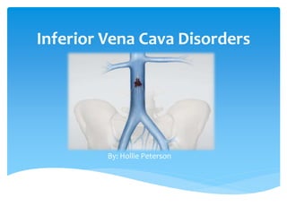
3. Largest vein of the body that drains the
LE & abdomen into the RA of the heart
The Inferior Vena Cava is a continuation of
the common femoral vein (CFV)
Right & left CFV join to form IVC
Travels cephalad and terminates in the RA
What is the Inferior Vena Cava?
5. Asymptomatic
The hepatic segment of
the IVC is absent and
the hepatic veins join and
drain directly into
the right atrium.
Absent Intrahepatic IVC
“Azygos Continuation”
Left sided IVC
Paired IVC (duplication
terminates at level of
renal veins)
IVC on left side only (can
terminate in left renal
vein or extend cranially
to drain into azygos vein
in the chest)
Anatomic Variants
“Azygos Vein”: a large vein on the right
side at the back of the thorax, draining
into the superior vena cava.
6. Neoplastic Obstruction
(Rare)
Typically: Intraluminal tumors arising from renal or hepatic veins
that secondarily obstruct or thrombose the IVC
Leimyosarcoma:
Rare intraluminal tumor that develops in the middle portion of
the IVC
Primarily found in women
Discovered by German doctor in 1871
By 2006, <300 cases have been reported
Unknown cause
Asymptomatic (due to deep origin)
Vague Symptoms: abdominal pain, palpable mass, LE
edema
Treatment: surgical removal of tumor & surrounding tissue.
(Resection necessary)
7. Acute Thrombosis
Obstruction of venous outflow within the deep or superficial vein
by thrombosis
Location: valve site, venous confluences
Patient history: Previous DVT, Post-op, Persistent swelling ( usually
unilateral ), clotting issues, localized pain/burning/itching,
symptoms of PE
Causes: Virchow’s Triad, Varicose veins, Extrinsic Compression
Once thrombosis forms it:
1) Stabilizes (adheres to vessel wall)
2) Propagate (“growth of thrombus” in size)
3) Shed/Embolize
(portion of thrombus breaks free & travels)
8. Theory of Virchow’s Triad
States that venous thrombus is caused by combination of:
A) Venous Stasis
Non-movement of blood that leads to coagulation
Platelets become trapped behind valve cusp (recirculation)
Results from: obesity, post-op, immobility, etc.
B) Hypercoaguble State
Increased activation of coagulation factors along with a
decrease of coagulation inhibitors
Results from: cancer, pregnancy, etc.
C) Endothelial Damage (Intimal Injury)
Results from: Surgery, trauma, central catheterization, IV
drug abuse
9. Theory of Virchow’s Triad
Combination of any of these can result in venous thrombosis
10. Extrinsic Compression
Paget-Syndrome
Refers to axillary-subclavian vein thrombosis associated with
strenuous + repetitive activity of upper extremity
Unusual cause of venous compression & intimal injury leading
to thrombosis
May-Thurner Syndrome (MTS)
Caused when the left iliac vein is compressed by the right iliac
artery, which increases risk of Deep Vein Thrombosis (DVT) in
left extremity
11. Pulmonary Embolism (PE)
The occlusion of a pulmonary artery by a thromboembolus
o Complication of venous thrombosis
o Third most common cause of death in the United States
Top “unexpected death” in ANY age group
o 1/3 patients are asmptomatic
o Source is typically iliofemoral thrombus
(LE deep veins involved 90% of time)
IVC RA RV
Pulmonary
Artery
12. What is an IVC filter?
A device that traps large clots from the lower extremity
o Consist of thin metal struts that converge together to form
a cone shape
13. IVC Filter Placement
Aim of procedure is to “catch” any thrombi floating in the blood
stream from the LE (legs + pelvis) that occasionally travel to the
lungs causing a PE
Who gets one?
Patients with a history/ at risk for developing blood clots in
the legs
Patients unable to tolerate blood thinning agents
Inserted via catheter (using the CFV, EIV, or jugular access)
Patient under local anesthesia
Hospital stay: 12-24 hours (back to normal activity in 1-2 days)
14. Retrieval of IVC Filter
A: Infrarenal IVC surrounded by scar tissue (tan color) on endoluminal surface
B: Filter is beginning to be sheathed
C: Filter is sheathed from top to bottom in preparation for removal
D: Laser sheath is activated and removal occurs by detaching filter from wall of IVC
15. Quiz
1. is a device used to traps large clots from the lower
extremity that consists of a metal frame that converges into a
cone shape.
1. The Theory of Virchow’s Triad states that a combination of
stasis, hypercoaguable state, and endothelial damage leads to
.
2. is considered to be the most
common cause of venous thrombosis.
IVC filter
thrombosis
Pulmonary Embolism (PE)