FtsK is a DNA translocase in E. coli that is involved in chromosome dimer resolution and segregation. Through single-molecule experiments tracking FtsK movement on DNA substrates containing the E. coli chromosome region near the dif site, the authors identified four zones where FtsK frequently reverses direction of translocation. Analysis of these zones identified the DNA sequence motif GNGNAGGG as a candidate sequence that causes FtsK to pause and reverse direction upon encountering it from the 3' end of the G-rich strand. FtsK paused for an average of 1.0 seconds at these reversal sites.
2. amplification, digestion with XmaI, and ligation with T4 DNA
ligase. The test sequence was cloned into the XbaI site of the
resultant plasmid (pJP1) as a duplex oligonucleotide containing
5-mer repeats of the sequence GGGCAGGGG (anti), CCCCT-
GCCC (iso), or GGAGGCGGG (scramble), spaced with four
intervening A, C, or T nucleotides and verified by sequencing (see
Supporting Text for exact sequence). To generate clones with a
single test sequence, we amplified pJP1 by using tailed PCR primers
containing an XbaI site and the sequence GGGCAGGGG or
CCCCTGCCC.
To produce fragments for tether assembly, plasmids described
above were digested with BamHI, dephosphorylated, and purified.
Products were then digested with KpnI and gel-purified. The 41-kb
lambda, 6-kb plasmid, and two molecular handle fragments were
then ligated together by using T4 DNA ligase.
Single-Molecule Experimental Procedures and Data Analysis. Trans-
locations were carried out with 10 g͞ml FtsK50C in 50 mM Tris,
pH 7.5͞5 mM MgCl2͞50 mM NaCl͞1 mM DTT͞3 mM ATP͞100
g/ml BSA. The DNA tethers were held at Ϸ40 pN of tension
between a 2.7-m anti-digoxygenin antibody-coated polystyrene
bead in an optical trap and a 3.2-m streptavidin-coated polysty-
rene bead immobilized on a glass pipette. Translocation of FtsK50C
particles was recorded on digital video at 30 Hz, and image analysis
was used to ascertain position as a function of time. Noise was
smoothed by using an eight-point moving average. A translocation
event is defined as the period of activity in which FtsK50C moves for
at least 0.5 m in one direction and at a minimum speed of 0.25
m͞sec. A turnaround occurs when a translocation event termi-
nates, and FtsK50C translocates in the opposite direction. A pause
is the time between two consecutive translocation events. Pause
duration analysis was done on turnaround zone II or I. All errors
are given as standard errors.
Informatics. We compiled a list of all octamers (words) occurring on
the leading strand of E. coli within 15 kb to the left of dif. For each
octamer, we identified and separately grouped all octamers found
in the same 15-kb region with 1, 2, or 3 mismatches (mismatch
words). We generated a list of candidates by comparing each
octamer to its corresponding mismatch words list. We first grouped
all of the mismatch words that had mismatches in the same
positions. Next, we determined all of the combinations of the
degenerate bases (N, R, S, W, etc.) for these mismatch positions.
The result was a list of octamer words with 0, 1, 2, or 3 degenerate
bases. The above process was repeated on the opposite strand of the
15-kb region to the right of dif. The list was filtered by requiring that
on the 15-kb regions to the left and right of dif, (i) the octamers have
a skew Ͼ50, (ii) the octamers occur within 1,000 bp of the
turnaround peak center (Fig. 1A, zones I, II, and IV), and (iii) the
observed sequence distribution was not present by chance alone (P
value Ͻ 0.01). A nucleic acids substitution-scoring matrix (see
Supporting Text) was used to cluster similar octamers into distinct
motifs. Two octamers were considered to be part of the same group
if any alignment between them gave a substitution score that was
above the arbitrarily selected threshold of 22. The motifs were
determined by manual observations for any group that had more
than five members.
Triplex Substrate Preparation and Displacement Assays. DNA sub-
strates for triplex displacement were prepared by first cloning a
DNA triplex-promoting site (15) into the EcoRV and PstI sites of
a derivative of pBS-SK(ϩ). Duplex oligos containing 5-mer repeats
of the anti, iso, or scramble sequences (described in Single-Molecule
DNA Substrate Preparation) or derivatives were cloned into the SpeI
site, and the final products were verified by sequencing. DNA
triplexes were assembled as described in ref. 15 and Supporting Text.
Triplex displacement reactions were carried out at 25°C in 50 mM
Tris, pH 7.5͞5 mM MgCl2͞3 mM ATP͞0.1 mg/ml BSA͞1 mM
DTT͞0–152 nM FtsK50C͞5 nM triplex substrate.
Results
Identification of FtsK Turnaround Regions. Previously, we used op-
tical tweezers to track the movement of FtsK50C particles on single
DNA molecules. We demonstrated that only a single motor is active
in the FtsK particle (ref. 5 and Supporting Text). We showed that
FtsK50C moves unidirectionally overall, punctuated by occasional
reversals, both on lambda DNA and on the 30-kb chromosomal
regions immediately to the left or right of dif (5). On a chromosomal
DNA molecule with dif at the center (c-tether), FtsK oscillated
around dif. We visually tracked FtsK50C particles as they oscillated
about dif on the c-tether to identify possible turnaround zones
(Movie 1, which is published as supporting information on the
PNAS web site). To improve the resolution of the assay, we kept the
DNA tether at a high force of Ϸ40 pN to reduce the size and
frequency of FtsK50C-induced loops (5, 10). The location of FtsK50C
as a function of DNA sequence could be measured within Ϯ750 bp,
with resolution limited by diffraction, the size of the FtsK50C
particle, and FtsK50C-induced looping of the DNA.
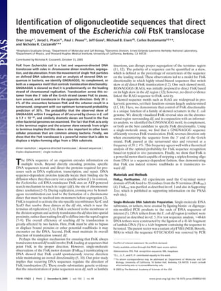
A histogram of FtsK50C turnaround locations shows that 88% of
turnarounds (486͞555 over three tethers) occur when FtsK50C is
translocating away from dif (Fig. 1A). This behavior is expected,
given FtsK’s biological role. There are several zones where FtsK50C
frequently reverses direction (Fig. 1A). Two of these zones are
located at 2,500 Ϯ 750 bp (I) and 500 Ϯ 750 bp (II) to the left of
dif. To the right of dif, the most prominent turnaround zone is
2,400 Ϯ 750 bp from dif (IV). A fourth peak (III) is located between
dif and zone IV. However, its location and magnitude varies among
the three tethers analyzed (Fig. 6, which is published as supporting
information on the PNAS web site), and therefore we excluded it
from further analysis. We also found that there is a sequence
dependence of the dwell time of FtsK50C, as FtsK50C pauses at the
turnaround zones (compare Fig. 1 A and B). The mean pause time
Fig. 1. FtsK pauses and turns around at specific points in the dif region of the
E. coli chromosome. (A) Histogram of turnaround locations shows that 88%
(486͞555) of turnarounds occur when FtsK translocates away from dif (n ϭ 555
over three tethers). Red indicates movement from right to left; blue represents
movement from left to right. Location and orientation of GNGNAGGG and
RGNAGGGS motifs are indicated above the observed turnaround zones (I–IV).
(B) Dwell time histogram of FtsK on the same tether as in A indicates that FtsK
pauses at the turnaround zones. (C) Histogram of pause time (n ϭ 193 on three
tethers) observed at turnaround zones I or II has a mean of 1.0 Ϯ 0.1 sec.
Levy et al. PNAS ͉ December 6, 2005 ͉ vol. 102 ͉ no. 49 ͉ 17619
BIOPHYSICS
3. is 1.0 Ϯ 0.1 sec, and the pause duration appears to be random (Fig.
1C). We conclude that there are specific DNA sequences in the
observed turnaround zones, the FtsK recognition sequences
(FRSs), which cause FtsK to reverse direction when encountering
the sequence in one orientation. A significant skew in the orien-
tation of the FRS toward dif can efficiently bias FtsK translocation
in the direction of dif. We discuss in the next section the implication
of these results for the identity of the FRS.
Bioinformatics Identifies Candidate FRSs. To develop a short list of
candidate FRSs, we used the experimentally identified turnaround
zones to constrain an informatics analysis of skewed sequences near
dif. Salzberg et al. (14) identified 150 octamers in the E. coli
chromosome that are significantly skewed and whose skew switches
strand at the origin and terminus of replication. We reasoned that
the direct observation of FtsK turnaround zones would help us
focus our informatics analysis by concentrating on sequences
present in these zones; however, we did not limit our analysis to
candidates that were identified by Salzberg’s study. An FRS should
display a significant skew on the E. coli chromosome and switch
strands at dif. The algorithm was initially executed against the 15-kb
regions to the left and right of dif, because these regions are covered
by the dif-centered DNA tether on which we observed directional
movement of FtsK (5). We modeled the FRS as an octamer to make
the search computationally more tractable (see Supporting Text).
Degeneracy in up to three positions was allowed; we presumed that
the FRS is a motif rather than a specific sequence.
We initially identified eight motifs that satisfied these criteria
(Table 1, which is published as supporting information on the PNAS
web site). We anticipated that FtsK activity extends beyond the
immediate dif proximal region, as was confirmed by Corre and
Louarn (16). Therefore, we extended the analysis to longer regions.
Only the GNGNAGGG candidate maintained a strong skew and
statistical significance within 100 kb and 1 megabase (Mb) from dif
(Fig. 2A; see also Fig. 7A, which is published as supporting
information on the PNAS web site). This motif has a skew of 86
(P ϭ 4.4 ϫ 10Ϫ11
) at 100 kb and a skew of 79 (P ϭ 1.7 ϫ 10Ϫ57
) at
1 Mb away from dif. RAG was not identified as a candidate motif
in this analysis, because there are no RAGs within turnaround zone
II. The other two major turnaround zones (I and IV) contain both
RAG and GNGNAGGG sequences (Fig. 1A). One of the major
reasons that RAG was proposed as an FRS is its very high skew of
97 (P ϭ 2.9 ϫ 10Ϫ17
) at 100 kb and 88 (P ϭ 1.9 ϫ 10Ϫ97
) at 1 Mb
from dif (12). However, as we show below, such a high skew is not
necessary to impart directionality.
The GNGNAGGG motif is randomly distributed on the leading
strand of the E. coli genome. The distribution of distances between
two consecutive GNGNAGGG sequences is consistent with an
exponential distribution with a mean of 3,753 bp (Fig. 7B). The
skew of the GNGNAGGG motif in the genic and intergenic regions
is similar (76 vs. 70, respectively). Finally, we verified that the
GNGNAGGG motif was not part of a larger consensus by using an
information content analysis (Fig. 7C and refs. 17 and 18).
Genetic and single-molecule experiments indicate that there
should also be skewed FRSs present on lambda DNA and possibly
lambdoid phages shown to have a conserved polarity with respect
to dif (5, 12, 19). Only one of the motifs in Table 1 showed a skew
in the direction we observed in vitro on lambda DNA, our top
candidate GNGNAGGG (skew ϭ 64). The relatively lower skew of
GNGNAGGG on lambda DNA is congruent with the observation
that FtsK can meander along lambda DNA in the ‘‘wrong’’ direction
before reversing and eventually translocating to the expected end of
the tether.
We tested whether the GNGNAGGG motif is conserved in five
other bacterial genomes that range from the very closely related
Salmonella enterica to Bacillus subtilis, which diverged from E. coli
at least 1 billion years ago (13, 20–24). In all six genomes examined,
the GNGNAGGG motif was preferentially skewed on the leading
strand (Fig. 8, which is published as supporting information on the
PNAS web site) and flipped abruptly at dif (Fig. 2B). In B. subtilis,
the skew is 83 (P ϭ 2.2 ϫ 10Ϫ6
) at 100 kb and 77 (P ϭ 6.5 ϫ 10Ϫ31
)
at 1 Mb away from dif. It is remarkable that statistically significant
and similar skew levels are found in organisms that diverged so long
ago. In summary, the observation of FtsK turnaround zones near
dif, in combination with the informatics analysis, strongly implicates
the GNGNAGGG motif as an FRS.
Direct Observation of FtsK Directionality Switch by an FRS. We used
single DNA molecules in an optical tweezers to directly test the
effect of our candidate sequences on FtsK translocation. We
constructed DNA tethers consisting of three segments: a 41-kb
fragment of lambda phage DNA, the test sequence, and a 6-kb
plasmid spacer (Fig. 3A). The lambda DNA promotes FtsK50C
translocation toward the sequence to be tested (5). Each test
sequence was engineered in two different orientations at the
junction between the lambda and plasmid DNA. In the anti
orientation, where FtsK approaches the test sequence from the 3Ј
end of the G-rich strand, we expected the FRS to reverse the
FtsK50C particles until an FRS in the lambda DNA portion caused
FtsK to reverse direction again. Therefore, the test sequence should
act as a gatekeeper between the lambda portion of the tether and
the plasmid. In the iso orientation, the test sequence is approached
from the 5Ј end of the G-rich strand, and we expected that the
FtsK50C particles would not recognize the sequence and translocate
past it.
We tested the candidate FRS (GGGCAGGGG), which is both
a GNGNAGGG and a RAG sequence, as a single copy and as a
multimer (5-mer) to enhance the turnaround effect. Individual
DNA molecules were scored by the percentage of FtsK50C particles
that reversed direction at the test sequence. The results show a
dramatic and orientation-dependent effect of the test sequence on
FtsK translocation. On both the iso 1-mer and 5-mer substrates,
Fig. 2. GNGNAGGG motif is the top FRS candidate. (A) The probability that
the skew of GNGNAGGG occurred by chance (P value) at 15 kb, 100 kb, and 1
Mb away from dif. This probability is extremely small (P ϭ 1.7 ϫ 10Ϫ57) at 1 Mb
away from dif. (B) Walk diagrams for GNGNAGGG motif display a clear skew
bias centered on dif for all six genomes examined. An eight-base window is
shifted across the genome, and the trace moves up by one when GNGNAGGG
is encountered and down by one when its complement occurs.
17620 ͉ www.pnas.org͞cgi͞doi͞10.1073͞pnas.0508932102 Levy et al.
4. FtsK50C reversed direction in only Ϸ10% of cases (Fig. 3B),
suggesting that this reversal frequency is a background signal
inherent in our assay. In contrast, FtsK50C displayed a dramatic
increase in turnaround frequency on the anti substrates that de-
pended on the number of sequences. The anti 1-mer and 5-mer
turnaround frequencies were 47 Ϯ 4% (77͞162 on 13 tethers) and
91 Ϯ 2% (150͞165 in 7 tethers), respectively (Fig. 3B). Fig. 3C shows
typical traces of single FtsK50C particles on anti 1-mer and anti
5-mer substrates (Movies 2 and 3, which are published as supporting
information on the PNAS web site). FtsK efficiently bounced off
the anti 5-mer substrate (compare Fig. 3 C and D). As predicted,
FtsK50C particles often oscillated between the anti orientation test
sequence and naturally occurring directionality sequences in the
lambda portion of the tether, just as FtsK50C oscillates around dif on
the E. coli chromosome (Movie 1). FtsK50C also fairly consistently
bounced off the 5-mer GGGCAGGG derivative (68 Ϯ 5%; 50͞73
on 11 tethers), which satisfies the GNGNAGGG motif but not the
RAG motif. The 5-mer sequence of GGCAGGGG, which is a
RAG but not a GNGNAGGG motif, was 2.2 times less efficient at
reversing FtsK direction (31 Ϯ 8%; 10͞32 on five tethers).
To demonstrate that a specific sequence is responsible for
reversing FtsK50C, rather than base composition or G content, we
used a 5-mer substrate in the anti orientation with the test sequence
partially scrambled (GGCGGGAGG). The scrambling is only
partial because the test sequence and the scrambled sequence share
the same base (G) in five positions (GGCGGGAGG). On the
partially scrambled 5-mer tether, FtsK reversed in 30 Ϯ 5% (23͞76
in 18 tethers) of observations, down from 91% on the anti 5-mer
(Fig. 3B). Even one copy of the anti 1-mer is significantly better at
turning FtsK50C around than five scrambled sequences (47% vs.
30%, respectively). Thus, we rule out the hypothesis that base
content or G content is the dominating factor. It is clear that a
number of octamers have a directing effect, although, thus far,
GNGNAGGG shows the strongest effect.
Our ability to track the translocase in real time as it moves over
an FRS allows us to measure the probability of recognition for the
FtsK translocase. When FtsK50C approached an FRS from the 3Ј
end of the G-rich strand, the turnaround percentage was 39 Ϯ 4%
(192 observations) on the c-tether and 47 Ϯ 4% (162 observations)
on the anti 1-mer tether. Assuming that each test sequence is acting
independently on the anti 5-mer tether, the turnaround percentage
is 38 Ϯ 5% per sequence. Thus, all our estimates of turnaround
probability are in remarkably good agreement.
Although it may seem surprising that the directionality signal is
not deterministic, an optimal physiological search strategy using
recognition sites with an imperfect skew requires a fractional
turnaround probability. Consider a DNA substrate with n direc-
tional sequences per kb with fractional skewness s (defined as
skew͞100). If an FtsK motor binds at distance D kb from dif and has
a velocity v, then what is the turnaround probability, p, that
minimizes the expected time to reach dif? p ϭ 1 is the worst possible
value, because the motor will oscillate forever between two oppos-
ing sequences. However, p ϭ 0 is clearly not optimal, because it is
equivalent to not having any directional sequences. Assuming that
the motor moves in a random direction after binding the DNA, then
the expected time to reach dif can be expressed as (see Supporting
Text):
T͑D͒ ϭ
D
v
ͫ1 ϩ
2͑1 Ϫ s͒
͑1 Ϫ p͒s Ϫ ͑1 Ϫ s͒
ͬϩ
1 Ϫ p
p͓͑1 Ϫ p͒s Ϫ ͑1 Ϫ s͔͒
1
nv
.
[1]
The expected time to reach dif is plotted in Fig. 4A for four
skew values. Exceedingly high or low turnaround probabilities
sharply increase the time required for the motor to reach dif. The
expected time to reach dif is minimized at the optimal turn-
around probability, which for a highly skewed sequence (s Х 1)
simplifies to:
Fig. 3. FtsK50C recognizes the GGGCAGGGG (FRS) when approaching from
the 3Ј end of the G-rich strand and reverses direction. (A) Schematic repre-
sentation of the tethers and optical tweezers experiment. Tethers are Ϸ48 kb
long, consisting of 41 kb from lambda phage, a 6-kb plasmid DNA as a spacer,
and the test sequence located between them. FtsK approaches from the
bottom. In the anti orientation, FtsK (blue circle) approaches the test sequence
(red arrow) from the 3Ј end of the G-rich strand, and in the iso orientation, it
approaches from the opposite side. (B) FtsK preferentially recognizes an FRS
in the anti orientation. Test sequences consist of one or five FRSs in the iso
orientation (iso 1-mer and iso 5-mer), one or five FRSs in the anti orientation
(anti 1-mer and anti 5-mer), five scrambled FRSs (GGAGGCGGG) in the anti
orientation (scrambled 5-mer), five RAG sequences (GGCAGGGG), and five
GNGNAGGG sequences (GGGCAGGG). (C) Representative trace of a single FtsK
translocation on anti 5-mer (Left) and anti 1-mer (Right). (D) Representative
trace for three separate FtsK translocation events on iso 5-mer (Left) and iso
1-mer (Right). Location of test sequence is indicated with a gray dotted line.
Fig. 4. Theoretical optimal turnaround probability for FtsK as it translocates
over an FRS is congruent with measured values. The expected time to reach dif
(Eq. 1) is plotted against the turnaround probability; assuming FtsK binds 100
kb away from dif, the FRS density is 1͞3 kbϪ1, and the skew is 80, 85, 90, or 95.
Squares show the minimum, and circles show the simplified optimal turn-
around probability as defined by Eq. 2.
Levy et al. PNAS ͉ December 6, 2005 ͉ vol. 102 ͉ no. 49 ͉ 17621
BIOPHYSICS
5. popt ϭ
1
1 ϩ ͱ2Dn͑1 Ϫ s͒
ϭ
1
1 ϩ ͱ2NϪ
. [2]
Here, NϪ ϭ Dn(1 Ϫ s) is the expected number of directional
sites between the motor and dif that are pointing in the opposing
direction. For reasonable in vivo values of D ϭ 100 kb, n ϭ (1͞3)
kbϪ1
, and s ϭ 0.90, the optimal probability is p ϭ 0.30, a
surprisingly low value but very close to the measured turnaround
probability of 0.39 Ϯ 0.04 for GNGNAGGG on its natural
substrate (Fig. 4A). The optimal turnaround probability as a
function of the expected number of FRSs in the anti orientation
(pointing away from dif) is provided in Fig. 9, which is published
as supporting information on the PNAS web site.
FtsK Acts as a Molecular Wirestripper. As FtsK draws DNA through
its stationary position in the closing septum, it must pass potential
roadblocks, such as bound RNA and proteins. To examine this
‘‘wirestripping’’ capability, we measured FtsK-mediated displace-
ment of a radiolabeled 21-nucleotide, parallel pyrimidine–purine–
pyrimidine triplex (Fig. 5A), which is a method that was used in the
past to study DNA translocation (15, 25). These structures form
only at acidic pH, because they require the protonation of cytosine
for formation but, after assembly, remain relatively stable at neutral
pH (26). Upon displacement at neutral pH, cytosines are rapidly
deprotonated, preventing reassembly of the triplex (15) and con-
stituting an irreversible signal for wirestripping and translocation
past a given sequence. Using this substrate, we found that FtsK
indeed disrupts a DNA triple helix in an ATP-dependent manner
(Fig. 5B).
We next showed that a 5-mer FRS in the anti orientation near the
DNA triplex protects the structure from FtsK-mediated wirestrip-
ping. A 5-mer repeat of the sequence GGGCAGGGG was placed
in either the iso or anti orientation between an Ϸ3-kb ‘‘antenna’’
sequence and a DNA triplex. FtsK should most often load within
the antenna and therefore must pass the GGGCAGGGG se-
quences to encounter the triplex. A 5-mer repeat of the partially
scrambled sequence, GGAGGCGGG, was also tested in the anti
orientation. On the iso substrate, FtsK displaced the triplex ap-
proximately twice as rapidly as on the anti substrate. On the
scrambled substrate, the rate of displacement was approximately
equal to that of the iso substrate (Fig. 5C). These results confirm in
bulk our single-molecule observations that GGGCAGGGG re-
verses FtsK in an orientation- and sequence-specific manner. The
fairly modest difference in triplex displacement rate between iso
and anti substrates is perhaps not surprising. In singulo, FtsK almost
always passes the sequence eventually, which would lead to a
displacement signal in the bulk assay. Furthermore, the natural
polarity of FRSs in the antenna region points toward the direc-
tionality patch and may continuously bounce FtsK back toward the
triplex.
To examine further the specificity of the FRS, we repeated the
assay with several test sequences containing point mutations.
Mutations of A to T in GGGCAGGGG completely abrogated
recognition by FtsK (Fig. 10A, which is published as supporting
information on the PNAS web site). To evaluate GNGNAGGG
and RAG motifs separately, we changed either the first or the last
G in the GGGCAGGGG sequence to T. A 5-mer repeat of the
sequence GGGCAGGGT, a GNGNAGGG but not a RAG se-
quence, behaved similarly to the complete GGGCAGGGG, as
evidenced by the fact that displacement rates were similar to those
using the full GGGCAGGGG substrates (Fig. 10B). The mutation
of the first G to a T tested a RAG sequence (GGCAGGGG) that
does not satisfy the GNGNAGGG motif. A 5-mer repeat of this
sequence was only slightly better than the iso 5-mer substrate,
suggesting less efficient recognition (Fig. 10C).
Discussion
FtsK is a fast and powerful DNA translocase that is responsible for
critical tasks in cell division: clearing DNA from the closing septum
and promotion of chromosome dimer resolution. FtsK must oper-
ate not just quickly but in the right direction; FtsK activity would be
counterproductive if it pushed the two dif sites away from each
other. Our previous single-molecule work with purified FtsK50C
demonstrated that the DNA sequence directs the translocase (5).
Here, we demonstrate that a specific instance of the GNGNAGGG
motif, its complement, or both are effective in specifying FtsK’s
directionality and that the skew of GNGNAGGG well explains
FtsK’s action in vivo. We found that FtsK also recognizes some
related sequences but not as efficiently as the GNGNAGGG
sequence we tested. Remarkably, despite moving at Ϸ5 kb͞sec,
FtsK50C can read the DNA sequence at a base pair level, as the
transversion of A to T abolishes recognition, indicating a major
groove interaction. To our knowledge, no other DNA translocase
Fig. 5. FtsK50C is a molecular wirestripper. (A) Schematic depicting FtsK50C
displacing a DNA triplex substrate. FtsK50C (blue circle) likely binds within the
3-kb duplex region because of relative size, and translocation into the DNA
triplex (jagged line) leads to triplex displacement. (B) Triplex displacement by
FtsK requires ATP hydrolysis. Varying amounts (0–152 nM) of FtsK50C were
incubated for 15 min at 25°C with 3 mM ATP (lanes 1–4), no ATP or 3 mM ATP␥S
with 152 nM FtsK50C controls (lanes 5 and 6), or no ATP and no FtsK control
(lane 7). Lane 8 is identical to lane 4, but the reaction mixture was heat-
denatured at 75°C for 5 min before loading. (C) Molecular wirestripping by
FtsK50C is controlled by its directionality sequence. Schematic diagram of DNA
triplex substrates indicating the location of directionality sequences (top).
DNA triplex displacement reactions were performed as described in Materials
and Methods, using substrates with 5-mer repeats of the sequence
GGGCAGGGG in iso orientation (black), anti orientation (red), or the partially
scrambled sequence GGAGGCGGG in the anti orientation (blue).
17622 ͉ www.pnas.org͞cgi͞doi͞10.1073͞pnas.0508932102 Levy et al.
6. is known to change direction when encountering a specific DNA
sequence.
We propose that FtsK’s directionality-sensing mechanism mon-
itors DNA sequence during translocation and only recognizes the
FRS when approaching it from the 3Ј end of the G-rich strand (anti
orientation). When FtsK encounters an FRS from the 5Ј end of the
G-rich strand (iso orientation), FtsK travels uninhibited, but in the
anti orientation, the motor is momentarily stalled, as evidenced by
the abrupt stop and pause observed at turnaround sites. The stalling
could be due to the inactivation of the ATPase domain or another
component that is important to power stroke generation. FtsK is
made up of at least two motors, and only one of the motors is active
at any one time (5). A turnaround occurs when translocation
resumes in the opposite direction by activating the reverse motor.
The pause time may reflect the time necessary to activate the motor
that carries FtsK in the opposite direction.
On the anti 5-mer tether, FtsK reversed 91% of the time, as
opposed to 11% on the iso 5-mer tether. The FRS acts as a one-way
door that preferentially allows FtsK to pass in one direction.
However, the turnaround process is not 100% effective. On the
chromosomal region surrounding dif, we determined that the
reversal percentage is 39 Ϯ 4% when approaching from the 3Ј end
of the G-rich strand. Interestingly, a similar recognition frequency
is observed for RecBCD. RecBCD requires Chi to generate a 3Ј
ssDNA tail for homologous recombination and not for direction-
ality (27). RecBCD recognizes Chi Ϸ30–40% of the time based on
bulk phase experiments that measure the attenuation of nuclease
activity. This estimate may be a lower bound, because it is based on
detection of Chi-containing ssDNA products, and, despite attenu-
ation of nuclease activity, the remaining level of activity may
nonetheless degrade some of this ssDNA, leading to an underes-
timate of recognition efficiency (S. Kowalczykowski, personal
communication).
A number of experimental and theoretical studies have been
performed to elucidate the optimal search strategy for a diffusional
search process such as that performed by restriction enzymes (1,
28). Less well understood is the process relevant for FtsK: namely,
a directional, ATP-powered search guided by skewed DNA se-
quences. We derived a mathematical expression describing the time
it would take to reach dif and the optimum turnaround probability
that would minimize the time to reach dif. We find that the optimal
turnaround probability is Ϸ0.30, very close to our measured value.
The turnaround probability can vary over a relatively broad range
of values, as evidenced by the wide minima region in Fig. 4A.
Nevertheless, a very low or high turnaround probability is clearly
undesirable, because it would greatly increase the time to reach dif.
DNA motifs with a skewed orientation toward dif are abundant
and conserved in many bacterial species (14). Despite many efforts,
their roles have largely remained unclear. The only skewed se-
quence whose function has been described thus far is the RecBCD
recognition sequence Chi. In this work, we have demonstrated a
unique function for skewed sequences: the control of FtsK direc-
tionality. Establishing and maintaining the observed GNG-
NAGGG skew level is energetically prohibitive. The comparison of
the six bacterial genomes suggests that their common ancestor had
already established the GNGNAGGG skew and that there might
have been a selective pressure to maintain it after divergence.
Given that the GNGNAGGG skew also switches at the origin of
replication in these diverse organisms, it seems likely that the skew
was not established for directing FtsK, whose only known roles are
near the termination of replication. The presence of the skew in
many organisms suggests that it is important in other basic cellular
processes that are common among bacteria. For example, Viollier
et al. (29) recently demonstrated in Caulobacter crescentus that a
nascent replicated chromosomal region is quickly condensed and
transported to a specific destination in the daughter cell. This
process occurs before septum formation, and thus FtsK activity can
be ruled out. The process that moves these chromosome loci must
be directional, because the DNA segments travel in a specific
direction and to a specific location. The exact proteins involved and
the mechanism of this process are poorly understood, but it is
possible that the FRS skew is involved. It has been speculated that
the skew could be acting as a barrier to horizontal gene transfer
between organisms that don’t share this skew, possibly due to FtsK
usage of the skew for directionality (30).
In addition to the challenge of determining in which direction to
translocate, FtsK must be able to bypass any bound proteins or
other roadblocks along the DNA. We show that FtsK can displace
a DNA triplex during translocation. FtsK can generate Ͼ60 pN of
force (5), and therefore it is likely that this wirestripping activity can
dislodge a wide variety of roadblocks in vivo. However, just as the
termination protein Tus specifically stops the activity of helicases,
there may be obstacles that specifically stop FtsK. The most obvious
candidate is XerD, with which FtsK interacts to activate chromo-
some dimer resolution.
We thank Ed Green and Steven E. Brenner for helpful discussions and
time on the multiprocessor cluster. Thomas H. Massey and David
Sherratt (both of University of Oxford, Oxford) kindly provided the
FtsK50C His-tagged construct. This work was supported by National
Institutes of Health Grants GM31655 (to N.R.C.), GM32543 (to C.B.),
GM31657 (to J.L.P.), and GM31655 (to P.J.P.); the Program in Math-
ematics and Molecular Biology; a Burroughs Wellcome Graduate Fel-
lowship (to O.L.); the Fannie and John Hertz Foundation (J.G.); and
U.S. Department of Energy Grants DE-AC03-76DF00098, GTL2BN,
and SNANOB (to C.B.).
1. Halford, S. E. & Marko, J. F. (2004) Nucleic Acids Res. 32, 3040–3052.
2. Blakely, G., Colloms, S., May, G., Burke, M. & Sherratt, D. J. (1991) New Biol. 3, 789–798.
3. Kuempel, P. L., Henson, J. M., Dircks, L., Tecklenburg, M. & Lim, D. F. (1991) New Biol.
3, 799–811.
4. Aussel, L., Barre, F. X., Aroyo, M., Stasiak, A., Stasiak, A. Z. & Sherratt, D. J. (2002) Cell
108, 195–205.
5. Pease, P. J., Levy, O., Cost, G. J., Gore, J., Ptacin, J. L., Sherratt, D. J., Bustamante, C. &
Cozzarelli, N. R. (2005) Science 307, 586–590.
6. Steiner, W., Liu, G., Donachie, W. D. & Kuempel, P. (1999) Mol. Microbiol. 31, 579–583.
7. Liu, G., Draper, G. C. & Donachie, W. D. (1998) Mol. Microbiol. 29, 893–903.
8. Capiaux, H., Lesterlin, C., Perals, K., Louarn, J. M. & Cornet F. (2002) EMBO Rep. 3,
532–536.
9. Massey, T. H., Aussel, L., Barre, F. X. & Sherratt, D. J. (2004) EMBO Rep. 5, 399–404.
10. Saleh, O. A., Perals, C., Barre, F. X. & Allemand, J. F. (2004) EMBO J. 23, 2430–2439.
11. Perals, K., Cornet, F., Merlet, Y., Delon, I. & Louarn, J. M. (2000) Mol. Microbiol. 36, 33–43.
12. Corre, J. & Louarn, J. M. (2002) J. Bacteriol. 184, 3801–3807.
13. Blattner, F. R., Plunkett, G., III, Bloch, C. A., Perna, N. T., Burland, V., Riley, M.,
Collado-Vides, J., Glasner, J. D., Rode, C. K., Mayhew, G.F., et al. (1997) Science 277,
1453–1474.
14. Salzberg, S. L., Salzberg, A. J., Kerlavage, A. R. & Tomb, J. F. (1998) Gene 14, 57–67.
15. Firman, K. & Szczelkun, M.D. (2000) EMBO J. 19, 2094–2102.
16. Corre, J. & Louarn, J. M. (2005) Mol. Microbiol. 56, 1539–1548.
17. Schneider, T. D. & Stephens, R. M. (1990) Nucleic Acids Res. 18, 6097–6100.
18. Crooks, G. E., Hon, G., Chandonia, J. M. & Brenner, S. E. (2004) Genome Res. 14,
1188–1190.
19. Campbell, A. M. (2002) Theor. Popul. Biol. 61, 503–507.
20. Fleischmann, R. D., Adams, M. D., White, O., Clayton, R. A., Kirkness, E. F., Kerlavage,
A. R., Bult, C. J., Tomb, J.-F., Dougherty, B. A., Merrick, J. M., et al. (1995) Science 269,
496–512.
21. Fraser, C. M., Casjens, S., Huang, W. M., Sutton, G. G., Clayton, R., Lathigra, R., White,
O., Ketchum, K. A., Dodson, R., Hickey E. K., et al. (1997) Nature 390, 580–586.
22. Kunst, F., Ogasawara, N., Moszer, I., Albertini, A. M., Alloni, G., Azevedo, V., Bertero,
M. G., Bessieres, P., Bolotin, A., Borchert, S., et al. (1997) Nature 390, 249–256.
23. Stover, C. K., Pham, X. Q., Erwin, A. L., Mizoguchi, S. D., Warrener, P., Hickey, M. J.,
Brinkman, F. S., Hufnagle, W. O., Kowalik, D. J., Lagrou, M., et al. (2000) Nature 406,
959–964.
24. McClelland, M., Sanderson, K. E., Clifton, S. W., Latreille, P., Porwollik, S., Sabo, A., Meyer,
R., Bieri, T., Ozersky, P., McLellan, M., et al. (2004) Nat. Genet. 36, 1268–1274.
25. Jaskelioff, M., Van Komen, S., Krebs, J. E., Sung, P. & Peterson, C. L. (2003) J. Biol. Chem.
278, 9212–9218.
26. Kopel, V., Pozner, A., Baran, N. & Manor, H. (1996) Nucleic Acids Res. 24, 330–335.
27. Bianco, P. R. & Kowalczykowski, S. C. (1997) Proc. Natl. Acad. Sci. USA 94, 6706–6711.
28. von Hippel, P. H. & Berg, O.G. (1989) J. Biol. Chem. 264, 675–678.
29. Viollier, P. H., Thanbichler, M., McGrath, P. T., West, L., Meewan, M., McAdams, H. H.
& Shapiro, L. (2004) Proc. Natl. Acad. Sci. USA 101, 9257–9262.
30. Lawrence, J. G. & Hendrickson, H. (2003) Mol. Microbiol. 50, 725–727.
Levy et al. PNAS ͉ December 6, 2005 ͉ vol. 102 ͉ no. 49 ͉ 17623
BIOPHYSICS