Contenu connexe Similaire à Bio194ResearchPaper (6) 2. Abstract: Lyme disease, caused by the bacterial spirochete B. burgdorferi and transmitted by
ticks, is a debilitating disease endemic to many parts of North America and Europe (Radolf,
2012). After a tick bites the mammalian host, the spirochetes spread from this initial infection
site to various tissues throughout the organism, causing symptoms that include arthritis, as well
as heart and neurological complications. B. burgdorferi’s infection of and dissemination
throughout the mammalian host is believed to be facilitated by bacterial surface proteins called
adhesins. Bmp (basic membrane protein) family proteins are adhesins produced on the surface
of B. burgdorferi and include the four paralogous proteins BmpA, BmpB, BmpC, and BmpD. It
has been hypothesized that these proteins bind to receptors in the extracellular matrix (ECM) of
mammalian cells, particularly the protein laminin. (Verma, 2009) However, we have previously
found that although BmpA enhances spirochetal binding to mammalian epithelial cells, it could
not facilitate spirochetal binding to laminin in vitro (Klein, 2014). It was thus hypothesized that
other ECM receptors besides laminin bind to BmpA and facilitate spirochetal binding to
mammalian cells. Results of this study found that purified recombinant BmpA can bind to
purified human type I collagen in vitro, suggesting that BmpA may play a potential role in
facilitating B. burgdorferi’s infection and colonization of the human mammalian host through
its binding to type I collagen in the ECM.
Introduction:
Lyme disease: In the United States alone, more than 30,000 Lyme disease cases are reported
annually to the Centers for Disease Control and Prevention
(CDC, 2013). They are most
commonly reported in the Northeast and parts of the Midwest, areas with ecological conditions
3. most conducive to its spread: high population density in close proximity to wooded areas with
large populations of deer and whitefooted mice, two of the reservoir organisms for its
pathogen. The Lyme disease pathogen is transmitted from its reservoir organisms to humans
through the tickvector Ixodes scapularis in the northeastern United States (Steere, 2004).
The disease itself manifests in various symptoms. The first symptom of Lyme disease to
appear, usually within a few days of initial infection, is the appearance of an erythema migrans
(EM), a slowly expanding bullseye shaped skin lesion at the initial site of infection. This is
usually accompanied by flulike symptoms, especially headache, fever, and muscle and joint
pain at the early stages of infection. If left untreated, acute neurological complications including
facial palsy, meningitis, encephalopathy, and polyneuropathy may develop within days to weeks
of the initial infection. Other symptoms such as atrioventricular (AV) nodal block and Lyme
arthritis, resulting in migratory musculoskeletal and joint pain, may also manifest at this stage of
infection. Chronic symptoms from persistent infection over years include permanent
neurological damage and arthritis. Since the species B. burgdorferi is responsible for Lyme
disease in North America, while different species are responsible for Lyme disease in Asia and
Europe, proteins produced by B. burgdorferi were studied (Steere, 2004).
The Borrelia burgdorferi bacteria: Borrelia burgdorferi as a member of the spirochete phylum
is long and corkscrewshaped, about 1020 microns long by only about 0.20.3 microns wide. It
is doublemembraned with rotating flagella in the perimplasmic space between its inner and
outer membranes that facilitate its motility (Rosa, 2005).
B. burgdorferi was one of the first organisms to have its genome sequenced and has had
many of its genes identified. Its genome consists of only one small linear chromosome in
4. addition to 9 circular and 12 linear plasmids, some of which are quite large and collectively
comprise 40% of the genome. As such, B. burgdorferi is a difficult species to genetically
manipulate. Analysis of its genome has provided valuable insights, such that and that it does not
encode any recognizable toxins. Instead, B. burgdorferi must rely on a mode of infection that
entails migration and dissemination through tissues, adhesion to host cells, and evasion of
immune clearance (Steere, 2004). Thus, protein adhesins produced on the surface of the
spirochete play a central role in allowing B. burgdorferi to establish and maintain Lyme
infection in the mammalian host.
Bmp family proteins initiate infection of Lyme disease: Basic membrane protein A (BmpA)
is an adhesin produced on the surface of B. burgdorferi. It has three paralogous proteins, or the
other members of the Bmp family: BmpB, BmpC, and BmpD, which are encoded by genes
located close to each other on the B. burgdorferi chromosome, but not in the same operon
(Ramamoorthy, 1996). Crother et al, using a method called hydrophobic antigen tissue Triton
extraction (HATTREX) to ascertain B. burgdorferi protein production over the course of
infection in a rabbit model, reported that BmpA was detected in the skin of the rabbit at 7 days
post infection, indicating that BmpA may play a role in the early stages of Lyme infection
(Crother, 2004).
In addition, in an experiment performed by Pal et al, a bmpA/B knockout mutant strain of B.
burgdorferi was generated to test its infectivity in the murine model. Interestingly, this mutant
strain could be detected in skin and bladder tissue, but not in joint tissue, while the wildtype
strain producing BmpA and BmpB was detected in skin, bladder, and joint tissue (Pal, 2007).
5. These results suggest that BmpA/BmpB possibly play a role in the colonization of joints and the
development of Lyme arthritis.
Bmp family proteins may bind to components of the mammalian ECM: Previously, Verma
et al, using in vitro ligand affinity blot analysis, reported that purified BmpA/B/C/D could bind
to purified laminin, a potential receptor protein component of the ECM of mammalian cells,
raising the hypothesis that the Bmp family proteins may promote spirochetal adhesion to
mammalian cells and allow B. burgdorferi to establish and maintain infection in the mammalian
host through their binding to laminin (Verma, 2009). We previously used radioactive assay to
show that BmpA was able to enhance spirochetal binding to human epithelial cells derived from
different tissues, indicating this protein as a potential adhesin. However, we also used flow
cytometry to find that BmpA was incapable of promoting B. burgdorferi binding to purified
laminin (Klein, 2014). These results raised the hypothesis that BmpA may be produced in a
different configuration on the surface of B. burgdorferi than in its purified form and facilitate B.
burgdorferi’s adhesion to mammalian host cells through binding to other ECM components
besides laminin. To test this hypothesis, E. coli were genetically engineered to produce
recombinant BmpA, which was purified and tested for its ability to bind to several mammalian
ECM components using qualitative and quantitative ELISA.
Materials and Methods:
Experiment qualitative ELISA with ECM components: In this experiment, a recombinant
glutathioneStransferase tagged (GST)/BmpA fusion protein, produced by E. coli, was tested
for its ability to bind to various mammalian ECM components that included human aorta elastin
6. (Eln), human placental type IV collagen (Col IV), human skin type I collagen (Col I), mouse
laminin (Ln), bovine plasma fibronectin (Fn), heparin (Hep), chondrointin4sulfate (C4S),
dermatan sulfate (DS), chondrointin6sulfate (C6S), and heparin sulfate (HS), with bovine
serum albumin (BSA) used as a control, using the method of qualitative ELISA. This qualitative
ELISA was performed using purified recombinant GST produced by E. coli as a negative
control against the GSTBmpA fusion protein.
This experiment was performed by diluting the ECM components previously described into
a coating buffer consisting of a 50 mM sodium bicarbonate and sodium carbonate solution at a
pH of 9.6, yielding a concentration of 10 μg ECM molecule/mL solution. 1 μg of the
ECMcoating buffer solution was coated onto each well of a NUNC Maxisorp ELISA plate
(eBioscience, San Diego, CA), such that each column of the plate corresponded to each ECM
component and the BSA control with the last column left blank, after which the plate was left to
incubate overnight at 4° C. The following day, each well in the plate was washed 4 times with
200 μL of wash buffer (phosphate buffer salinePBS with 0.5% Tween 20 at pH 7.5). The
plate’s wells were then blocked with 100 μL of blocking buffer (5% BSA in PBS solution) and
incubated for 1 hour at room temperature. Next, the wells were washed 4 times with 200 μL of
wash buffer and 50 μL of a 1 μM solution of either GSTBmpA or GST control was added to
each well, with GSTBmpA placed in the top 4 rows and GST placed in the bottom 4 rows, to
test the ability of these proteins to bind to the ECM components coated on each well of the
plate. The plate was again incubated for 1 hour at room temperature and its wells washed 4
times with 200 μL of wash buffer before 50 μL of primary antibody (goat antiGST tag;
SigmaAldrich, St Louis, MO; 1:200), capable of binding GST, was added. Following the
7. addition of the primary antibody, the plate was once again incubated for 1 hour at room
temperature and its wells washed 4 times with 200 μL of wash buffer before 50 μL of secondary
antibody (HRPconjugated antigoat IgG; SigmaAldrich, St. Louis, MO; 1:1000), capable of
binding the primary antibody, was added. Subsequently, the plate was incubated for 1 hour at
room temperature and its wells washed 4 times with 200 μL of wash buffer as was previously
done, and of 100 μL of tetramethyl benzidine (TMB, HRP substrate) solution (KPL,
Gaithersburg, MD), reacting with the secondary antibody to create a bluecolored solution, was
added to each well and incubated for 5 minutes. The reaction was stopped by adding 100 μL of
5% hydro sulfuric acid to each well. The plate was read at OD405 nm using a 96well Spectra
MAX 250 reader (Molecular Devices, Sunnyvale, CA) to determine the absorption of light
given off by light waves of 405 nm, which in turn indicated the concentration of GSTantibody
complexes bound to the ECM components in each well. A similar procedure for ELISA has
been described previously by Lin et al (Lin, 2015).
Experiment quantitative ELISA with type I and IV collagen: The recombinant GST
tagBmpA fusion protein, produced by E. coli, was further tested for its ability to bind to the
mammalian ECM components type I or IV collagen, using the method of quantitative ELISA.
This quantitative ELISA was performed using purified recombinant GST produced by E. coli as
a negative control against the GSTBmpA fusion protein as in the previous experiment.
As in the previous experiment, this experiment was performed by diluting collagen into a
coating buffer consisting of a 50 mM sodium bicarbonate and sodium carbonate solution at a
pH of 9.6, yielding a concentration of 10 μg ECM molecule/mL solution. 50 μL of the
collagencoating buffer solution was coated onto each well of NUNC Maxisorp ELISA plate,
8. with Col I placed into the first 6 columns and Col IV placed into the last 6 columns, after which
the plate was left to incubate overnight at 4° C. The following day, the plate’s wells were
washed 4 times with 200 μL of wash buffer, blocked with 100 μL of blocking buffer, and
incubated for 1 hour at room temperature. The wells were washed again 4 times with 200 μL of
wash buffer and 100 μL of different concentrations of GSTBmpA or GST (2, 1, 0.5, 0.25,
0.125, 0.0625, 0.03125 μM) was added to the wells in each row of the plate, with 100 μL of
BSA control added to the wells in the final row. The plate was incubated overnight at 4° C, and
the following day each well was washed 4 times with 200 μL of wash buffer before 50 μL of
primary antibody, capable of binding GST, was added, as in the previous experiment. Following
the addition of the primary antibody, the plate was incubated for 1 hour at room temperature
and each well was washed 4 times with 200 μL of wash buffer before 50 μL of secondary
antibody, capable of binding the primary antibody, was added, as in the previous experiment.
Afterwards, as in the previous experiment, the plate was incubated for 1 more hour at room
temperature, each well was again washed 4 times with 200 μL of wash buffer, and of 100 μL of
TMB, HRP substrate solution, reacting with the secondary antibody to create a bluecolored
solution, was added to each well and incubated for 5 minutes. As in the previous experiment,
the reaction was stopped by adding 100 μL of 5% hydro sulfuric acid to each well and the plate
was read at OD405 nm using a 96well Spectra MAX 250 reader to determine the absorption of
light given off by light waves of 405 nm, which in turn indicated the concentration of
GSTantibody complexes bound to the collagen in each well, indicating the amount of
GSTBmpA or GST control proteins in different concentrations bound to type I or IV collagen.
9. As with the previous experiment, a similar procedure for ELISA has been described previously
by Lin et al (Lin, 2015).
Results:
Recombinant version of BmpA binds to collagen and laminin:
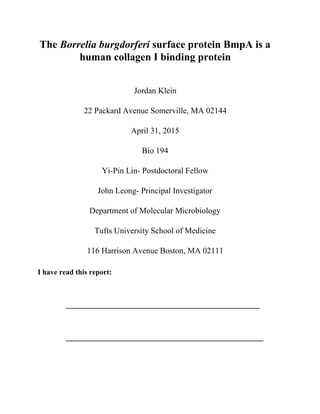
Figure 1. BmpA binds to laminin and type I collagen. One µM recombinant
GSTtagged BmpA or GST (as a negative control) was added to quadruplicate
wells coated with elastin (Eln), type IV collagen (Col IV), type I collagen (Col I),
laminin (Ln), fibronectin (Fn), heparin (Hep), chondroitin4sulfate (C4S),
dermatan sulfate (DS), chondroitin6sulfate (C6S), heparan sulfate (HS), and BSA
as a negative control. Bound protein was measured by ELISA (see Materials and
Methods) and mean OD405 ± standard deviation was determined. Asterisks
indicate that binding of GSTBmpA statistically significantly different than GST
binding to laminin or type I collagen (p≤0.01). Shown is a representative of three
independently performed experiments.
We have previously found that BmpA was able to enhance spirochetal binding to mammalian
epithelial cells, suggesting that BmpA is an adhesin (Klein, 2014). It has also been reported by
Verma et al. that the purified recombinant version of BmpA is able to bind to the purified ECM
component protein laminin, but we further found that BmpA could not promote the adhesion of
B. burgdorferi to purified laminin (Verma, 2009; Klein, 2014). This raised the possibility that
ECM components other than laminin may mediate the adhesion of spirochetes to mammalian
10. cells. To test this hypothesis, GST tagged BmpA (GSTBmpA) was applied to ELISA plate
wells coated with different ECM components, and the ECMbinding activity of BmpA was
determined by qualitative ELISA. As shown in figure 1, GSTBmpA was not statistically
significantly better able than the GST control to bind to Eln, Fn, Hep, C4S, DS, C6S, HS, nor to
the BSA control (Fig. 1). However, GSTBmpA was calculated to be statistically significantly
better able than the GST negative control to bind to laminin (Fig. 1; p=0.0051), consistent with
previous findings showing the lamininbinding activity of purified recombinant BmpA (Verma,
2009). However, as has been demonstrated previously, BmpA may be produced in a different
configuration in its purified form than on the surface of B. burgdorferi, so this does not refute
other findings that BmpA does not promote laminin binding by B. burgdorferi (Klein, 2014).
Interestingly, GSTBmpA was also calculated to be statistically significantly better able than the
GST negative control to bind to type I collagen (Fig. 1; p<0.0001), suggesting that BmpA is
also a collagenbinding protein.
12. bind in the qualitative ELISA experiment, was also included in this experiment as a negative
control. As shown in Figure 2, both GSTBmpA and GST were incapable of binding to type IV
collagen (Fig. 2 bottom panel), which is in agreement with the results obtained from the
qualitative ELISA experiment (Fig. 1). While the type I collagen binding of GST was detected,
the binding action was not saturable when greater amounts of GST were applied and thus not
dose dependent (Fig. 2 top panel). Further, GSTBmpA displayed saturable type I
collagenbinding activity at greater concentrations (Fig. 2 top pane; KD=0.65 ± 0.17 μM),
indicating that BmpA binds to type I collagen in a dose dependent manner.
Discussion: As observed in Fig. 1 from the qualitative ELISA with ECM components
experiment, GSTBmpA was statistically significantly better able than GST to bind to Col I and
Ln, while it was not statistically significantly better able to bind to any of the other ECM
components studied, nor to the BSA control. Verma et al. had previously found that purified
laminin binds to purified BmpA in vitro (Verma, 2009). However, we previously observed that
BmpA produced by B. burgdorferi promotes spirochetal adhesion to mammalian epithelial cells
but not to purified laminin, suggesting that the observed activity of purified recombinant
proteins may not always reflect their function when they are produced on the surface of bacteria
(Klein, 2014). Similarly, the recombinant proteins Leptospira immunoglobulinlike proteins
(Lig), the ECMbinding outer surface protein from another pathogenic spirochete, Leptospira
interrogans, displayed elastinbinding activity in its purified form, but this activity could not be
observed when Lig was produced on the surface of L. interrogans (Lin, 2009; Figueira, 2011).
The inconsistency of the ECMbinding activity of Lig when it was produced in different
13. conditions further supports the hypothesis that the structure of proteins may be different when
they are produced on the bacterial surface than in their purified form, which in turn impacts
their function.
In this study, we have identified type I collagen purified from human skin as another
potential receptor of BmpA. However, Verma et al. had reported that BmpA does not bind to
type I collagen extracted from mouse tissue (Verma, 2009). The inconsistency of these two
studies leads to the hypothesis that BmpA may possess speciesspecific type I collagenbinding
activity. B. burgdorferi can be carried by different mammalian hosts, i.e. human, dog, mouse,
rat, rabbit. and the possibility of hostspecific type I collagenbinding activity illustrated from
our findings might be able to illuminate the determinants of the promotion of spirochetal
colonization in distinct mammalian host environments (Radolf, 2012). Furthermore, as we
previously found that despite being able to bind in its purified form, BmpA as produced on the
surface of B. burgdorferi could not bind to laminin, we will need to similarly examine whether
BmpA as produced on the surface or B. burgdorferi can still bind type I collagen (Verma, 2009;
Klein, 2014).
To further study the mechanism of BmpA as a collagen I binding protein and its apparent
speciesspecific action, certain experiments can be performed. One such experiment, studying
the mechanism of action of BmpA as a type I collagen binding protein as produced on the
surface B. burgdorferi, could entail an otherwise nonadherent strain of B. burgdorferi, such as
B31A, being engineered to produce BmpA on its surface and tested for its ability to bind to
purified type I collagen in vitro using the method of flow cytometry, similar to experiments that
we had previously performed with laminin (Klein, 2014). Further experiments can also be
14. conducted to study the mechanism of the species specific binding of BmpA to type I collagen
through the testing of the ability of purified BmpA to bind to purified type I collagen from a
variety of mammalian species, such as human, rabbit, mouse, and rat, in vitro, using the method
of qualitative ELISA as previously performed in this experiment. Following this experiment, to
further study the mechanism of BmpA as a species specific type I collagen binding protein in its
configuration as produced on the surface of B. burgdorferi, an otherwise nonadherent strain of
B. burgdorferi, such as B31A, could be engineered to produce BmpA on its surface and tested
for its ability to bind to purified type I collagen from a variety of mammalian species, including
human, rabbit, mouse, and rat, in vitro, using flow cytometry as in previous experiments we
have performed, as described previously (Klein, 2014).
Literature Cited:
1. "Press Release." Centers for Disease Control and Prevention. Centers for Disease Control and
Prevention, 19 Aug. 2013.
<http://www.cdc.gov/media/releases/2013/p0819lymedisease.html>.
2. Crother TR, Champion CI, Whitelegge JP, Aguilera R, Wu XY, Blanco DR, Miller JN, Lovett
MA (2004) Temporal Analysis of the Antigenic Composition of Borrelia burgdorferi during
Infection in Rabbit Skin. Infect Immun 72(9): 50635072
3. Figueira CP, Croda J, Choy HA, Haake DA, Reis MG, Ko AI, Picardeau M (2011)
Heterologous expression of pathogenspecific genes ligA and ligB in the saprophyte Leptospira
biflexa confers enhanced adhesion to cultured cells and fibronectin. BMC Microbiology 11:129
15. 4. Klein JD, Lin YP, Leong JM (2014) Borrelia burgdorferi BmpA promotes bacterial adhesion to
mammalian cellsBio 93 final research paper. Unpublished data
5. Lin YP, Bhowmick R, Coburn J, Leong JM (2015) Host cell heparan sulfate
glycosaminoglycans are ligands for OspFrelated proteins of the Lyme disease spirochete. Cell
Microbiol 10.1111/cmi.12448
6. Lin YP, Lee DW, McDonough SP, Nicholson LK, Sharma Y, Chang YF (2009) Repeated
domains of leptospira immunoglobulinlike proteins interact with elastin and tropoelastin. J Biol
Chem 284(29):1938091
7. Pal U, Wang P, Bao F, Yang X, Samanta S, Schoen R, Wormser GP, Schwartz I, Fikrig E
(2007) Borrelia burgdorferi basic membrane proteins A and B participate in the genesis of
Lyme arthritis. J Exp Med 205(1): 133141
8. Radolf JD, Caimano MJ, Stevenson B, Hu LT (2012) Of ticks, mice and men: understanding the
dualhost lifestyle of Lyme disease spirochaetes. Nat Rev Micobiol 10: 8799
9. Ramamoorthy R, Povinelli L, Philipp MT (1996) Molecular Characterization, Genomic
Arrangement, and Expression of bmpD, a New Member of the bmp Class of Genes Encoding
Membrane Proteins of Borrelia burgdorferi. Infect Immun 64(4): 12591264
10. Rosa PA, Tilly K, Stewart PE (2005) The burgeoning molecular genetics of the Lyme disease
spirochaete. Nat Rev Microbiol 3: 129143
11. Steere AC, Coburn J, Glickstein L (2004) The emergence of Lyme disease. J Clin Invest 113(8):
10931101
12. Verma A, Brissette CA, Bowman A, Stevenson B (2009) Borrelia burgdorferi BmpA Is a
LamininBinding Protein. Infect Immun 77(11): 49404946