
Recommandé
Contenu connexe
Similaire à Hypertensive disorder during pregnancy.pptx
Similaire à Hypertensive disorder during pregnancy.pptx (20)
Plus de MesfinShifara
Plus de MesfinShifara (20)
Dernier
Dernier (20)
Hypertensive disorder during pregnancy.pptx
- 1. 1
- 2. 2
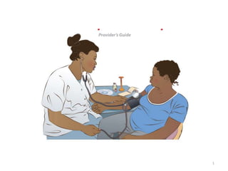
- 3. Hypertension: • A systolic blood pressure ≥140 mmHg, diastolic blood pressure ≥90 mmHg or both in two occasions taken 4 hours or more apart • A single blood pressure recording ≥160/110 mmHg. • A rise of 20 mm Hg MAP over the previous reading, or when the MAP is 105 mm Hg or more should be considered as significant. 3
- 4. Proteinuria 1. Two urine dipstick measurements of at least 1+(30 mg/dL) taken 6 hours apart 2. at least 300 mg of protein in a 24-hour urine sample 3. a urinary protein/creatinine ratio of 0.3 or greater. 4
- 6. Classification 1. Gestational hypertension 2. Preeclampsia 3. Eclampsia 4. Chronic hypertension 5. Superimposed preeclampsia 5.1 Superimposed pre-eclampsia without severe features 5.2 Superimposed pre-eclampsia with severe features 6
- 7. Rest and Digest 1. 7
- 8. Rest and Digest 2. 8
- 9. Rest and Digest 3. 9
- 10. Rest and Digest 4. 10
- 11. Rest and Digest 5. 11
- 12. Rest and Digest 6. 12
- 13. 13
- 14. Abnormal trophoblastic invasion 14 • Normally migration of endovascular trophoblast into the walls of the spiral arterioles produce low- resistance, low-pressure, high-flow uteroplacental bed. • In preeclampsia, trophoblastic invasion is restricted to decidua segment of uteroplacental arteries. • No myometrial spiral arterial invasion.
- 15. 15
- 16. . . .cont • Myometrial musculoelastic architecture remains intact • Decreased blood supply to placenta • This results placental endothelial injury & ischemia 16
- 17. Vascular endothelial damage • Placental ischemia leads to production of placental factor (sFlt-1). • sFlt-1 antagonize VEGF and PLGF in the systemic circulation by binding them. • This results systemic endothelial cell dysfunction. 17
- 18. 18
- 19. Endothelial cell activation causes 19 • Increased capillary permeability • Vasospasm • PGI2 production by endothelial cells decreases & TXA2 by platelet increases. • Endothelin-1 (ET-1) production increases. • NO production by endothelium decreases
- 21. 21
- 22. Genetic predisposition Genetic conflict theory Fetal and maternal genes conflict each other to control nutrient transfer to the fetus The conflict hypothesis suggests that placental factors (fetal genes) act to increase maternal BP, whereas maternal factors act to reduce BP When uteroplacental blood supply is inadequate, fetal rescue strategy to increase non-placental resistance→ SVR →endothelial cell dysfunction 22
- 23. Immunologic factors The formation of blocking antibodies is impaired for placental antigenic activity previous pregnancy has immunologic effect large area of antigenic site multipara with a new partner is at risk 23
- 24. Immunologic factors Maternal tolerance of fetus • Mechanisms to prevent rejection of fetus, a ’’foreign’’ tissue • Expansion of regulatory T cells (immunosuppressive in pregnancy during normal pregnancy • Th2 cells promote humoral immunity, whereas Th1 cells stimulate inflammatory cytokine secretion • Normal pregnancy: Th1 to Th2 switch so that type 2 activity is increased in relation to type 1(Th2 bias) • Preeclampsia: beginning in the early second trimester in women who develop preeclampsia, Th1 action is increased 24
- 25. Kidney 25 • GFR decrease by 25% Glomerular endotheliosis Renal hypoperfusion Decreased plasma volume • Acute tubular necrosis
- 26. Liver 26 • Hepatocyte infarction & necrosis is accompanied by periportal hemorrhage. • Bleeding may extend to collect beneath hepatic capsule- subcapsular hematoma • Liver enzymes increases in the systemic circulation
- 27. Brain 27 • Intracerebral bleeding due to the hypertension • Edema, hypermia, ischemia, thrombosis focal or diffuse • Amaurosis –occipital lobe vasogenic edema --10% follows convulsion --resolve within 4hr-8days
- 28. Others 28 Lung –capillary oncotic pressure decreases -endothelial integrity disrupts -hydrostatic pressure increases- predispose to non-cardiogenic pulmonary edema Eye – retinal vasospasm, retinal edema, retinal detachment -blindness is uncommon
- 29. 29
- 30. 1. Gestational hypertension • Hypertension without proteinuria or other features of preeclampsia developing after the 20th week of pregnancy or within 48 hours of delivery in a previously normotensive woman 30
- 31. Management of GHTN Manage as an outpatient: • Monitor BP, urine (for proteinuria) and fetal condition weekly – If blood pressure worsens or the woman develops features of pre-eclampsia, manage as pre-eclampsia • Counsel the woman and her family about danger signs indicating severe pre-eclampsia or eclampsia • If all observations remain stable, allow to proceed with spontaneous labor and childbirth • If spontaneous labor has not occurred before term, induce labor at term 31
- 32. Preeclampsia Definition • A new onset of hypertension and proteinuria after 20 weeks of gestation in a previously normotensive woman. Sub-classification 1. Pre-eclampsia without severe features 2. Pre-eclampsia with severe features 32
- 33. Risk factors of PE First pregnancy Age <18 or >35 years Multiple gestation History of hypertension Renal disease Diabetes Obesity Family history of pre-eclampsia 33
- 34. From research • The risk increases in those who have limited sperm exposure with the same partner before conception protective effect • Abortions • Previous success pregnancy with same partner 34
- 35. 35
- 36. Preeclampsia Pre-eclampsia with severe features Headache & blurred vision Oliguria (<400 ml/24 hours) Epigastric or right upper quadrant pain, Difficulty of breathing (pulmonary edema) Low platelet count (<100,000/µl) Liver enzymes elevated more than twice the upper limit of normal range, serum creatinine higher than 1.1 mg/dl or a doubling (or higher) of the baseline serum creatinine concentration in the absence of other renal disease. 36
- 37. • AST/ALT/: >70IU/L • Creatinine: >1mg/dl • LDH >600 IU/L) and • bilirubin (>1.2 mg/dL) • A serum uric acid level ≥ 4.5 mg/dL indicates the presence of preeclampsia. • Blood urea level remains normal or slightly raised 37
- 38. Treatment of PE without severe features Management varies depending on the gestational age 1. GA<37 Weeks a. Outpatient b. Admission 2. GA≥37 Weeks 38
- 39. GA≤37wks as outpatient Twice weekly outpatient follow-up Monitor BP, fetal condition, CBC, liver and renal function tests twice weekly Counsel about the danger signs associated with features of severe pre-eclampsia Encourage the woman to eat a normal diet Orient on fetal movement counting (kick chart) daily Do not give anticonvulsants or anti-hypertensives unless clinically indicated Delivery at 37 completed weeks 39
- 40. GAl≤37wks as Inpatient If outpatient mgt’s not possible or close observation preferred or preeclampsia worse rapidly: Admit to hospital Urine output & weight (daily), FHB & kick chart daily BP, Urine protein, fetal condition twice weekly If the diastolic DBP decrease to normal levels or her condition remains stable manage as outpatient If clinical features worsen (urinary protein level increased), manage as severe preeclampsia Do not give diuretics 40
- 41. 2.GA ≥37complete wks Delivery is recommended Anticonvulsant during labor 41
- 42. B. TREATMENT OF PE WITH SEVERE FEATURES The steps of management include: 1. General measures: 2. Prevent convulsion 3. Control hypertension 4. Delivery / expectant management in selected cases 42
- 43. 1.General Measures Admit the patient urgently Manage in left lateral position Setup IV line & infuse maintenance fluids(?) maintain urine output at >30ml/hr & maintain a strict fluid balance chart Prepare equipment for convulsion management at bed side Never leave the patient alone Monitor vital signs, FHB & reflexes Auscultate the lung bases for fine crepitation. If they occur, withhold fluids & administer a diuretic (furosemide 40 mg IV stat) 43
- 44. 2. Anticonvulsant therapy 44 • Magnesium sulfate (MgSO4) schedules for severe pre- eclampsia and eclampsia Loading dose MgSO4 20% solution, 4g IV over 5 minutes (mix 8 ml of 50% Mgso4 solution with 12ml of D5W or 09% normal saline to make 20% solution ) Follow promptly with 10g of 50% MgSO4 solution, 5g in each buttock as deep IM injection with 1mL of 2% lidocaine in the same syringe. Warn the woman that a feeling of warmth will be felt when Mgso4 is given. If convulsion recurs after 15 minutes, give 2g MgSO4 (20% solution) IV over 5 minutes (?)
- 45. 2. Anticonvulsant therapy Maintenance dose 5g MgSO4 (50% solution) + 1 mL lidocaine 2% IM every 4 hours into alternate buttocks. Continue treatment with MgSO4 for 24 hours after delivery or the last convulsion, whichever occurs last 45
- 46. 2. Anticonvulsant therapy Diazepam: • increases the risk of respiratory depression and newborn apnea, in babies who may already be suffering from the effects of utero-placental ischemia & pre-term birth. • The effect may last several days. • Loading dose – Diazepam10 mg IV slowly over 2 minutes – If convulsion recur, repeat the same dose • Maintenance dose – Diazepam 40 mg in 500 ml IV fluids (N/S or Ringer's lactate) no of drops titrated to keep the woman sedated but arousable 46
- 48. 48
- 49. 49
- 50. 3.Control hypertension • anti-hypertensives if the SBP is ≥160 mmHg and/or DBP ≥110 mmHg Drug of choice for acute control 1. Hydralazine Give 5 mg IV slowly every 20 minutes until blood pressure is lowered (to DBP 90-110 mmHg). • The maximum dose is 20 mg per 24 hours 50
- 51. 51 Source: Williams obstetric 26th edition
- 52. 3.Control hypertension 2. Labetalol Oral treatment: Administer 200 mg; repeat dose after one hour until the treatment goal is achieved The maximum dose is 1200mg in 24 hours Intravenous treatment: Administer 10 mg IV The dose can be doubled to 20mg, 40mg and then 80mg with 10-minute intervals until desired effect obtained The maximum total dose is 300 mg; then switch to oral treatment 52
- 53. 3.Control hypertension 3. Nifedipine administer 10 mg orally Repeat dose after 30 minutes if response is inadequate until optimal blood pressure is reached The maximum total dose is 30 mg in the acute treatment setting For maintenance therapy10-20 mg PO bid is given. 4. Alpha methyldopa Administer 250-750 mg every six to eight hours. The maximum dose is 3000 mg per 24 hours. 53
- 54. 4. Planning delivery Gestational age < 28 weeks Termination of pregnancy Gestational age ≥ 28 weeks and <34 weeks: Expectant management is recommended, provided that there is no indication for delivery (what are they ?) 54
- 55. 4. Planning delivery Indications for delivery during Expectant management : Failure to control HTN with two AHTN drugs with a maximum dose in 48 hours Persistent maternal severity symptoms HEELP Syndrome and Eclampsia Pulmonary edema or left ventricular failure IUFD and DIC Severe renal dysfunction 55
- 56. 4. Planning delivery For expectant management: Transfer to maternity ward Follow vital signs every 4 hours CBC, every other day Liver enzymes, creatinine, Fetal surveillance twice weekly Fetal kick count daily Administer Dexamethasone 6 mg IM every 12 hours for 2 days or Betamethasone 12 mg daily for 2 days 56
- 57. 4. Planning delivery Gestation 34 to 37 Weeks Expectant management if there is stable feto- maternal condition (no uncontrolled maternal hypertension, worsening maternal status and fetal distress ) Gestation after 37 completed Weeks regardless of severity features, giving birth is recommended. 57
- 58. 4. Planning delivery • MODE OF DELIVERY: depends on GA, fetal condition, presentation, cervical condition & maternal condition Indication for C/S: – Unfavorable cervix (firm, thick, closed) esp. in seriously ill patients – Poor progress of labor – Patient has not entered active labor within 8 hrs of induction of labor – If there is evidence of fetal distress, or other obstetric indications 58
- 59. 4. Planning delivery POSTPARTUM MANAGEMENT • Watch closely for at least 2 hours after delivery for complications such as shock, PPH & eclampsia. • Anticonvulsive therapy should be maintained for 24 hrs to 48 hrs after delivery or the last convulsion, whichever occurs last. • Continue anti-hypertensive therapy as long as the blood pressure is ≥ 110mmhg • Continue to monitor urine output & check for coagulation failure, LFT and RFT • Postnatal follow-up 59
- 60. Eclampsia • Generalized convulsion and / or coma in a woman with preeclampsia where the convulsion or coma is not attributed to other causes. 60
- 61. Eclampsia Cont… Stages of fit: 1. Premonitory stage: lasts about 10 -20 seconds Patient is restless, twitching of facial muscles, eye roll, respiration becomes spasmodic 2. Tonic stage: Lasting about 10–20 seconds, general muscle rigidity, and whole body goes in to tonic spasms, Backaches, features distorted by grimace, tongue may be bitten, breathing ceases and pt becomes cyanosed 61 6/10/2023 Obstetric II for midwives by Helen Dec 2007EC
- 62. Eclampsia Cont… 3. Clonic stage: Lasting about 60 – 90 minutes. Convulsive movements frothy saliva fills the mouth, may be stained. The woman becomes unconscious. 4. Stage of coma: Snoring( large volume of air through narrowed space) breathing continued and may be persistent for minutes or hours. Further convulsive movements sometimes recur with or without pt gaining from consciousnes. 62 6/10/2023 Obstetric II for midwives by Helen Dec 2007EC
- 63. Eclampsia MANAGEMENT Check airway Aspirate (suction) the mouth & throat as necessary & ensure open airway. Place an oral airway. Check breathing If breathing, give oxygen by mask at 6 litters per minute If not breathing, ventilate using bag and face mask. Check circulation Set up IV line Maintain intravascular volume and replace ongoing losses. Avoid fluid overload 63
- 64. Eclampsia ANTI-HYPERTENSIVE THERAPY : As stated in the management of sever preeclampsia with severe features FLUID BALANCE – Keeping strict input & output record is essential and determine serum electrolytes. – For unconscious patient, 5% DW & RL are infused for maintenance of nutrition & fluid balance during 24 hrs. – Replace extra fluid loss through vomiting, diarrhea, sweating or blood loss. – NPO until conscious 64
- 65. Eclampsia DELIVERY • Delivery should take place as soon as the woman's condition has stabilized, regardless of the gestational age i.e within 12 hours of onset of convulsions 65
- 66. Chronic hypertension • Hypertension that antedates pregnancy or is present before the 20th week of pregnancy or persists after 12 weeks postpartum Management: • If the woman was on an antihypertensive medication before pregnancy and her BP is well-controlled, continue the same medication if safe in pregnancy 66
- 67. CHRONIC HYPERTENSION • If the SBP≥160 mmHg or/and or DBP≥ 110 mmHg treat with antihypertensive medications. • If proteinuria or other signs and symptoms of pre- eclampsia are present, consider superimposed pre- eclampsia and manage as pre-eclampsia. • Monitor fetal growth and induce labor at term if there are no complications, • If fetal growth restriction is severe and pregnancy dating is accurate deliver 67
- 68. Superimposed preeclampsia • If proteinuria or other features of pre-eclampsia develop in a patient with chronic hypertension Classified as 1. Superimposed pre-eclampsia without severe features. 2. Superimposed pre-eclampsia with severe features 68
- 69. Atypical preeclampsia • Defined as the development of preeclampsia (even eclampsia) without fulfilling the standard definition or criteria: – Early onset preeclampsia/eclampsia ≤ 20 weeks – Late postpartum preeclampsia, eclampsia more than 48 hours postpartum – Women with gestational hypertension or gestational proteinuria presenting with symptoms of preeclampsia, ELLP. • Women with atypical preeclampsia and who have other diagnostic criteria of severe preeclampsia should be treated as if they have severe preeclampsia 69
- 70. 70
- 71. Algorithm for the Mx of HELLP syndrome.
- 72. Prevention Low-Dose Aspirin • Early successes of 60/81-mg or 50-150mg aspirin • Reduce the incidence of preeclampsia were attributed to selective thromboxane suppression with resultant dominance of endothelial prostacyclin
- 73. 73 Tried in trials and results have revealed minimal to no benefit
- 74. Thank you 74