Contenu connexe Similaire à Sinusitis (20) 1. Maxillary sinus disease:
diagnosis and treatment
G. W. Bell,1
B. B. Joshi2
and R. I. Macleod3
Verifiable CPD paper
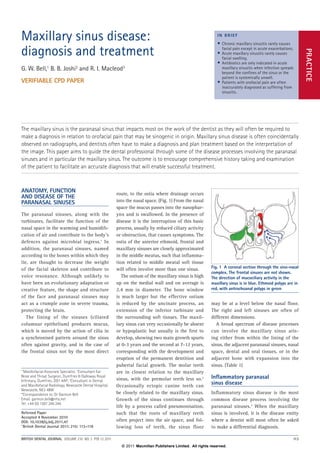
route, to the ostia where drainage occurs
into the nasal space. (Fig. 1) From the nasal
space the mucus passes into the nasophar‑
ynx and is swallowed. In the presence of
disease it is the interruption of this basic
process, usually by reduced ciliary activity
or obstruction, that causes symptoms. The
ostia of the anterior ethmoid, frontal and
maxillary sinuses are closely approximated
in the middle meatus, such that inflamma‑
tion related to middle meatal soft tissue
will often involve more than one sinus.
The ostium of the maxillary sinus is high
up on the medial wall and on average is
2.4 mm in diameter. The bone window
is much larger but the effective ostium
is reduced by the uncinate process, an
extension of the inferior turbinate and
the surrounding soft tissues. The maxil‑
lary sinus can very occasionally be absent
or hypoplastic but usually is the first to
develop, showing two main growth spurts
at 0‑3 years and the second at 7‑12 years,
corresponding with the development and
eruption of the permanent dentition and
pubertal facial growth. The molar teeth
are in closest relation to the maxillary
sinus, with the premolar teeth less so.2
Occasionally ectopic canine teeth can
be closely related to the maxillary sinus.
Growth of the sinus continues through
life by a process called pneumonisation,
such that the roots of maxillary teeth
often project into the air space, and fol‑
lowing loss of teeth, the sinus floor
Anatomy, function
and disease of the
paranasal sinuses
The paranasal sinuses, along with the
turbinates, facilitate the function of the
nasal space in the warming and humidifi‑
cation of air and contribute to the body’s
defences against microbial ingress.1
In
addition, the paranasal sinuses, named
according to the bones within which they
lie, are thought to decrease the weight
of the facial skeleton and contribute to
voice resonance. Although unlikely to
have been an evolutionary adaptation or
creative feature, the shape and structure
of the face and paranasal sinuses may
act as a crumple zone in severe trauma,
protecting the brain.
The lining of the sinuses (ciliated
columnar epithelium) produces mucus,
which is moved by the action of cilia in
a synchronised pattern around the sinus
often against gravity, and in the case of
the frontal sinus not by the most direct
The maxillary sinus is the paranasal sinus that impacts most on the work of the dentist as they will often be required to
make a diagnosis in relation to orofacial pain that may be sinogenic in origin. Maxillary sinus disease is often coincidentally
observed on radiographs, and dentists often have to make a diagnosis and plan treatment based on the interpretation of
the image. This paper aims to guide the dental professional through some of the disease processes involving the paranasal
sinuses and in particular the maxillary sinus. The outcome is to encourage comprehensive history taking and examination
of the patient to facilitate an accurate diagnosis that will enable successful treatment.
may be at a level below the nasal floor.
The right and left sinuses are often of
different dimensions.
A broad spectrum of disease processes
can involve the maxillary sinus aris‑
ing either from within the lining of the
sinus, the adjacent paranasal sinuses, nasal
space, dental and oral tissues, or in the
adjacent bone with expansion into the
sinus. (Table 1)
Inflammatory paranasal
sinus disease
Inflammatory sinus disease is the most
common disease process involving the
paranasal sinuses.3
When the maxillary
sinus is involved, it is the disease entity
where a dentist will most often be asked
to make a differential diagnosis.
1*
Maxillofacial Associate Specialist, 2
Consultant Ear
Nose and Throat Surgeon, Dumfries & Galloway Royal
Infirmary, Dumfries, DG1 4AP; 3
Consultant in Dental
and Maxillofacial Radiology, Newcastle Dental Hospital,
Newcastle, NE2 4BW
*Correspondence to: Dr Garmon Bell
Email: garmon.bell@nhs.net
Tel: +44 (0) 1387 246 246
Refereed Paper
Accepted 4 November 2010
DOI: 10.1038/sj.bdj.2011.47
©British Dental Journal 2011; 210: 113–118
• Chronic maxillary sinusitis rarely causes
facial pain except in acute exacerbations.
• Acute maxillary sinusitis rarely causes
facial swelling.
• Antibiotics are only indicated in acute
maxillary sinusitis when infection spreads
beyond the confines of the sinus or the
patient is systemically unwell.
• Patients with orofacial pain are often
inaccurately diagnosed as suffering from
sinusitis.
in b r ief
practice
Fig. 1 A coronal section through the sino-nasal
complex. The frontal sinuses are not shown.
The direction of mucociliary activity in the
maxillary sinus is in blue. Ethmoid polyps are in
red, with antrochoanal polyps in green
british dental journal VOLUME 210 NO. 3 FEB 12 2011 113
© 2011 Macmillan Publishers Limited. All rights reserved.
2. practice
Most inflammatory paranasal sinus
disease causing symptoms of pain occurs
within one week following an upper respi‑
ratory tract infection, and is usually viral
in origin. The term biphasic illness is occa‑
sionally used to describe a patient recover‑
ing from what is usually a head cold only
to become unwell a few days later with
facial pain, nasal congestion and discharge.
This presentation is what is termed acute
rhinosinal disease, and once the diagnosis
has been made the aim of treatment is to
relieve symptoms. The duration of the ill‑
ness is usually not influenced by treatment
and can last up to four weeks.
Chronic rhinosinal disease is the term
generally used to describe nasal conges‑
tion or discharge that persists for eight to
12 weeks. Chronic disease rarely causes
symptoms of pain except during acute
exacerbations, and dentists are unlikely
to be presented with a patient who has
orofacial pain because of chronic sinus
disease. Chronic rhinosinus disease is usu‑
ally bacterial rather than viral.4
Stasis in
the maxillary sinus following acute infec‑
tion as a result of reduced ciliary activity
can predispose to bacterial infection. The
maxillary sinus is predisposed to stasis due
to the ostium being situated high up on
the medial wall. However, stasis within
the maxillary sinus can also occur due
to nasal polyps, most commonly ethmoid
polyps, which effectively block the middle
meatus and the drainage of the sinuses.5
(Fig. 1) Stasis can also occur secondary
to anatomical variation such as a devi‑
ated nasal septum or a concha bullosa,
a bulky pneumatised middle turbinate,
both of which impede drainage from the
middle meatus.
The diagnostic issue that dentists often
encounter is to determine the cause of oro‑
facial pain. Interested readers are directed
to the comprehensive review of orofacial
pain by Scully and Felix in a previous
publication in this journal.6
Acute sinusi‑
tis that follows an upper respiratory tract
infection may cause facial pain, whereas
chronic sinusitis is unlikely to. Pain due
to tension headache, migraine, atypical
facial pain or temporomandibular disorder
is often mistaken for sinusitis simply on
the basis that the patient locates the source
of the pain to the sinonasal region, with
the distinction between acute or chronic
disease having not been made.7
There is
no single diagnostic sign or symptom that
will differentiate between acute sinusitis
and acute dental pain. Rather, a diagnosis
is reached based on a combination of clini‑
cal and, if appropriate, radiological signs,
along with patient symptoms. (Table 2)
Orofacial pain without nasal obstruction,
nasal discharge or impaired smell sense
is unlikely to be sinogenic. In a specialist
setting using fibre-optic techniques, direct
examination of the osteomeatal complex
may show pus, which confirms a diagnosis
of sinusitis. (Fig. 2) A diagnosis of sinusitis
in the absence of inflammation or pus in the
middle meatus would be incorrect. Acute
sinusitis generally does not cause facial
swelling. Exceptions are acute infection
involving the anterior ethmoids or frontal
sinuses which can cause medial canthal
swelling or glabellar swelling respectively;
Table 1 Disease processes involving the maxillary sinus
Inflammation Infection Bacterial/viral/fungal
Allergic Polyps
Fungal
Neoplastic Benign Inverted papilloma
Osteoma
Adenomatoid odontogenic tumour
Keratocystic odontogenic tumour
Neurofibroma
Angiofibroma
Cylindrinoma
Malignant epithelial Squamous cell carcinoma and subtypes
Adenocarcinoma
Adenoid cystic carcinoma
Acinic cell carcinoma
Mucosal melanoma
Malignant non-epithelial Soft tissue sarcoma
Neurogenic sarcoma
Angiosarcoma
Leiomyosarcoma
Rhabdomyosarcoma
Fibrosarcoma
Chondrosarcoma
Osteosarcoma
Haemangiopericytoma
Malignant lymphoreticular Lymphoma
Plasmacytoma
Metastatic
Odontogenic Infection Pulpal/periapical
Periodontal
Oroantral fistula
Cystic Radicular
Dentigerous
(Benign neoplastic lesions)
Fibrocemento osseous
Foreign body Teeth/implants/restorative materials
Mucocele (Multifactorial aetiology)
Granulomatous
Vasculitis
Wegener’s granulomatous
Churg-Strauss syndrome
Silent sinus syndrome (Aetiology yet to be fully determined)
114 british dental journal VOLUME 210 NO. 3 FEB 12 2011
© 2011 Macmillan Publishers Limited. All rights reserved.
3. practice
spray and pump delivered devices are
also available.
When infection is spreading beyond the
confines of the sinuses or the patient is
pyrexial, with a pussy nasal discharge, as
a first-line measure amoxicillin remains
the antibiotic of choice. However, for
those patients in whom there is a poor
clinical response to amoxicillin, a recent
generation cephalosporin antibiotic may
be indicated. For those patients allergic
to penicillins, doxycycline or clarithro‑
mycin may be prescribed.11
If a patient is
clinically unwell, or shows signs of orbital
involvement, urgent referral to a hospital
setting is recommended.
Although general dental practition‑
ers will not be prescribing nasal ster‑
oids or antihistamines, these items are
sometimes prescribed for acute sinusi‑
tis but have no clinical role in reducing
patient symptoms.
Chronic rhinosinus disease will gener‑
ally not cause facial pain and the dental
surgeon is unlikely to be making a diag‑
nosis of chronic maxillary sinusitis when
a patient presents with orofacial pain.
However, the dental team should have a
basic knowledge of therapies for chronic
rhinosinal disease.3
Treatment is usu‑
ally decided on the presence or absence
of nasal polyps, which can impede sinus
drainage. (Fig. 1) Treatment of chronic
disease, namely nasal congestion and dis‑
charge, is the same as for acute disease,
with nasal irrigation and nasal decongest‑
ants in the first instance. Nasal decongest‑
ants may be used for a prolonged period
on the basis that their use is restricted
to once daily.12
When polyps are present,
steroids, either topical or systemic, may
be prescribed.13
Chronic disease or recur‑
rent acute disease that does not respond
to conventional medical therapy may
require surgery. Following assessment by
an ear, nose and throat surgeon, treatment
aimed at restoring normal mucociliary
function and clearance of the sinuses may
be undertaken.14
This can involve treat‑
ing a deviated nasal septum, removing
polyps, removing or trimming turbinates,
or increasing the size of the ostium from
the maxillary sinus and removal of excess
tissue in the middle meatus. This treat‑
ment is now regularly undertaken with
the use of fibre-optic devices because of
and the very occasional case of acute max‑
illary sinusitis when the antrum is large
and there is expansion of the thin lateral
wall into the oral cavity. Acute buccal sul‑
cus or facial swelling should be regarded
as odontogenic until proven otherwise.
Treatment of acute sinusitis is based on
relief of symptoms and does not involve
antibiotics unless the patient is pyrexial
or there is evidence of spread of infection
beyond the confines of the sinus.8
Rather,
treatment is based on topical nasal decon‑
gestants and saline irrigation of the nasal
cavity. Topical decongestants such as ephe‑
drine or xylometazoline constrict the nasal
lining, widening the paranasal sinus ostia,
facilitating drainage by ciliary activity. Most
decongestants are now provided in a spray
delivery device and are easily administered.
Droplet preparations require more care‑
ful administration to be effective. (Fig. 3)
Excessive decongestant usage will cause
localised nasal discomfort. Generally, nasal
decongestants should not be used for more
than 7 days because of rebound mucosal
swelling when the medication is stopped.9
Saline irrigation of the nasal cavity is the
equivalent of a warm salty mouthwash in
that it shifts surface debris and will facilitate
sinus drainage.10
In practice this is delivered
using a 10 ml or 20 ml syringe with the
patient vigorously washing out their nasal
cavity while leaning over a sink. Proprietary
Table 2 Symptoms and signs of acute sinogenic and dental pain
Specific sinogenic Unilateral nasal obstruction
Unilateral nasal discharge
Observation of pus in middle meatus (specialist setting)
Concurrent or recent upper respiratory tract infection
Increased pain on vertical change in head position
Shared sinogenic and dental Increased pain with changes in atmospheric pressure
Unilateral maxillary pain
Disturbance of sleep
Facial swelling (rare cases of acute ethmoid or frontal sinusitis)
Buccal sulcus swelling (very rare cases of maxillary sinusitis when
the antrum is large)
Specific dental Increased pain with temperature changes when eating or drinking
Tooth mobility
Painful fractured, carious or heavily restored tooth
Buccal sulcus swelling adjacent to tooth that is cause of symptoms
Specific dental or periapical radiographic signs
Fig. 2 Endoscopic view of the left nasal
space with pus arising from the left maxillary
ostium because of chronic sinusitis. A is
the deviated nasal septum. B is the middle
turbinate. C is pus within the middle meatus.
D is the inferior turbinate
Fig. 3 Illustration of correct application
technique of nasal drops to ensure accurate
delivery to the middle meatus. Incorrect
application results in the medicament running
along the nasal floor into the pharynx
bypassing the middle meatus. This technique
is not necessary for spray delivery devices
british dental journal VOLUME 210 NO. 3 FEB 12 2011 115
© 2011 Macmillan Publishers Limited. All rights reserved.
4. practice
reduced morbidity as compared to more
open traditional surgical techniques.
Retention cysts may occur within the
maxillary sinus and arise from inflam‑
mation of the sinus lining, such that
the secretory duct becomes obstructed,
and have been observed in up to 14% of
people living in industrialised environ‑
ments. Retention cysts commonly occur
on the floor of the maxillary sinus, are
frequent coincidental findings on dental
radiographs and cross-sectional imaging,
and are often mistaken for sinister sinus
disease or attributed to a dental aetiology.
(Fig. 4) No treatment is required.15
Mucoceles arise when the drainage of the
sinus is occluded such that mucus collects
and can completely fill the sinus. They can
occur in any of the paranasal sinuses, but
mostly the frontal. The maxillary sinus is
involved in only 10% of cases. Mucoceles
can also lead to bone expansion due to the
pressure effect.16
Mucociliary function is impaired when
the paranasal sinuses are exposed to high
dose radiation as in radiotherapy, such that
the patient may be predisposed to chronic
rhinosinal disease.17
The same event may
occur in patients with cystic fibrosis due to
thick mucoid secretions and recurrent infec‑
tions with scarring of the sinus lining.18
Maxillary sinus disease
of dental origin
Approximately 10‑12% of cases of inflam‑
matory maxillary sinus disease are of dental
origin.19
Most relate to pulpal necrosis and
periapical disease, but also advanced peri‑
odontal disease, and oro-antral communi‑
cations following dento-alveolar surgery.
Extruded pulp space filling materials will
act as local irritants when displaced into
the maxillary sinus, and have predisposed
to fungal infections such as aspergillo‑
sis.20
During endodontic treatment sodium
hypochlorite solution may be inadvertently
passed through into the maxillary sinus.
Most patients will simply experience a taste
of bleach in the nasopharynx, but a few
will experience a localised inflammatory
response. Increasingly dental implants are
being displaced into the maxillary sinus
where they will act as local irritants in the
same way that displaced teeth or roots will.
Where possible, displaced foreign bodies
should be removed from the maxillary
sinus, which increasingly is being performed
using endoscopic techniques. When an oro‑
antral fistula is treated it is often necessary
to treat concurrent chronic sinus infection,
as failure to do so will result in failure of
treatment. Therefore, when sinus drainage
is impaired through concurrent rhinosinal
disease, irrigation of the sinus with removal
of diseased tissue may be insufficient and
middle meatal surgery may also be neces‑
sary before normal mucociliary clearance
can be re-established.
Dental disease extending into the maxil‑
lary sinus is uncommon. However, odon‑
togenic cysts and tumours will often expand
into the maxillary sinus. (Fig. 5) Equally,
changes within the maxillary sinus related
to inflammation may occasionally be mis‑
interpreted as odontogenic disease when
viewed on radiographs. (Fig. 6) Dentists may
occasionally experience difficulty interpret‑
ing radiographs to determine whether or
not disease or changes arise from within
the sinus or are of odontogenic origin. The
presence or absence of the maxillary sinus
floor, periodontal ligament space or lamina
dura, trabecular pattern of bone and blood
vessel channels within the lateral maxil‑
lary wall will aid a differential diagnosis.
(Table 3) (Figs 7, 8)
While radiographic techniques available
to the dental practitioner may occasionally
show disease within the maxillary sinus, the
taking of dental radiographs, particularly
dental panoramic tomograms, in cases of
Table 3 Radiographic features of the healthy maxillary sinus, soft tissue sinus disease
and odontogenic lesions
Healthy maxillary sinus Trabecular pattern of bone of lateral wall is visible superimposed on
radiolucency of sinus
Sinus floor and walls are imaged as a thin continuous white line
(corticated outline)
Small blood vessels are visible as fine radiolucent channels as they
traverse the lateral sinus wall
Soft tissue lesion of sinus Lesion is viewed as radio-opaque when compared to air of sinus,
without a corticated margin
In benign disease the walls of the maxillary sinus are imaged as a thin
continuous white line
In malignant, infective or expansile disease the walls of the sinus are
resorbed such that the corticated outline is discontinuous
In malignant or rapidly expansile disease the roots of the maxillary teeth
may be resorbed
Odontogenic lesion Dental disease, namely caries or resorption, is visible
The periodontal ligament space is widened apically or discontinuous.
The lamina dura may be missing.
As periapical tissues or cysts expand into the sinus space the corticated
outline of the sinus floor is elevated (the antral halo effect)
When superimposed on the air space of the sinus there is limited or no
trabecular pattern and limited blood vessel channels
Fig. 4 Periapical radiograph showing a dome
shape, non-corticated radiopacity in the
floor of the maxillary sinus. The sinus floor is
intact and a trabecular pattern of bone with
blood vessels is observed superimposed on the
radiopacity. This is a retention cyst
Fig. 5 Sectional dental panoramic tomogram
showing a corticated, unilocular radiolucency
extending into the right maxillary sinus.
There is widening of the periodontal ligament
space over the UR5 tooth. The trabecular
pattern of bone does not extend over the
radiolucency and blood vessel channels are
not seen. This is a radicular cyst arising from
the UR5 tooth
116 british dental journal VOLUME 210 NO. 3 FEB 12 2011
© 2011 Macmillan Publishers Limited. All rights reserved.
5. practice
acute sinusitis is contra-indicated, unless it
is to exclude a dental cause for the patient’s
symptoms. Dental practitioners who have
access to cone beam computed tomography
will often intentionally or unintentionally
also obtain images of the paranasal sinuses
and have a legal responsibility to ensure
that any abnormalities are accurately
reported and acted upon.21
Malignant disease
of the maxillary sinus
Malignancy arising within the paranasal
sinuses is relatively rare, constituting 1.0%
of all malignancies, with approximately
80% of these malignancies arising in the
maxillary sinus with a lesser prevalence in
the ethmoid sinus. Malignant disease of the
sphenoid and frontal sinuses is very rare.22
Almost 80% of malignancies are squamous
cell carcinomas, with acinic cell carcino‑
mas causing 10%. (Table 1) Metastatic dis‑
ease presents in the bone and expands into
the sinus space.
Malignant disease of the paranasal
sinuses unfortunately often presents at a
late stage when the tumour has become
large enough to cause symptoms. The
Fig. 6 Sectional dental panoramic tomogram
showing a diseased UL6 tooth with periapical
bone loss. The non-corticated, dome-shaped
radiopacity within the maxillary sinus is a
retention cyst and not related to the dental
disease. The bone density of the hard palate is
superimposed on the maxillary sinus
Fig. 7 Periapical radiograph showing dental
disease. The large radiolucency shows a
trabecular pattern with blood vessel channels
excluding an odontogenic cyst, demonstrating
an enlarged maxillary sinus. The UL5 tooth
has a periapical lesion demonstrating an
antral halo as the periosteum of the sinus
floor is elevated. Note the lack of lamina
dura on the UL5
Fig. 8 Periapical radiograph showing a
radiolucency above the apex of the extracted
UR5, superimposed within the corticated
outline of the maxillary sinus. Note the
intact lamina dura of the tooth socket, the
presence of a trabecular pattern of bone and
fine channels caused by the presence of blood
vessels. This is an antral locule rather than a
periapical lesion from the UR5
Table 4 Some signs and symptoms that may be suspicions for maxillary sinus malignancy
Unilateral orofacial pain
Unilateral firm, non-infective facial swelling
Nasal obstruction
Epistaxis
Diplopia
Trismus
Non-healing extraction site
Infra-orbital parasthesia
Mobility of teeth in the absence of periodontal or periapical disease
Resorption of roots of teeth
Spontaneous oro-antral fistula formation
Loss of radiopaque outline of maxillary sinus
Fig. 9 A non-healing upper right extraction
site with a spindle cell squamous carcinoma
arising from the maxillary sinus
Fig. 10 Left sided nasal obstruction with
epistaxis in a patient with an advanced
squamous cell carcinoma of the left maxillary
sinus. Examination also showed lateral
expansion of the alveolus
british dental journal VOLUME 210 NO. 3 FEB 12 2011 117
© 2011 Macmillan Publishers Limited. All rights reserved.
6. practice
mucosa of the paranasal sinuses is not
as easily accessible as the oral mucosa
for routine inspection and early mucosal
abnormalities are not seen or investigated.
The dental professional can play a role
in the diagnosis of a patient with maxil‑
lary sinus malignancy. A combination of
patient symptoms and clinical signs should
arouse suspicion of maxillary sinus malig‑
nancy, warranting immediate referral to an
appropriate specialist. (Table 4) (Figs 9‑11)
Unfortunately, it has been known for
patients to be treated for long periods on
the assumption that their symptoms arise
from chronic inflammatory rhinosinal dis‑
ease, only for a diagnosis of malignancy
to be made at a later date.
Malignancy of the maxillary sinus is
managed by multidisciplinary teams with
input from the surgical specialties of oral
and maxillofacial, ear, nose and throat,
and plastic and reconstructive surgery. It
is not the intention of this paper to out‑
line treatment for sinonasal malignancy
and interested readers are referred to more
comprehensive texts.22
Fungal disease of the maxillary sinus
Most fungal disease of the maxillary
sinus involves the organism Aspergillus
which lives within moulds and spores
and is regularly inhaled into the respira‑
tory system. When infection occurs with
Aspergillus in relation to dental foreign
materials, the infection is normally con‑
tained within the confines of the maxil‑
lary sinus.20
Foci of infection may lead to
dystrophic calcification and the forma‑
tion of rhinoliths, which may be seen on
dental radiographs. (Fig. 12) Large rhino‑
liths are known as fungal balls. Treatment
is normally surgical with removal of
any predisposing cause, and this is also
increasingly being provided endoscopi‑
cally with the aim of restoring normal
mucociliary function.
However, in the immuno-compromised
patient, as in poorly controlled diabetes
mellitus, HIV infection and chemotherapy,
fungal infections such as aspergillosis or
mucormycosis may extend beyond the con‑
fines of the sinus into the orbit, temporal
fossa or oral cavity producing symptoms
and signs suggestive of malignant disease.
Occasionally disease extends to the brain.23
While most of these patients will be clini‑
cally unwell and already within a hospital
setting, the vigilant dental professional has
a role to play in identifying early signs or
symptoms. (Table 4)
Conclusion
The aim of this paper has been to enlighten
the dental professional in the disease
processes that may involve the maxillary
sinus. Of importance is the accurate diag‑
nosis of acute rhinosinusitis in treating
orofacial pain and the shift away from
routine antibiotic therapy. When viewed
together, specific signs and symptoms
that can be suggestive of maxillary sinus
malignancy are emphasised. The vari‑
ous radiographic features that guide the
dental professional when viewing radio‑
graphs; and determining whether or not
changes are related to disease or health,
and whether the disease is dental or sino‑
genic have been outlined.
1. Paranasal sinus anatomy and function. http://www.
utmb.edu/otoref/grnds/paranasal-sinus-2002-01/
paranasal-sinus-2002-01.htm. Accessed 22
November 2010.
2. Eberhardt J A, Torabinejad M, Christiansen E L.
A computed tomographic study of the
distances between the maxillary sinus floor
and the apices of the maxillary posterior teeth.
Oral Surg Oral Med Oral Pathol 1992;
73: 345–346.
3. Schaefer S D. Rhinology and sinus disease: a
problem-oriented approach. St Louis:
Mosby, 1998.
4. Erkan M, Aslan T, Ozcan M, Koç N. Bacteriology of
antrum in adults with chronic maxillary sinusitis.
Laryngoscope 1994; 104: 321–324.
5. Bachert C, Hörmann K, Mösges R et al. An update
on the diagnosis and treatment of sinusitis and
nasal polyposis. Allergy 2003; 58: 176–191.
6. Scully C, Felix D H. Oral medicine – update for the
dental practitioner: orofacial pain. Br Dent J 2006;
200: 75–83.
7. Jones N S. Sinus headaches: avoiding over-
and mis-diagnosis. Expert Rev Neurother 2009;
9: 439–444.
8. Chan Y, Kuhn F A. An update on the classifications,
diagnosis and treatement of rhinosinusitis.
Curr Opin Otolaryngol Head Neck Surg 2009;
17: 204–208.
9. Graf P. Long-term use of oxy- and xylometazoline
nasal sprays induces rebound swelling, tolerance,
and nasal hyperreactivity. Rhinology 1996;
34: 9–13.
10. Harvey R, Hannan S A, Badia L, Scadding G. Nasal
saline irrigations for symptoms of chronic sinusitis.
Cochrane Database Syst Rev 2007: CD006394.
11. Sinus and Allergy Health Partnership.
Antimicrobial treatment guidelines for acute
bacterial rhinosinusitis. Otolaryngol Head Neck Surg
2000; 123: 5–31.
12. Yoo J K, Seikaly H, Calhoun K H. Extended use of
topical nasal decongestants. Laryngoscope 1997;
107: 40–43.
13. Lund V J, Black J H, Szabó L Z, Schrewelius C,
Akerlund A. Efficacy and tolerability of budesonide
aqueous nasal spray in chronic rhinosinusitis
patients. Rhinology 2004; 42: 57–62.
14. Khalil H, Nunez D A. Functional endoscopic sinus
surgery for chronic rhinosinusitis. Cochrane
Database Syst Rev 2006: CD004458.
15. Wang J H, Jang Y J, Lee B J. Natural course of
retention cysts of the maxillary sinus: long-term
follow-up results. Laryngoscope 2007;
117: 341–344.
16. Ling F T K. Mucoceles of the paranasal sinuses.
http://drfling.hyperphp.com/Notes/Mucoceles%20
of%20the%20Paranasal%20Sinuses.pdf. Accessed
22 November 2010.
17. Kamel R, Al-Badawy S, Khairy A, Kandil T, Sabry
A. Nasal and paranasal sinus changes after
radiotherapy for nasopharyngeal carcinoma. Acta
Otolaryngol 2004; 124: 532–535.
18. Gysin C, Alothman G A, Papsin B C. Sinonasal
disease in cystic fibrosis: clinical characteristics,
diagnosis, and management. Pediatr Pulmonol
2000; 30: 481–489.
19. Mehra P, Jeong D. Maxillary sinusitis of
odontogenic origin. Curr Allergy Asthma Rep 2009;
9: 238–243.
20. Burnham R, Bridle C. Aspergillosis of the maxillary
sinus secondary to a foreign body (amalgam) in the
maxillary antrum. Br J Oral Maxillofac Surg 2009;
47: 313–315.
21. Department of Health. The Ionising Radiation
(Medical Exposure) Regulations 2000. Department
of Health, 2007. http://www.dh.gov.uk/prod_con-
sum_dh/groups/dh_digitalassets/@dh/@en/
documents/digitalasset/dh_064707.pdf. Accessed
22 November 2010.
22. Katzenmeyer K, Pou A. Neoplasms of the nose and
paranasal sinuses. http://www.otohns.net/default.
asp?id=14054.
23. Sarti E J, Blaugrund S M, Tang Lin P, Camins M B.
Paranasal sinus disease with intracranial extension:
aspergillosis versus malignancy. Laryngoscope
1988; 98: 632–635.
Fig. 11 A dental panoramic tomograph
demonstrating complete loss of bone in
the left maxillary alveolus due to antral
carcinoma. The UL8 tooth is embedded within
the tumour mass. The soft tissue shadow
demonstrates the tumour extension into the
oral cavity
Fig. 12 Periapical radiograph of posterior
maxilla showing multiple spheroidal
calcifications (antroliths) within a thickened
antral lining. These are almost certainly an
example of dystrophic calcification within
chronically inflamed tissue
118 british dental journal VOLUME 210 NO. 3 FEB 12 2011
© 2011 Macmillan Publishers Limited. All rights reserved.
View publication statsView publication stats