SlideShare une entreprise Scribd logo
1  sur  8
Télécharger pour lire hors ligne
Nanomed. J., 2(4): 261-268, Autumn 2015
261
A comparative study about toxicity of CdSe quantum dots on reproductive
system development of mice and controlling this toxicity by ZnS coverage
1*
A. Valipoor; 2
Gh. Amiri; 3
K. Parivar; 4
M. Modaresi; 5
J. Taheri; 5
A. Kazemi; 6
M. Abasi; 7
A. Mirzakhani
1
Department of Physiology, Faculty of Basic Sciences, Shahrekord Branch, Islamic Azad University,
Shahrekord, Iran.
2
Department of Physic, Falavarjan Branch, Islamic Azad University, Isfahan, Iran
3
Department of Biology, Science and Research Branch, Islamic Azad University, Tehran, Iran
4
Department of Physiology, Islamic Azad University, Khorasgan Branch, Isfahan, Iran
5
Department of Chemistry, Islamic Azad University, Shahrekord, Iran .
6
Department of Matematic, Islamic Azad University, Shahrekord, Iran
7
Department of Mechanical Engineering, Payame Noor University, Tehran, Iran
ABSTRACT:
Objective(s): Medicinal benefits of quantum dots have been proved in recent years but there is little known about their
toxicity especially in vivo toxicity. In order to use quantum dots in medical applications, studies ontheir in vivo toxicity
is important.
Materials and Methods: CdSe:ZnS quantum dots were injected in 10, 20, and 40 mg/kg doses to male mice10 days later,
mice were sacrificed and five micron slides were prepared structural and optical properties of quantum dots were
evaluated using XRD.
Results: Histological studies of testis tissue showed high toxic effect of CdSe:ZnS in 40 mg/kg group. Histological
studies of epididymis did not show any effect of quantum dots in terms of morphology and tube structure. Mean
concentration of LH and testosterone and testis weight showed considerable changes in mice injected with 40 mg/kg
dose of CdSe:ZnS compared to control group. However, FSH and body weight did not show any difference with control
group.
Conclusion: Although it has been reported that CdSe is highly protected from the environment by its shell, but this
study showed high toxicity for CdSe:ZnS when it is used in vivo which could be suggested that shell could contribute
to increased toxicity of quantum dots. Considering lack of any previous study on this subject, our study could
potentially be used as an basis for further extensive studies investigating the effects of quantum dots toxicity on
development of male sexual system.
Keywords: CdSe:ZnS, In vivo toxicity, Quantum dots, ZnS shell
Nanomed. J., 2(4): 261-268, Autumn 2015
DOI: 10.7508/nmj. 2015.04.003
*Corresponding Author Email:valipoor.akram@gmail.com
Tel: (+98) 381-2227864
Note. This manuscript was submitted on July 20, 2015;
approved on September 6, 2015
Received; 20 July 2015 Accepted; 6 September 2015
INTRODUCTION
Nowadays, nanotechnology is widely known and
has many applications including in medicine (3). With
advances in nanotechnology, many nanoparticles have
been used widely in biology and medicine. Cytotoxicity
of these particles is an important factor in their use in
medicine and hence has received a great deal of
attention in recent years (2, 27).
Among nanoparticles, quantum dots (QDs) are
considered as extremely interesting materials for the
development of photovoltaic devices because of their
potential in medical diagnosis, cell imaging, and as
biological tracers in human diseases diagnosis (3, 19,
27). But, in spite of obvious advantages of QDs,
concerns regarding human exposure to these particles
and its effect on human health have increased.
Nanomed. J., 2(4): 261-268, Autumn 2015
262
A. Valipoor et al.
Therefore, studies have been conducted on reaction
of QDs in vivo and in vitro but there are still limited
data on this category, which has led to not using dots
in medications as therapeutic or diagnostic tools. So,
further studies are needed to investigate QDs in the
environments similar to human body (4, 9, 18(.Among
semiconductor nanocrystal QDs, the CdSe core
nanocrystals QDs have shown the best optical
properties in light stability and radiative quantum
efficiency in optical tests (4, 7, 19) Coolen et al study
demonstrated that CdSe:ZnS nanocrystal QDs are the
most widely used QDs )7).
Most studies reported CdSe, which is a semiconductor
matter, as core of nanocrystals QDs covered by another
semiconductor matter (mainly ZnS). On the other hand,
in vitro studies have shown an approximately complete
control of CdSe-induced cytotoxicity by ZnS coverage
(7,11,12,23,29).
Therefore, CdSe and CdSe:ZnS nanocrystal QDs were
selected for this study. Because of their importance as
semiconductor and in biology and medicine, which has
been highlighted in different studies, the synthesized
QDs were characterized and their in vivo toxicity effects
on male sexual system were also investigated.
MATERIALS AND METHODS
Materials
All chemicals and solvents were purchased form
Merck and were of analytical grades.
Preparation of CdSe and CdSe:ZnS QDs
Quantum dots were synthesized by chemical
precipitation method. Briefly, three solutions of
cadmiumchloride (CdCl2
-4H2
O), mercaptoethanol (ME)
and sodium selenite (Na2
SeO3
-5H2
O) were prepared in
the distilled deionized water, under vigorous stirring.
At first, CdCl2
solution was poured into a three-spout
balloon container and simultaneously ME solution was
added to the same balloon. Finally, Na2
SeO3
solution
was added under nitrogen . The resulting solution was
mixed withdeionized water and thencentrifuged in order
to remove any impurity aggregate. Then, the
precipitated sample was dried at room temperature.All
processes were done at room temperature (1).
The crystal structure and optical properties of
CdSe:ZnS QDs were characterized by XRD (X-Ray
Diffraction, Bruker D8ADVANCE λ = 0.154 nm Cu Kα
radiation) and UV-Vis spectrophotometer (Ultra Violet
Visible, UV-2600 Shimadzu,Japan). ScanningTunneling
Microscope (STM) (NATSICO, Iran) was used to
investigate particle size distribution.
Breeding and treatment of animals
Male Balb/Cmice were purchased fromRaziVaccine
and Serum Research Institute (Hesarak, Iran). Some
male mice (about 20 days old) were kept for 10 days in
natural day light and at temperature 22-24°C. Then,
male mice were divided in 7 groups of 6 mice: control,
three groupd injected with CdSe at 10, 20, and 40 mg/
kg and three groups injected with CdSe:ZnS QDs at 10,
20, and 40 mg/kg doses. Either CdSe or CdSe:ZnS
nanoparticles were prepared in normal saline solution
and single-dose was injected intraperitoneally at 10,
20, and 40 mg/kg doses. Control group received only
normal saline solution. The protocol of study was
approved by the Ethics Committee of the University.
Tissue preparing
10 Days after CdSe and CdSe:ZnS injection, all
control and treatment groups were anesthetized and
testis and epididymis organs were rapidly removed,
weighed, and preserved in formaldehyde as fixative
agent. Five micron slides were dehydrated and prepared
in paraffin. Then, the slides were stained using
hematoxylin-eosin. Morphological structure of
seminiferous tubes, and mean number of
spermatogonia, spermatocytes, spermatids, and
matured sperms in testis were studied, and epithelial
height, connective tissue, smooth muscle and sperm
density were measured in epididymis.
Hormones measurements
Serum FSH and LH were measured by double
antibody radioimmunoassay as previously described
(20, 21). Serum testosterone was assayed using a
competitive chemiluminescent immunoassay kit (DRG
Co, Germany; Pishtaz Teb, Iran).
(testis and body weight and the number of cells in
seminiferous tubes of various groups) were analyzed
using SPSS 16 (by one way ANOVA followed by
Tukey’s post hoc test.). Data were represented as
means± standard error. Differences were considered
significant at p<0.05 and p<0.01.
RESULTS
QDs characterization by XRD, STM and UV-Vis
analysis
The structure of the CdSe QDs was investigated
by XRD. Fig 1 illustrates the XRD pattern of the CdSe
Nanomed. J., 2(4): 261-268, Autumn 2015
263
QDs. As seen, the sample has a single phase and also
a cubic crystal structure.
According to the standard Joint Committee on Powder
Diffraction Standards card No. 19-0191, the diffraction
peaks correspond to the 111, 220 and 311 crystal
planes. The mean size of the particles was determined
by Debye-Scherer formula. It was calculated as 2.4 nm
for CdSe QDs (1).
Dose-finding study
there was no study onin vivo toxicity of QDs, we
selected three doses of 125, 250, and 500 mg/kg
according to an in vitro study conducted by Ming-
Shu Hsieh (13). In the first stage, CdSe and CdSe:ZnS
nanoparticles were prepared in normal saline solution
and were intraperitoneally injected into 36 mice. All
mice died in less than 24 hours after the injection. The
remarkable finding was that in vivo administration of
ZnS as the shell increased toxicity of CdSe and mice
showed greater responses to CdSe:ZnS compared to
CdSe. These results were contrary to what has been
reported for the cytotoxicity of CdSe:ZnS where the
ZnS shell would minimize the cell toxicity of the CdSe
core. The CdSe:ZnS-treated mice died within a short
time after ip injection of the QDsand even in some
cassesmice died almost immediately after the injection.
In the next step, DMSA-coated CdSe nanoparticles
were injected to groups of 8 mice each intraperitoneally
at 125 and 250 mg/kg doses. The results showed that
all mice injected with DMSA-coated CdSe at 250 mg/
kg dose and 80% of mice injected with DMSA-coated
CdSe at 125 mg/kg dose died within one week post-
injection. Then, CdSe and CdSe:ZnS were
intraperitoneally injected to groups of 16 mice each at
75 and 100 mg/kg doses. In CdSe:ZnS groups at 75 and
100mg/kg doses50%and 83% ofmicedied, respectively
whereas in CdSe groups at 75 and 100 mg/kg doses
50% and 66% of mice died within one week,
respectively. Finally, lower doses of CdSe and
CdSe:ZnS were selected and injected to group of 4
mice each. In CdSe groups, no mortality was observed
during 20 days post-injection at all three doses of 10,
20 and 40 mg/kg. However, in CdSe:ZnS groups, while
no mortality was observed at 10 mg/kg group but 12.5
and 20% mortality was observed, respectively.
According to the obtained results in dose-finding
study, 10 mg/kg dose was determined as a safe dose
and 20 and 40 mg/kg doses were determined as doses
with toxicity effects.
Histological study of testis
The seminiferous tubules are in different
spermatogenic stages in control group, and in mice
treated with all three doses of CdSe and CdSe:ZnS QDs.
Spermatozoids were observed in lumen tubules of the
control group, but in the group treated with 40 mg/kg
CdSe:ZnS QDs, abnormal growth of seminiferous
tubes, impaired spermatogenesis, reduction in number
of spermatogonia, spermatocyst 1, spermatides and an
obvious decrease in matured sperms of lumen were
observed (Table 2). On the other hand, degeneration
of the interstitial tissue and blood vessels and reduction
in thickness of the lamina propria were observed as
illustrated in Fig 1.
Nanomed. J., 2(4): 261-268, Autumn 2015
264
Phytotoxicity of methylene blue
Fig. 1. Microscopic images of testis slides, 10 days post-
injection (H & E, 400×). A control group;B1, C1 and D1
groups treated with doses of 10, 20, 40 mg/kg CdSe,
respectively; B2, C2 and D2 groups treated with doses of 10,
20, 40 mg/kg CdSe:ZnS, respectively. Sz: spermatozoa, Lc:
Leydig cells, Lp: Lamina propria, Spg: Espermatogoni, Spc:
Espermatocyte, Spt: Espermatid
Histological study of epididymis
Qualitative studies using optical microscope
showed that epithelia, lumen, connective tissue and
smooth muscle of epididymis were similar in groups
treated with the QDs and control, but spermatozoa
volume in lumen epididymis obviously decreased in
mice treated with CdSe:ZnS at 40 mg/kg (Fig 2).
Nanomed. J., 2(4): 261-268, Autumn 2015
265
Fig. 2. Microscopic images of epididymis slides, 10 days post-
injection (H & E, 400×). A control group;B1, C1 and D1
groups treated with doses of 10, 20, 40 mg/kg CdSe,
respectively; B2, C2 and D2 groups treated with doses of 10,
20, 40 mg/kg of CdSe:ZnS, respectively. SPM: Sperm mass,
SMM: Smooth muscle, SC: Stereocilia, PC: Principal cells, SCE:
Stereociliated simple columnar epithelium, LE: Lumen
epididymis, LCT: Loose connective tissue
Changes in body and testis weight, and hormones
level
Body weight did not change significantly in
treatment groups but testis weight decreased
significantly (P<0.01) by CdSe:ZnS at 40 mg/kg. FSH
did not change by treatments with QDs (Table 2) but
testosterone decreased significantly and LH increased
significantly in mice treated with CdSe:ZnS at 40 mg/
kg (Fig 3, 4, 5).
Fig. 3. Mean of concentration LH in 10 days post-injection
(**p < 0.01, *p < 0.05 )
Fig. 4. Mean of concentration testosterone in 10 days post-
injection (**p < 0.01, *p <0.05)
Fig. 5. Mean of concentration FSH in 10 day post-injection
(**p < 0.01, *p < 0.05 )
Nanomed. J., 2(4): 261-268, Autumn 2015
266
A. Valipoor et al.
Table 1. Average and mean comparison of sperm stem cell numbers in one tubule 10 days post-injection (p < 0.01)
Mean of spermatidMean of spermatocyte IMean of spermatogoniaNanoparticle dosesgroups
111.95 ± 33.6344.15 ± 9.3534.55 ± 6.390 mg/kgControl
100.45 ± 18.64
100.85 ± 24.96
99.10 ± 24.68
45.05 ± 9.76
46.10 ± 8.58
45.70 ± 7.01
35.50 ± 7.55
35.95 ± 6.76
34.30 ± 7.55
10 mg/kg
20 mg/kg
40 mg/kg
CdSe
113.65 ± 23.29
109.15 ± 20.72
83.00** ± 23.44
45.25 ± 8.45
43.80 ± 8.04
29.60** ± 10.76
33.60 ± 8.94
32.80 ± 6.67
18.85** ± 6.94
10 mg/kg
20 mg/kg
40 mg/kg
CdSe:ZnS
Table 2. Average and mean comparison of body and testis in 10 dayspost-injection (**p < 0.01, *p < 0.05 )
Mean of testis weightMean of body weightQDs dosesgroups
.093 ± .0127.17 ± 1.30 mg/kgControl
.087 ± .01
.087 ±.01
.083 ± .02
25.98 ± 1.6
27.67 ± 3.9
26.83 ± 1.4
10 mg/kg
20 mg/kg
40 mg/kg
CdSe
.087 ± .01
.107 ± .02
.055** ± .01
27.33 ± 2.8
26.83 ± 2.1
27.67 ± 1.3
10 mg/kg
20 mg/kg
40 mg/kg
CdSe:ZnS
Discussion
Recently, there are a lot of reports on the use of
nanoparticulate systems in biomedical applications (14,
30(.Among these nanoparticles, crystals with nanometer
size, which are called mainly QDs, have been studied
extensively for molecular imaging and targeted therapy.
Because QDS are fluorescent nanoparticles with high
sensitivityusing them as probe is a promising technique
for biological applications. Due to large area provided
by QDs, they are appropriate as targeting ligand to direct
them to site of action (13, 21). Moreover, because of
high light stability which provides long time observation
of biological molecules, QDs could be the main hope as
label for biological systems imaging. Therefore, the use
of QDs as bright and photostable probes for long-term
fluorescence imaging is gaining more interest (13, 27).
Studies about the physicochemical properties of QDs
and their use in vivo are important. There are a few
studies about the effects of QDs on testis. In some
studies, exposing young mice to DE(Diesel Exhaust) and
silver induced degeneration of Leydig cells action and
hence fluctuation of testosterone level (11, 15,32).
Guo et al reported reduction in density and movement
of spermatozoids, increase in spermatozoids
abnormalities, and apoptosis of cells in mice injected by
TiO2
nanoparticles. Histopathological studies of testis
and epididymis did not show any change (12).
In another study by Wen-Qing, gold nanoparticles (14
nm) with two ω-methoxy and ω-aminothyl polyethylene
glycol covers passed testis blood barrier, entered to germ
cells and increased testosterone (33). Recently, effects
ofnanoparticlessmallerthan 100 nm(like DEP) (Diesel
Exhaust Particles)have been studied. Results
indicated separation of cells of seminiferous
epithelium(26, 33).
Sleiman et al examined the adverse productive toxic
effects ofAgNPs in male Wistar rats exposed toAgNP
(Silver Nanoparticles) during the prepubertal period
and sacrificed at postnatal days 53 and 90. The weight
of the animals at the postnatal day 90 did not change
markedly, but growth was less in the group treated
with AgNP at 50 μg/kg from the postnatal day 34 to
53. AgNP exposure produced a delay in puberty in
both treated groups. Decreased sperm reserves in
the epididymis and diminished sperm transit time were
observed at the postnatal day 53, while a reduction in
sperm production occurred at the postnatal day 90.
The morphology of the seminiferous epithelium was
markedly altered and data demonstrated that
prepubertal exposure to AgNP altered reproductive
development in prepubertal male Wistar rats, as
evidenced by impairment in spermatogenesis and a
lower sperm count in adulthood (27).
Mathias et al showed AgNPs reduced the acrosome
and plasma membrane integrities, reduced the
mitochondrial activity and increased the abnormalities
of the sperm in both treatment groups.AgNP exposure
also delayed the onset of puberty, although no
changes in body growth were observed in the
treatment groups. The animals did not show changes
in sexual behavior or serum hormone concentrations
Nanomed. J., 2(4): 261-268, Autumn 2015
267
(18). In another study,AgNPs(Silver Nanoparticles) at
low dose (1 mg/kg) were intravenously injected into
male CD1 mice over 12 days. Treatment resulted in no
changes in body and testis weights, sperm
concentration and motility, fertility indices, or follicle-
stimulating hormone and luteinizing hormone serum
concentrations; however, serum and intratesticular
testosterone concentrations were significantly
increased 15 days after initial treatment. Histologic
evaluation revealed significant changes in epithelium
morphology, germ cell apoptosis, and Leydig cell size.
Additionally, gene expression analysis revealed that
Cyp11a1 and Hsd3b1 mRNA were up regulated
significantly in treated animals. These data suggest
thatAgNPs do not impair spermatogonial stem cells in
vivo since treatment did not result in significant
decreases in testis weight and sperm concentrations.
However,AgNPs appear to affect Leydig cell function,
leading to increase in testicular and serum testosterone
levels (11). In vitro studies revealed high
concentrations of TiO2
nanoparticles affected the
viability and proliferation of mouse Leydig cells, but
not the gene expression associated with
spermatogenesis(20). And gold nanoparticles
decreased the motility of human sperm. Also, silver,
aluminum, and MoO3
nanoparticles were toxic to mouse
spermatogonia stem cells. Silica nanoparticles inhibited
the differentiation of mouse ES cells and C60 –based
nanoparticles inhibited the differentiation of mouse
midbrain cells, CB (Carbon Black)decreased the
viability of mouse Leydig cells and CdSe QDs inhibited
the pre- and post-implantation development of mouse
embryos (13, 23, 31, 32). In vitro studies have shown
cytotoxic effect of TiO2
and CB (Carbon
Black)nanoparticles on viability of mice Leydig cells,
as well (9, 12, 15,33).Also injection ofTiO2
,gold, diesel
exhaust (DE) and C60 nanoparticles to pregnant
mothers caused cytotoxic effects on spermatogenesis
and testis tissuein their male children (20, 26, 30, 32,33).
In the present study, CdSe and CdSe:ZnS with 2-3 nm
size awhich were prepared by sedimentation method
was investigated in vivo. Histopathological studies of
testis tissue showed toxic effect of these nanoparticles
at 40 mg/kg dose. Moreover, study of the testis slides
showed reduction in spermatogonia, spermatocytes,
spermatid, and mature sperm in seminiferous tubular,
degeneration of interstitial tissue and reduction in
Leydig cells number. Histological study of epididymis
tissue showed intensive reduction in sperm volume of
epididymis at 40 mg/kg. According to Table 4, FSH
level did not change by treatments with QDs but
testosterone decreased significantly and LH increased
significantly in group received CdSe:ZnS at 40 mg/kg
dose, which is in agreement with the findings of testis
weight and histopathology of mature group which
include significant reduction in weight, degeneration
of the interstitial tissue and Leydig cells by CdSe:ZnS
at 40 mg/kg. Significant increase in LH could be due to
a feedback mechanism in response to degeneration of
interstitial tissue and considerable reduction of
testosterone. Obviously, considering small size of these
particles which provide them with easy passing from
blood barrier of testis, we could infer that passing of
this nanoparticles from brain blood barrier and
enforcing direct toxicity on brain tissue and
hypothalamus is possible but the first former
suggestion is more probable because FSH level was
not affected. In this study, cytotoxic effect of CdSe
and the role of ZnS cover in controlling the degree of
toxicityon sexual hormones, testis, and body weight of
animals was investigated. In view of the results
obtained in the present study, QDs can pass testis
blood barrier and enforce effects on reproduction
system. Although epithelium epididymis did not show
any special histopathological changes in CdSe-treated
mice by optical microscope but lumen content showed
many failures occurring in testis tissue.
CONCLUSIONS
According to histopathological results of this
study, CdSe:ZnS nanoparticles can pass testis blood
barrier and enforce direct destruction of germinal cells
and spermatogenesis. It seems that despite in vitro
findings, including the role of ZnS for controlling the
toxicity as the shell in embryonic culture environment,
in vivo use of ZnS has increased toxicity of CdSe, which
can be due to the increase in CdSe:ZnS stability or
independent in vivo toxicity of ZnS itself. Although
most of in vitro studies have offered cadmium ions
release as the main reason for QDs toxicity, other
mechanisms seem to contribute mainly to this toxicity,
among which QDs stability plays an important role.
ACKNOWLEDGEMENT
Authors are thankful to dr amiri for his kind support
and Iran nanotechnology initiative for the financial
support.
REFERENCE
1. Amiri Gh. Fatahian S, Mahmoudi S , Preparation and Optical
properties assessment of CdSe quantum dot, Dig Nanomater
Bios. 2011; 4(2): 1161.
2. Bae PK , Kim KN, Lee SJ, Chang HJ, Lee CK, Park JK. The
modification of quantum dot probes used for the targeted
Nanomed. J., 2(4): 261-268, Autumn 2015
268
A. Valipoor et al.
imaging of his-tagged fusion proteins. Biomaterials. 2008;
30(5): 836-842.
3. Chang SQ , Dai YD, Kang B, Han W, Mao L, Chen D. UV-
enhanced cytotoxicity of thiol-capped CdTe quantum dots
in human pancreatic carcinoma cells. Toxicol Lett. 2009;
188(2): 104-111.
4. Chan WC, Maxwell DJ, Gao X, Bailey RE, Han M, Nie S.
Luminescent quantum dots for multiplexed biological
detection and imaging. Curr Opin Biotechnol. 2002; 13(1):
40–46.
5. Chan WH , Shiao NH. Cytotoxic effect of CdSe quantum dots
on mouse embryonic development. Mol Sci. 2008; 29(2):
259-266.
6. Clift MJ , Stone V. Quantum Dots: An Insight and Perspective
of Their Biological Interaction and How This Relates to
Their Relevance for Clinical Use. Theranostics. 2012; 2(7):
668-680.
7. Coolen L, Brokmann, Hermier. JP. Chapter 24-Quantum
Optics with Single CdSe/ZnS Colloidal Nanocrystals.
Handbook of Self Assembled Semiconductor Nanostructures
for Novel Devices in Photonics and Electronics. 2008; 708–
748.
8. Drbohlavova J , Adam V, Kizek R, Hubalek J. Quantum Dots-
Characterization: Preparation and Usage in Biological
Systems. Mol Sci. 2009; 10: 656–673.
9. Ema M , Kobayashi N, Naya M, Hanai S, Nakanishi J.
Reproductive and developmental toxicity studies of
manufactured nanomaterials. Reprod Toxicol. 2010; 30(3):
343–352.
10. Galeone A, Vecchio G, Malvindi MA, Brunetti V, Cingolani
R, Pompa PP. In vivo assessment of CdSe-ZnS quantum
dots: coating dependent bioaccumulation and genotoxicity.
Nanoscale. 2013; 4(20): 6401-6407.
11. Garcia TX, Costa GM, França LR, Hofmann MC. Sub-acute
intravenous administration of silver nanoparticles in male
mice alters Leydig cell function and testosterone levels.
Reprod Toxicol. 2014; 45: 59-70.
12. Guo LL, Liu XH, Qin DX, Gao L, Zhang HM, Liu JY, Cui
YG. Effects of nanosized titanium dioxide on the reproductive
system of male mice. Nan Ke Xue. 2009; 15(6): 517-522.
13. Hsieh MS, Shiao NH, Chan WH. Cytotoxic Effects of CdSe
Quantum Dots on Maturation of Mouse Oocytes and
Fertilization and Fetal Development. Mol Sci. 2009; 10(5):
2122 - 2135.
14. Jamiesona T, Bakhshia R, Petrovaa D, Pococka R and et al.
Biological applications of quantum dots. Biomaterials. 2007;
28(3): 4717-4732.
15. Komatsu T , Tabata M, Kubo-Irie M, Shimizu T, Suzuki K,
Nihei Y, Takeda K. The effects of nanoparticles on mouse
testis Leydig cells in vitro. Toxicol In Vitro. 2008; 22(8):
1825-1831.
16. Li C , Taneda S, Taya K, Watanabe G, Li X, Fujitani Y,
Nakajima T, Suzuki AK. Effects of in utero exposure to
nanoparticle-rich diesel exhaust on testicular function in
immature male rats. Toxicol Lett. 2009; 185(1): 1–8.
17. Li KG , Chen JT, Bai SS, Wen X, Song SY, Yu Q, Li J, Wang
YQ. Intracellular oxidative stress and cadmium ions release
induce cytotoxicity of unmodified cadmium sulfide quantum
dots. Toxicol In Vitro. 2009; 23(6): 1007-1013.
18. Mathias FT , Romano RM, Kizys MM, Kasamatsu T,
Giannocco G, Chiamolera MI, Dias-da-Silva MR, Romano
MA. Daily exposure to silver nanoparticles during
prepubertal development decreases adult sperm and
reproductive parameters. Nanotoxicology. 2014; 9(1): 64-
70.
19. Muller L, Gasser M, Raemy DO, Herzog F, Brandenberger C,
Schmid O, Gehr P, Rothen-Rutishauser B, Clift MJD. Realistic
exposure methods for investigating the interaction of
nanoparticles with the lung at the air-liquid interface in vitro.
Theranostics. 2011; 1(1): 30-64.
20. Ono N, Oshio S, Niwata Y, Yoshida S, Tsukue N, Sugawara I,
Takano H, Takeda K. Prenatal exposure to diesel exhaust
impairs mouse spermatogenesis. Inhal Toxicol. 2007; 19:
275-281.
21. Pan Z, Mora-Sero I, Shen Q, Zhang H, Li Y, Zhao K, Wang
J, Zhong X, Bisquert J. High Efficiency “Green” Quantum
Dot Solar Cells. Am Chem Soc. 136(25):9203-10.
22. Pinaud F , Michalet X, Bentolila LA, Tsay JM, Doose S, Li
JJ, Iyer G, Weiss S. Advances in fluorescence imaging with
quantum dot bio-probes. Biomaterials. 2006; 27(9): 1679-
1687.
23. Roberts JR, Antonini JM, Porter DW, Chapman RS, Scabilloni
JF, Young SH, Schwegler-Berry D, Castranova V, Mercer RR.
Lung toxicity and biodistribution of Cd/Se-ZnS quantum dots
with different surface functional groups after pulmonary
exposure in rats. Part Fibre Toxicol. 2013; 10.
24. Rzigalinski BA, Strobl JS. Cadmium-containing nanoparticles:
Perspectives on pharmacology and toxicology of quantum
dots. Toxicol Appl Pharmacol. 2009; 238(3): 280-288.
25. Smith AM , Duan H, MohsAM, Nie S. Bioconjugated quantum
dots for in vivo molecular and cellular imaging. Adv Drug
Deliv Rev. 2008; 60(11): 1226-1240.
26. Shimizu M, Tainaka H, Oba T, Mizuo K, Umezawa M,
Takeda K. Maternal exposure to nanoparticle titanium
dioxide during the prenatal period alters gene expression
related to brain development in the mouse. Part Fibre Toxicol.
2009; 6(20): 1-25.
27. Sleiman HK, Romano RM, Oliveira CA, Romano MA.
Effects of prepubertal exposure to silver nanoparticles on
reproductive parameters in adult male Wistar rats. Toxicol
Environ Health A. 2013; 76(17): 1023-32.
28. Soenen SJ, Manshian B, Aubert T, Himmelreich U, Demeester
J, De Smedt SC, Hens Z, Braeckmans K. Cytotoxicity of
cadmium-free quantum dots and their use for cell bioimaging.
Chem Res Toxicol. 2014; 27(6): 1050-9.
29. Su Y, He Y, Lu H, Sai L, Li Q, Li W, Wang L, Shen P, Huang
Q, Fan C. The cytotoxicity of cadmium based, aqueous phase-
Synthesized quantum dots and its modulation by surface
coating. Biomaterials. 2009; 30(1): 19–25.
30. Takeda K, Suzuki K, Ishihara A, Kubo-Irie M, Fujimoto R,
Tabata M, et al. Nanoparticles transferred from pregnant
mice to their offspring can damage the genital and cranial
nerve system. Health Sci. 2009; 55(1): 95-102.
31. Wiwantitkit V, Sereemaspum A, Rojanathanes R. Effect of
gold nanoparticles on spermatozoa: the first world report.
Fertil Steril. 2009; 91(1): 7-8.
32. Yoshida S1, Hiyoshi K, Oshio S, Takano H, Takeda K,
Ichinose T. Effects of fetal exposure to carbon nanoparticles
on reproductive function in offspring . Fertil Steril. 2010,
93(5): 1695-1699.
33. Yoshida S , Hiyoshi K, Ichinose T, Takano H, Oshio S,
Sugawara I, Takeda K, Shibamoto T. Effect of nanoparticles
on the male reproductive system of mice. Androl. 2008;
32(4): 337-342.
How to cite this article:
Valipoor A, Amiri GH, Parivar K, Modaresi M, Taheri J, Kazemi A, Abasi M, Mirzakhani A. A comparative study about toxicity of CdSe
quantum dots on reproductive system development of mice and controlling this toxicity by ZnS coverage. Nanomed. J., 2015; 2(4): 261-268.

Contenu connexe

Tendances

Lack of association between CD45 C77G polymorphism and multiple sclerosis in ...
Lack of association between CD45 C77G polymorphism and multiple sclerosis in ...Lack of association between CD45 C77G polymorphism and multiple sclerosis in ...
Lack of association between CD45 C77G polymorphism and multiple sclerosis in ...ijtsrd
 
Study of Glutathione Peroxidase GPX Activity Among Betel Quid Chewers of Indi...
Study of Glutathione Peroxidase GPX Activity Among Betel Quid Chewers of Indi...Study of Glutathione Peroxidase GPX Activity Among Betel Quid Chewers of Indi...
Study of Glutathione Peroxidase GPX Activity Among Betel Quid Chewers of Indi...ijtsrd
 
Rapid In Vivo Assessment of Bioactivity in Zebrafish: High Content Data for P...
Rapid In Vivo Assessment of Bioactivity in Zebrafish: High Content Data for P...Rapid In Vivo Assessment of Bioactivity in Zebrafish: High Content Data for P...
Rapid In Vivo Assessment of Bioactivity in Zebrafish: High Content Data for P...OSU_Superfund
 
Design study w1526455 finished
Design study w1526455 finishedDesign study w1526455 finished
Design study w1526455 finishedConnor Downing
 
Applying cheminformatics and bioinformatics approaches to neglected tropical ...
Applying cheminformatics and bioinformatics approaches to neglected tropical ...Applying cheminformatics and bioinformatics approaches to neglected tropical ...
Applying cheminformatics and bioinformatics approaches to neglected tropical ...Sean Ekins
 
Sensing metabolites for the monitoring of tissue engineered construct cellula...
Sensing metabolites for the monitoring of tissue engineered construct cellula...Sensing metabolites for the monitoring of tissue engineered construct cellula...
Sensing metabolites for the monitoring of tissue engineered construct cellula...Antoine DEGOIX
 
ORS poster 2017
ORS poster 2017 ORS poster 2017
ORS poster 2017 Jess Rames
 
Thesis Poster
Thesis PosterThesis Poster
Thesis PosterTravis Tu
 
Innovatival Functionalized Nanomaterials for Biomedicalm Sensing and Composit...
Innovatival Functionalized Nanomaterials for Biomedicalm Sensing and Composit...Innovatival Functionalized Nanomaterials for Biomedicalm Sensing and Composit...
Innovatival Functionalized Nanomaterials for Biomedicalm Sensing and Composit...researchcenterm
 
EGRupdatedCVwithCL052015
EGRupdatedCVwithCL052015EGRupdatedCVwithCL052015
EGRupdatedCVwithCL052015Erik Rogers
 

Tendances (20)

16 cytotherapy 2017
16 cytotherapy 201716 cytotherapy 2017
16 cytotherapy 2017
 
Lee2014
Lee2014Lee2014
Lee2014
 
my paper 2015
my paper 2015my paper 2015
my paper 2015
 
2007 sdarticlenon-biotin
2007 sdarticlenon-biotin2007 sdarticlenon-biotin
2007 sdarticlenon-biotin
 
Lack of association between CD45 C77G polymorphism and multiple sclerosis in ...
Lack of association between CD45 C77G polymorphism and multiple sclerosis in ...Lack of association between CD45 C77G polymorphism and multiple sclerosis in ...
Lack of association between CD45 C77G polymorphism and multiple sclerosis in ...
 
Study of Glutathione Peroxidase GPX Activity Among Betel Quid Chewers of Indi...
Study of Glutathione Peroxidase GPX Activity Among Betel Quid Chewers of Indi...Study of Glutathione Peroxidase GPX Activity Among Betel Quid Chewers of Indi...
Study of Glutathione Peroxidase GPX Activity Among Betel Quid Chewers of Indi...
 
Rapid In Vivo Assessment of Bioactivity in Zebrafish: High Content Data for P...
Rapid In Vivo Assessment of Bioactivity in Zebrafish: High Content Data for P...Rapid In Vivo Assessment of Bioactivity in Zebrafish: High Content Data for P...
Rapid In Vivo Assessment of Bioactivity in Zebrafish: High Content Data for P...
 
Introduction to alternatives to animal testing in toxicology
Introduction to alternatives to animal testing in toxicologyIntroduction to alternatives to animal testing in toxicology
Introduction to alternatives to animal testing in toxicology
 
KalacovD-Research
KalacovD-ResearchKalacovD-Research
KalacovD-Research
 
MEDI 409
MEDI 409MEDI 409
MEDI 409
 
Design study w1526455 finished
Design study w1526455 finishedDesign study w1526455 finished
Design study w1526455 finished
 
Applying cheminformatics and bioinformatics approaches to neglected tropical ...
Applying cheminformatics and bioinformatics approaches to neglected tropical ...Applying cheminformatics and bioinformatics approaches to neglected tropical ...
Applying cheminformatics and bioinformatics approaches to neglected tropical ...
 
Sensing metabolites for the monitoring of tissue engineered construct cellula...
Sensing metabolites for the monitoring of tissue engineered construct cellula...Sensing metabolites for the monitoring of tissue engineered construct cellula...
Sensing metabolites for the monitoring of tissue engineered construct cellula...
 
Poster
PosterPoster
Poster
 
ORS poster 2017
ORS poster 2017 ORS poster 2017
ORS poster 2017
 
Thesis Poster
Thesis PosterThesis Poster
Thesis Poster
 
Zebrafish as a model system to study toxicology
Zebrafish as a model system to study toxicologyZebrafish as a model system to study toxicology
Zebrafish as a model system to study toxicology
 
Innovatival Functionalized Nanomaterials for Biomedicalm Sensing and Composit...
Innovatival Functionalized Nanomaterials for Biomedicalm Sensing and Composit...Innovatival Functionalized Nanomaterials for Biomedicalm Sensing and Composit...
Innovatival Functionalized Nanomaterials for Biomedicalm Sensing and Composit...
 
Weber-Thesis
Weber-ThesisWeber-Thesis
Weber-Thesis
 
EGRupdatedCVwithCL052015
EGRupdatedCVwithCL052015EGRupdatedCVwithCL052015
EGRupdatedCVwithCL052015
 

Similaire à A comparative study about toxicity of CdSe quantum dots on reproductive system development of mice and controlling this toxicity by ZnS coverage

The interaction of QDs with RAW264.7 cells_ nanoparticle quantification, upta...
The interaction of QDs with RAW264.7 cells_ nanoparticle quantification, upta...The interaction of QDs with RAW264.7 cells_ nanoparticle quantification, upta...
The interaction of QDs with RAW264.7 cells_ nanoparticle quantification, upta...Olga Gladkovskaya
 
Subacute dermal toxicity investigation of nanosilver on serum chemical biomar...
Subacute dermal toxicity investigation of nanosilver on serum chemical biomar...Subacute dermal toxicity investigation of nanosilver on serum chemical biomar...
Subacute dermal toxicity investigation of nanosilver on serum chemical biomar...Nanomedicine Journal (NMJ)
 
Impact of carbon nanomaterials on the formation of multicellular spheroids by...
Impact of carbon nanomaterials on the formation of multicellular spheroids by...Impact of carbon nanomaterials on the formation of multicellular spheroids by...
Impact of carbon nanomaterials on the formation of multicellular spheroids by...Татьяна Гергелюк
 
The acute liver injury in rat caused by gold nanoparticles
The acute liver injury in rat caused by gold nanoparticlesThe acute liver injury in rat caused by gold nanoparticles
The acute liver injury in rat caused by gold nanoparticlesNanomedicine Journal (NMJ)
 
A systematic review of gold nanoparticles as novel cancer therapeutics
A systematic review of gold nanoparticles as novel cancer therapeuticsA systematic review of gold nanoparticles as novel cancer therapeutics
A systematic review of gold nanoparticles as novel cancer therapeuticsNanomedicine Journal (NMJ)
 
Nanotechnology; its significance in cancer and photodynamic therapy
Nanotechnology; its significance in cancer and photodynamic therapyNanotechnology; its significance in cancer and photodynamic therapy
Nanotechnology; its significance in cancer and photodynamic therapyNanomedicine Journal (NMJ)
 
2014 increasing survival study of kidney hek-293 t
2014 increasing survival study of kidney hek-293 t2014 increasing survival study of kidney hek-293 t
2014 increasing survival study of kidney hek-293 tOscar Moreno
 
Quantum dots and application in medical science
Quantum dots and application in medical scienceQuantum dots and application in medical science
Quantum dots and application in medical sciencekeyhan *
 
Dose-dependent hepatotoxicity effects of Zinc oxide nanoparticles
Dose-dependent hepatotoxicity effects of Zinc oxide nanoparticlesDose-dependent hepatotoxicity effects of Zinc oxide nanoparticles
Dose-dependent hepatotoxicity effects of Zinc oxide nanoparticlesNanomedicine Journal (NMJ)
 
Troy University Surface of Membrane Cells Summary.pdf
Troy University Surface of Membrane Cells Summary.pdfTroy University Surface of Membrane Cells Summary.pdf
Troy University Surface of Membrane Cells Summary.pdfsdfghj21
 
Methods For Improving The Cellular Uptake Of Dna Origami...
Methods For Improving The Cellular Uptake Of Dna Origami...Methods For Improving The Cellular Uptake Of Dna Origami...
Methods For Improving The Cellular Uptake Of Dna Origami...Christina Santos
 
Effects of intraperitoneal injection of gold nanoparticles in male mice
Effects of intraperitoneal injection of gold nanoparticles in male miceEffects of intraperitoneal injection of gold nanoparticles in male mice
Effects of intraperitoneal injection of gold nanoparticles in male miceNanomedicine Journal (NMJ)
 

Similaire à A comparative study about toxicity of CdSe quantum dots on reproductive system development of mice and controlling this toxicity by ZnS coverage (20)

The interaction of QDs with RAW264.7 cells_ nanoparticle quantification, upta...
The interaction of QDs with RAW264.7 cells_ nanoparticle quantification, upta...The interaction of QDs with RAW264.7 cells_ nanoparticle quantification, upta...
The interaction of QDs with RAW264.7 cells_ nanoparticle quantification, upta...
 
Subacute dermal toxicity investigation of nanosilver on serum chemical biomar...
Subacute dermal toxicity investigation of nanosilver on serum chemical biomar...Subacute dermal toxicity investigation of nanosilver on serum chemical biomar...
Subacute dermal toxicity investigation of nanosilver on serum chemical biomar...
 
Impact of carbon nanomaterials on the formation of multicellular spheroids by...
Impact of carbon nanomaterials on the formation of multicellular spheroids by...Impact of carbon nanomaterials on the formation of multicellular spheroids by...
Impact of carbon nanomaterials on the formation of multicellular spheroids by...
 
Impact of carbon nanomaterials on the formation of multicellular spheroids by...
Impact of carbon nanomaterials on the formation of multicellular spheroids by...Impact of carbon nanomaterials on the formation of multicellular spheroids by...
Impact of carbon nanomaterials on the formation of multicellular spheroids by...
 
The acute liver injury in rat caused by gold nanoparticles
The acute liver injury in rat caused by gold nanoparticlesThe acute liver injury in rat caused by gold nanoparticles
The acute liver injury in rat caused by gold nanoparticles
 
01 nmj 1-4
01   nmj 1-401   nmj 1-4
01 nmj 1-4
 
A systematic review of gold nanoparticles as novel cancer therapeutics
A systematic review of gold nanoparticles as novel cancer therapeuticsA systematic review of gold nanoparticles as novel cancer therapeutics
A systematic review of gold nanoparticles as novel cancer therapeutics
 
Nanotechnology; its significance in cancer and photodynamic therapy
Nanotechnology; its significance in cancer and photodynamic therapyNanotechnology; its significance in cancer and photodynamic therapy
Nanotechnology; its significance in cancer and photodynamic therapy
 
1 nmj 2-3
1   nmj 2-31   nmj 2-3
1 nmj 2-3
 
Chapter 3ppthorti
Chapter  3ppthortiChapter  3ppthorti
Chapter 3ppthorti
 
2014 increasing survival study of kidney hek-293 t
2014 increasing survival study of kidney hek-293 t2014 increasing survival study of kidney hek-293 t
2014 increasing survival study of kidney hek-293 t
 
Breakthroughs_of_using_Photodynamic_Therapy_and_Gold_Nanoparticles_in_Cancer_...
Breakthroughs_of_using_Photodynamic_Therapy_and_Gold_Nanoparticles_in_Cancer_...Breakthroughs_of_using_Photodynamic_Therapy_and_Gold_Nanoparticles_in_Cancer_...
Breakthroughs_of_using_Photodynamic_Therapy_and_Gold_Nanoparticles_in_Cancer_...
 
Quantum dots and application in medical science
Quantum dots and application in medical scienceQuantum dots and application in medical science
Quantum dots and application in medical science
 
Dose-dependent hepatotoxicity effects of Zinc oxide nanoparticles
Dose-dependent hepatotoxicity effects of Zinc oxide nanoparticlesDose-dependent hepatotoxicity effects of Zinc oxide nanoparticles
Dose-dependent hepatotoxicity effects of Zinc oxide nanoparticles
 
Effects of Losartan on Ischemia/Reperfusion–Induced Testicular Injury in a Ra...
Effects of Losartan on Ischemia/Reperfusion–Induced Testicular Injury in a Ra...Effects of Losartan on Ischemia/Reperfusion–Induced Testicular Injury in a Ra...
Effects of Losartan on Ischemia/Reperfusion–Induced Testicular Injury in a Ra...
 
Troy University Surface of Membrane Cells Summary.pdf
Troy University Surface of Membrane Cells Summary.pdfTroy University Surface of Membrane Cells Summary.pdf
Troy University Surface of Membrane Cells Summary.pdf
 
Methods For Improving The Cellular Uptake Of Dna Origami...
Methods For Improving The Cellular Uptake Of Dna Origami...Methods For Improving The Cellular Uptake Of Dna Origami...
Methods For Improving The Cellular Uptake Of Dna Origami...
 
Effects of intraperitoneal injection of gold nanoparticles in male mice
Effects of intraperitoneal injection of gold nanoparticles in male miceEffects of intraperitoneal injection of gold nanoparticles in male mice
Effects of intraperitoneal injection of gold nanoparticles in male mice
 
Sam Case Study
Sam Case StudySam Case Study
Sam Case Study
 
Effect of cadmium chloride on fresh water fish heteropneustes fossilis and re...
Effect of cadmium chloride on fresh water fish heteropneustes fossilis and re...Effect of cadmium chloride on fresh water fish heteropneustes fossilis and re...
Effect of cadmium chloride on fresh water fish heteropneustes fossilis and re...
 

Plus de Nanomedicine Journal (NMJ)

Evaluation of the effect of crocetin on antitumor activity of doxorubicin enc...
Evaluation of the effect of crocetin on antitumor activity of doxorubicin enc...Evaluation of the effect of crocetin on antitumor activity of doxorubicin enc...
Evaluation of the effect of crocetin on antitumor activity of doxorubicin enc...Nanomedicine Journal (NMJ)
 
Effects of combination of magnesium and zinc oxide nanoparticles and heat on ...
Effects of combination of magnesium and zinc oxide nanoparticles and heat on ...Effects of combination of magnesium and zinc oxide nanoparticles and heat on ...
Effects of combination of magnesium and zinc oxide nanoparticles and heat on ...Nanomedicine Journal (NMJ)
 
Preparation and evaluation of electrospun nanofibers containing pectin and ti...
Preparation and evaluation of electrospun nanofibers containing pectin and ti...Preparation and evaluation of electrospun nanofibers containing pectin and ti...
Preparation and evaluation of electrospun nanofibers containing pectin and ti...Nanomedicine Journal (NMJ)
 
The combined effects of Aloe vera gel and silver nanoparticles on wound heali...
The combined effects of Aloe vera gel and silver nanoparticles on wound heali...The combined effects of Aloe vera gel and silver nanoparticles on wound heali...
The combined effects of Aloe vera gel and silver nanoparticles on wound heali...Nanomedicine Journal (NMJ)
 
Simultaneous loading of 5-florouracil and SPIONs in HSA nanoparticles: Optimi...
Simultaneous loading of 5-florouracil and SPIONs in HSA nanoparticles: Optimi...Simultaneous loading of 5-florouracil and SPIONs in HSA nanoparticles: Optimi...
Simultaneous loading of 5-florouracil and SPIONs in HSA nanoparticles: Optimi...Nanomedicine Journal (NMJ)
 
Antimicrobial and cytotoxicity effect of silver nanoparticle synthesized by C...
Antimicrobial and cytotoxicity effect of silver nanoparticle synthesized by C...Antimicrobial and cytotoxicity effect of silver nanoparticle synthesized by C...
Antimicrobial and cytotoxicity effect of silver nanoparticle synthesized by C...Nanomedicine Journal (NMJ)
 
Investigation of the effect of different parameters on the phase inversion te...
Investigation of the effect of different parameters on the phase inversion te...Investigation of the effect of different parameters on the phase inversion te...
Investigation of the effect of different parameters on the phase inversion te...Nanomedicine Journal (NMJ)
 
Mechanism of oxidative stress involved in the toxicity of ZnO nanoparticles a...
Mechanism of oxidative stress involved in the toxicity of ZnO nanoparticles a...Mechanism of oxidative stress involved in the toxicity of ZnO nanoparticles a...
Mechanism of oxidative stress involved in the toxicity of ZnO nanoparticles a...Nanomedicine Journal (NMJ)
 
Combined effects of PEGylation and particle size on uptake of PLGA particles ...
Combined effects of PEGylation and particle size on uptake of PLGA particles ...Combined effects of PEGylation and particle size on uptake of PLGA particles ...
Combined effects of PEGylation and particle size on uptake of PLGA particles ...Nanomedicine Journal (NMJ)
 
Synthesis of silver nanoparticles and its synergistic effects in combination ...
Synthesis of silver nanoparticles and its synergistic effects in combination ...Synthesis of silver nanoparticles and its synergistic effects in combination ...
Synthesis of silver nanoparticles and its synergistic effects in combination ...Nanomedicine Journal (NMJ)
 
Synthesis of graphene oxide-TiO2 nanocomposite as an adsorbent for the enrich...
Synthesis of graphene oxide-TiO2 nanocomposite as an adsorbent for the enrich...Synthesis of graphene oxide-TiO2 nanocomposite as an adsorbent for the enrich...
Synthesis of graphene oxide-TiO2 nanocomposite as an adsorbent for the enrich...Nanomedicine Journal (NMJ)
 
Preparation and evaluation of vitamin A nanosuspension as a novel ocular drug...
Preparation and evaluation of vitamin A nanosuspension as a novel ocular drug...Preparation and evaluation of vitamin A nanosuspension as a novel ocular drug...
Preparation and evaluation of vitamin A nanosuspension as a novel ocular drug...Nanomedicine Journal (NMJ)
 
Functionalization of carbon nanotubes and its application in nanomedicine: A ...
Functionalization of carbon nanotubes and its application in nanomedicine: A ...Functionalization of carbon nanotubes and its application in nanomedicine: A ...
Functionalization of carbon nanotubes and its application in nanomedicine: A ...Nanomedicine Journal (NMJ)
 
The role of surface charge of ISCOMATRIX nanoparticles on the type of immune ...
The role of surface charge of ISCOMATRIX nanoparticles on the type of immune ...The role of surface charge of ISCOMATRIX nanoparticles on the type of immune ...
The role of surface charge of ISCOMATRIX nanoparticles on the type of immune ...Nanomedicine Journal (NMJ)
 
Preparation of protein-loaded PLGA-PVP blend nanoparticles by nanoprecipitati...
Preparation of protein-loaded PLGA-PVP blend nanoparticles by nanoprecipitati...Preparation of protein-loaded PLGA-PVP blend nanoparticles by nanoprecipitati...
Preparation of protein-loaded PLGA-PVP blend nanoparticles by nanoprecipitati...Nanomedicine Journal (NMJ)
 
Effect of gold nanoparticles on postoperative peritoneal adhesions in rats
Effect of gold nanoparticles on postoperative peritoneal adhesions in ratsEffect of gold nanoparticles on postoperative peritoneal adhesions in rats
Effect of gold nanoparticles on postoperative peritoneal adhesions in ratsNanomedicine Journal (NMJ)
 
Preparation and characterization of Sr-Ti-hardystonite (Sr-Ti-HT) nanocomposi...
Preparation and characterization of Sr-Ti-hardystonite (Sr-Ti-HT) nanocomposi...Preparation and characterization of Sr-Ti-hardystonite (Sr-Ti-HT) nanocomposi...
Preparation and characterization of Sr-Ti-hardystonite (Sr-Ti-HT) nanocomposi...Nanomedicine Journal (NMJ)
 
Determination of acute toxicity and the effects of sub-acute concentrations o...
Determination of acute toxicity and the effects of sub-acute concentrations o...Determination of acute toxicity and the effects of sub-acute concentrations o...
Determination of acute toxicity and the effects of sub-acute concentrations o...Nanomedicine Journal (NMJ)
 
Polymeric composite membranes for temperature and pH-responsive delivery of d...
Polymeric composite membranes for temperature and pH-responsive delivery of d...Polymeric composite membranes for temperature and pH-responsive delivery of d...
Polymeric composite membranes for temperature and pH-responsive delivery of d...Nanomedicine Journal (NMJ)
 

Plus de Nanomedicine Journal (NMJ) (20)

Evaluation of the effect of crocetin on antitumor activity of doxorubicin enc...
Evaluation of the effect of crocetin on antitumor activity of doxorubicin enc...Evaluation of the effect of crocetin on antitumor activity of doxorubicin enc...
Evaluation of the effect of crocetin on antitumor activity of doxorubicin enc...
 
Nmj61931451593800
Nmj61931451593800Nmj61931451593800
Nmj61931451593800
 
Effects of combination of magnesium and zinc oxide nanoparticles and heat on ...
Effects of combination of magnesium and zinc oxide nanoparticles and heat on ...Effects of combination of magnesium and zinc oxide nanoparticles and heat on ...
Effects of combination of magnesium and zinc oxide nanoparticles and heat on ...
 
Preparation and evaluation of electrospun nanofibers containing pectin and ti...
Preparation and evaluation of electrospun nanofibers containing pectin and ti...Preparation and evaluation of electrospun nanofibers containing pectin and ti...
Preparation and evaluation of electrospun nanofibers containing pectin and ti...
 
The combined effects of Aloe vera gel and silver nanoparticles on wound heali...
The combined effects of Aloe vera gel and silver nanoparticles on wound heali...The combined effects of Aloe vera gel and silver nanoparticles on wound heali...
The combined effects of Aloe vera gel and silver nanoparticles on wound heali...
 
Simultaneous loading of 5-florouracil and SPIONs in HSA nanoparticles: Optimi...
Simultaneous loading of 5-florouracil and SPIONs in HSA nanoparticles: Optimi...Simultaneous loading of 5-florouracil and SPIONs in HSA nanoparticles: Optimi...
Simultaneous loading of 5-florouracil and SPIONs in HSA nanoparticles: Optimi...
 
Antimicrobial and cytotoxicity effect of silver nanoparticle synthesized by C...
Antimicrobial and cytotoxicity effect of silver nanoparticle synthesized by C...Antimicrobial and cytotoxicity effect of silver nanoparticle synthesized by C...
Antimicrobial and cytotoxicity effect of silver nanoparticle synthesized by C...
 
Investigation of the effect of different parameters on the phase inversion te...
Investigation of the effect of different parameters on the phase inversion te...Investigation of the effect of different parameters on the phase inversion te...
Investigation of the effect of different parameters on the phase inversion te...
 
Mechanism of oxidative stress involved in the toxicity of ZnO nanoparticles a...
Mechanism of oxidative stress involved in the toxicity of ZnO nanoparticles a...Mechanism of oxidative stress involved in the toxicity of ZnO nanoparticles a...
Mechanism of oxidative stress involved in the toxicity of ZnO nanoparticles a...
 
Combined effects of PEGylation and particle size on uptake of PLGA particles ...
Combined effects of PEGylation and particle size on uptake of PLGA particles ...Combined effects of PEGylation and particle size on uptake of PLGA particles ...
Combined effects of PEGylation and particle size on uptake of PLGA particles ...
 
Synthesis of silver nanoparticles and its synergistic effects in combination ...
Synthesis of silver nanoparticles and its synergistic effects in combination ...Synthesis of silver nanoparticles and its synergistic effects in combination ...
Synthesis of silver nanoparticles and its synergistic effects in combination ...
 
Synthesis of graphene oxide-TiO2 nanocomposite as an adsorbent for the enrich...
Synthesis of graphene oxide-TiO2 nanocomposite as an adsorbent for the enrich...Synthesis of graphene oxide-TiO2 nanocomposite as an adsorbent for the enrich...
Synthesis of graphene oxide-TiO2 nanocomposite as an adsorbent for the enrich...
 
Preparation and evaluation of vitamin A nanosuspension as a novel ocular drug...
Preparation and evaluation of vitamin A nanosuspension as a novel ocular drug...Preparation and evaluation of vitamin A nanosuspension as a novel ocular drug...
Preparation and evaluation of vitamin A nanosuspension as a novel ocular drug...
 
Functionalization of carbon nanotubes and its application in nanomedicine: A ...
Functionalization of carbon nanotubes and its application in nanomedicine: A ...Functionalization of carbon nanotubes and its application in nanomedicine: A ...
Functionalization of carbon nanotubes and its application in nanomedicine: A ...
 
The role of surface charge of ISCOMATRIX nanoparticles on the type of immune ...
The role of surface charge of ISCOMATRIX nanoparticles on the type of immune ...The role of surface charge of ISCOMATRIX nanoparticles on the type of immune ...
The role of surface charge of ISCOMATRIX nanoparticles on the type of immune ...
 
Preparation of protein-loaded PLGA-PVP blend nanoparticles by nanoprecipitati...
Preparation of protein-loaded PLGA-PVP blend nanoparticles by nanoprecipitati...Preparation of protein-loaded PLGA-PVP blend nanoparticles by nanoprecipitati...
Preparation of protein-loaded PLGA-PVP blend nanoparticles by nanoprecipitati...
 
Effect of gold nanoparticles on postoperative peritoneal adhesions in rats
Effect of gold nanoparticles on postoperative peritoneal adhesions in ratsEffect of gold nanoparticles on postoperative peritoneal adhesions in rats
Effect of gold nanoparticles on postoperative peritoneal adhesions in rats
 
Preparation and characterization of Sr-Ti-hardystonite (Sr-Ti-HT) nanocomposi...
Preparation and characterization of Sr-Ti-hardystonite (Sr-Ti-HT) nanocomposi...Preparation and characterization of Sr-Ti-hardystonite (Sr-Ti-HT) nanocomposi...
Preparation and characterization of Sr-Ti-hardystonite (Sr-Ti-HT) nanocomposi...
 
Determination of acute toxicity and the effects of sub-acute concentrations o...
Determination of acute toxicity and the effects of sub-acute concentrations o...Determination of acute toxicity and the effects of sub-acute concentrations o...
Determination of acute toxicity and the effects of sub-acute concentrations o...
 
Polymeric composite membranes for temperature and pH-responsive delivery of d...
Polymeric composite membranes for temperature and pH-responsive delivery of d...Polymeric composite membranes for temperature and pH-responsive delivery of d...
Polymeric composite membranes for temperature and pH-responsive delivery of d...
 

Dernier

PROCESS RECORDING FORMAT.docx
PROCESS      RECORDING        FORMAT.docxPROCESS      RECORDING        FORMAT.docx
PROCESS RECORDING FORMAT.docxPoojaSen20
 
Presentation by Andreas Schleicher Tackling the School Absenteeism Crisis 30 ...
Presentation by Andreas Schleicher Tackling the School Absenteeism Crisis 30 ...Presentation by Andreas Schleicher Tackling the School Absenteeism Crisis 30 ...
Presentation by Andreas Schleicher Tackling the School Absenteeism Crisis 30 ...EduSkills OECD
 
How to Give a Domain for a Field in Odoo 17
How to Give a Domain for a Field in Odoo 17How to Give a Domain for a Field in Odoo 17
How to Give a Domain for a Field in Odoo 17Celine George
 
1029-Danh muc Sach Giao Khoa khoi 6.pdf
1029-Danh muc Sach Giao Khoa khoi  6.pdf1029-Danh muc Sach Giao Khoa khoi  6.pdf
1029-Danh muc Sach Giao Khoa khoi 6.pdfQucHHunhnh
 
Application orientated numerical on hev.ppt
Application orientated numerical on hev.pptApplication orientated numerical on hev.ppt
Application orientated numerical on hev.pptRamjanShidvankar
 
On National Teacher Day, meet the 2024-25 Kenan Fellows
On National Teacher Day, meet the 2024-25 Kenan FellowsOn National Teacher Day, meet the 2024-25 Kenan Fellows
On National Teacher Day, meet the 2024-25 Kenan FellowsMebane Rash
 
The basics of sentences session 2pptx copy.pptx
The basics of sentences session 2pptx copy.pptxThe basics of sentences session 2pptx copy.pptx
The basics of sentences session 2pptx copy.pptxheathfieldcps1
 
Seal of Good Local Governance (SGLG) 2024Final.pptx
Seal of Good Local Governance (SGLG) 2024Final.pptxSeal of Good Local Governance (SGLG) 2024Final.pptx
Seal of Good Local Governance (SGLG) 2024Final.pptxnegromaestrong
 
Measures of Central Tendency: Mean, Median and Mode
Measures of Central Tendency: Mean, Median and ModeMeasures of Central Tendency: Mean, Median and Mode
Measures of Central Tendency: Mean, Median and ModeThiyagu K
 
Explore beautiful and ugly buildings. Mathematics helps us create beautiful d...
Explore beautiful and ugly buildings. Mathematics helps us create beautiful d...Explore beautiful and ugly buildings. Mathematics helps us create beautiful d...
Explore beautiful and ugly buildings. Mathematics helps us create beautiful d...christianmathematics
 
Micro-Scholarship, What it is, How can it help me.pdf
Micro-Scholarship, What it is, How can it help me.pdfMicro-Scholarship, What it is, How can it help me.pdf
Micro-Scholarship, What it is, How can it help me.pdfPoh-Sun Goh
 
Beyond the EU: DORA and NIS 2 Directive's Global Impact
Beyond the EU: DORA and NIS 2 Directive's Global ImpactBeyond the EU: DORA and NIS 2 Directive's Global Impact
Beyond the EU: DORA and NIS 2 Directive's Global ImpactPECB
 
Class 11th Physics NEET formula sheet pdf
Class 11th Physics NEET formula sheet pdfClass 11th Physics NEET formula sheet pdf
Class 11th Physics NEET formula sheet pdfAyushMahapatra5
 
Energy Resources. ( B. Pharmacy, 1st Year, Sem-II) Natural Resources
Energy Resources. ( B. Pharmacy, 1st Year, Sem-II) Natural ResourcesEnergy Resources. ( B. Pharmacy, 1st Year, Sem-II) Natural Resources
Energy Resources. ( B. Pharmacy, 1st Year, Sem-II) Natural ResourcesShubhangi Sonawane
 
2024-NATIONAL-LEARNING-CAMP-AND-OTHER.pptx
2024-NATIONAL-LEARNING-CAMP-AND-OTHER.pptx2024-NATIONAL-LEARNING-CAMP-AND-OTHER.pptx
2024-NATIONAL-LEARNING-CAMP-AND-OTHER.pptxMaritesTamaniVerdade
 
Holdier Curriculum Vitae (April 2024).pdf
Holdier Curriculum Vitae (April 2024).pdfHoldier Curriculum Vitae (April 2024).pdf
Holdier Curriculum Vitae (April 2024).pdfagholdier
 
Unit-IV; Professional Sales Representative (PSR).pptx
Unit-IV; Professional Sales Representative (PSR).pptxUnit-IV; Professional Sales Representative (PSR).pptx
Unit-IV; Professional Sales Representative (PSR).pptxVishalSingh1417
 
Introduction to Nonprofit Accounting: The Basics
Introduction to Nonprofit Accounting: The BasicsIntroduction to Nonprofit Accounting: The Basics
Introduction to Nonprofit Accounting: The BasicsTechSoup
 
The basics of sentences session 3pptx.pptx
The basics of sentences session 3pptx.pptxThe basics of sentences session 3pptx.pptx
The basics of sentences session 3pptx.pptxheathfieldcps1
 

Dernier (20)

PROCESS RECORDING FORMAT.docx
PROCESS      RECORDING        FORMAT.docxPROCESS      RECORDING        FORMAT.docx
PROCESS RECORDING FORMAT.docx
 
Presentation by Andreas Schleicher Tackling the School Absenteeism Crisis 30 ...
Presentation by Andreas Schleicher Tackling the School Absenteeism Crisis 30 ...Presentation by Andreas Schleicher Tackling the School Absenteeism Crisis 30 ...
Presentation by Andreas Schleicher Tackling the School Absenteeism Crisis 30 ...
 
How to Give a Domain for a Field in Odoo 17
How to Give a Domain for a Field in Odoo 17How to Give a Domain for a Field in Odoo 17
How to Give a Domain for a Field in Odoo 17
 
1029-Danh muc Sach Giao Khoa khoi 6.pdf
1029-Danh muc Sach Giao Khoa khoi  6.pdf1029-Danh muc Sach Giao Khoa khoi  6.pdf
1029-Danh muc Sach Giao Khoa khoi 6.pdf
 
Application orientated numerical on hev.ppt
Application orientated numerical on hev.pptApplication orientated numerical on hev.ppt
Application orientated numerical on hev.ppt
 
On National Teacher Day, meet the 2024-25 Kenan Fellows
On National Teacher Day, meet the 2024-25 Kenan FellowsOn National Teacher Day, meet the 2024-25 Kenan Fellows
On National Teacher Day, meet the 2024-25 Kenan Fellows
 
The basics of sentences session 2pptx copy.pptx
The basics of sentences session 2pptx copy.pptxThe basics of sentences session 2pptx copy.pptx
The basics of sentences session 2pptx copy.pptx
 
Seal of Good Local Governance (SGLG) 2024Final.pptx
Seal of Good Local Governance (SGLG) 2024Final.pptxSeal of Good Local Governance (SGLG) 2024Final.pptx
Seal of Good Local Governance (SGLG) 2024Final.pptx
 
Measures of Central Tendency: Mean, Median and Mode
Measures of Central Tendency: Mean, Median and ModeMeasures of Central Tendency: Mean, Median and Mode
Measures of Central Tendency: Mean, Median and Mode
 
Explore beautiful and ugly buildings. Mathematics helps us create beautiful d...
Explore beautiful and ugly buildings. Mathematics helps us create beautiful d...Explore beautiful and ugly buildings. Mathematics helps us create beautiful d...
Explore beautiful and ugly buildings. Mathematics helps us create beautiful d...
 
Micro-Scholarship, What it is, How can it help me.pdf
Micro-Scholarship, What it is, How can it help me.pdfMicro-Scholarship, What it is, How can it help me.pdf
Micro-Scholarship, What it is, How can it help me.pdf
 
Beyond the EU: DORA and NIS 2 Directive's Global Impact
Beyond the EU: DORA and NIS 2 Directive's Global ImpactBeyond the EU: DORA and NIS 2 Directive's Global Impact
Beyond the EU: DORA and NIS 2 Directive's Global Impact
 
INDIA QUIZ 2024 RLAC DELHI UNIVERSITY.pptx
INDIA QUIZ 2024 RLAC DELHI UNIVERSITY.pptxINDIA QUIZ 2024 RLAC DELHI UNIVERSITY.pptx
INDIA QUIZ 2024 RLAC DELHI UNIVERSITY.pptx
 
Class 11th Physics NEET formula sheet pdf
Class 11th Physics NEET formula sheet pdfClass 11th Physics NEET formula sheet pdf
Class 11th Physics NEET formula sheet pdf
 
Energy Resources. ( B. Pharmacy, 1st Year, Sem-II) Natural Resources
Energy Resources. ( B. Pharmacy, 1st Year, Sem-II) Natural ResourcesEnergy Resources. ( B. Pharmacy, 1st Year, Sem-II) Natural Resources
Energy Resources. ( B. Pharmacy, 1st Year, Sem-II) Natural Resources
 
2024-NATIONAL-LEARNING-CAMP-AND-OTHER.pptx
2024-NATIONAL-LEARNING-CAMP-AND-OTHER.pptx2024-NATIONAL-LEARNING-CAMP-AND-OTHER.pptx
2024-NATIONAL-LEARNING-CAMP-AND-OTHER.pptx
 
Holdier Curriculum Vitae (April 2024).pdf
Holdier Curriculum Vitae (April 2024).pdfHoldier Curriculum Vitae (April 2024).pdf
Holdier Curriculum Vitae (April 2024).pdf
 
Unit-IV; Professional Sales Representative (PSR).pptx
Unit-IV; Professional Sales Representative (PSR).pptxUnit-IV; Professional Sales Representative (PSR).pptx
Unit-IV; Professional Sales Representative (PSR).pptx
 
Introduction to Nonprofit Accounting: The Basics
Introduction to Nonprofit Accounting: The BasicsIntroduction to Nonprofit Accounting: The Basics
Introduction to Nonprofit Accounting: The Basics
 
The basics of sentences session 3pptx.pptx
The basics of sentences session 3pptx.pptxThe basics of sentences session 3pptx.pptx
The basics of sentences session 3pptx.pptx
 

A comparative study about toxicity of CdSe quantum dots on reproductive system development of mice and controlling this toxicity by ZnS coverage

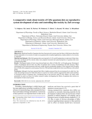
  • 1. Nanomed. J., 2(4): 261-268, Autumn 2015 261 A comparative study about toxicity of CdSe quantum dots on reproductive system development of mice and controlling this toxicity by ZnS coverage 1* A. Valipoor; 2 Gh. Amiri; 3 K. Parivar; 4 M. Modaresi; 5 J. Taheri; 5 A. Kazemi; 6 M. Abasi; 7 A. Mirzakhani 1 Department of Physiology, Faculty of Basic Sciences, Shahrekord Branch, Islamic Azad University, Shahrekord, Iran. 2 Department of Physic, Falavarjan Branch, Islamic Azad University, Isfahan, Iran 3 Department of Biology, Science and Research Branch, Islamic Azad University, Tehran, Iran 4 Department of Physiology, Islamic Azad University, Khorasgan Branch, Isfahan, Iran 5 Department of Chemistry, Islamic Azad University, Shahrekord, Iran . 6 Department of Matematic, Islamic Azad University, Shahrekord, Iran 7 Department of Mechanical Engineering, Payame Noor University, Tehran, Iran ABSTRACT: Objective(s): Medicinal benefits of quantum dots have been proved in recent years but there is little known about their toxicity especially in vivo toxicity. In order to use quantum dots in medical applications, studies ontheir in vivo toxicity is important. Materials and Methods: CdSe:ZnS quantum dots were injected in 10, 20, and 40 mg/kg doses to male mice10 days later, mice were sacrificed and five micron slides were prepared structural and optical properties of quantum dots were evaluated using XRD. Results: Histological studies of testis tissue showed high toxic effect of CdSe:ZnS in 40 mg/kg group. Histological studies of epididymis did not show any effect of quantum dots in terms of morphology and tube structure. Mean concentration of LH and testosterone and testis weight showed considerable changes in mice injected with 40 mg/kg dose of CdSe:ZnS compared to control group. However, FSH and body weight did not show any difference with control group. Conclusion: Although it has been reported that CdSe is highly protected from the environment by its shell, but this study showed high toxicity for CdSe:ZnS when it is used in vivo which could be suggested that shell could contribute to increased toxicity of quantum dots. Considering lack of any previous study on this subject, our study could potentially be used as an basis for further extensive studies investigating the effects of quantum dots toxicity on development of male sexual system. Keywords: CdSe:ZnS, In vivo toxicity, Quantum dots, ZnS shell Nanomed. J., 2(4): 261-268, Autumn 2015 DOI: 10.7508/nmj. 2015.04.003 *Corresponding Author Email:valipoor.akram@gmail.com Tel: (+98) 381-2227864 Note. This manuscript was submitted on July 20, 2015; approved on September 6, 2015 Received; 20 July 2015 Accepted; 6 September 2015 INTRODUCTION Nowadays, nanotechnology is widely known and has many applications including in medicine (3). With advances in nanotechnology, many nanoparticles have been used widely in biology and medicine. Cytotoxicity of these particles is an important factor in their use in medicine and hence has received a great deal of attention in recent years (2, 27). Among nanoparticles, quantum dots (QDs) are considered as extremely interesting materials for the development of photovoltaic devices because of their potential in medical diagnosis, cell imaging, and as biological tracers in human diseases diagnosis (3, 19, 27). But, in spite of obvious advantages of QDs, concerns regarding human exposure to these particles and its effect on human health have increased.
  • 2. Nanomed. J., 2(4): 261-268, Autumn 2015 262 A. Valipoor et al. Therefore, studies have been conducted on reaction of QDs in vivo and in vitro but there are still limited data on this category, which has led to not using dots in medications as therapeutic or diagnostic tools. So, further studies are needed to investigate QDs in the environments similar to human body (4, 9, 18(.Among semiconductor nanocrystal QDs, the CdSe core nanocrystals QDs have shown the best optical properties in light stability and radiative quantum efficiency in optical tests (4, 7, 19) Coolen et al study demonstrated that CdSe:ZnS nanocrystal QDs are the most widely used QDs )7). Most studies reported CdSe, which is a semiconductor matter, as core of nanocrystals QDs covered by another semiconductor matter (mainly ZnS). On the other hand, in vitro studies have shown an approximately complete control of CdSe-induced cytotoxicity by ZnS coverage (7,11,12,23,29). Therefore, CdSe and CdSe:ZnS nanocrystal QDs were selected for this study. Because of their importance as semiconductor and in biology and medicine, which has been highlighted in different studies, the synthesized QDs were characterized and their in vivo toxicity effects on male sexual system were also investigated. MATERIALS AND METHODS Materials All chemicals and solvents were purchased form Merck and were of analytical grades. Preparation of CdSe and CdSe:ZnS QDs Quantum dots were synthesized by chemical precipitation method. Briefly, three solutions of cadmiumchloride (CdCl2 -4H2 O), mercaptoethanol (ME) and sodium selenite (Na2 SeO3 -5H2 O) were prepared in the distilled deionized water, under vigorous stirring. At first, CdCl2 solution was poured into a three-spout balloon container and simultaneously ME solution was added to the same balloon. Finally, Na2 SeO3 solution was added under nitrogen . The resulting solution was mixed withdeionized water and thencentrifuged in order to remove any impurity aggregate. Then, the precipitated sample was dried at room temperature.All processes were done at room temperature (1). The crystal structure and optical properties of CdSe:ZnS QDs were characterized by XRD (X-Ray Diffraction, Bruker D8ADVANCE λ = 0.154 nm Cu Kα radiation) and UV-Vis spectrophotometer (Ultra Violet Visible, UV-2600 Shimadzu,Japan). ScanningTunneling Microscope (STM) (NATSICO, Iran) was used to investigate particle size distribution. Breeding and treatment of animals Male Balb/Cmice were purchased fromRaziVaccine and Serum Research Institute (Hesarak, Iran). Some male mice (about 20 days old) were kept for 10 days in natural day light and at temperature 22-24°C. Then, male mice were divided in 7 groups of 6 mice: control, three groupd injected with CdSe at 10, 20, and 40 mg/ kg and three groups injected with CdSe:ZnS QDs at 10, 20, and 40 mg/kg doses. Either CdSe or CdSe:ZnS nanoparticles were prepared in normal saline solution and single-dose was injected intraperitoneally at 10, 20, and 40 mg/kg doses. Control group received only normal saline solution. The protocol of study was approved by the Ethics Committee of the University. Tissue preparing 10 Days after CdSe and CdSe:ZnS injection, all control and treatment groups were anesthetized and testis and epididymis organs were rapidly removed, weighed, and preserved in formaldehyde as fixative agent. Five micron slides were dehydrated and prepared in paraffin. Then, the slides were stained using hematoxylin-eosin. Morphological structure of seminiferous tubes, and mean number of spermatogonia, spermatocytes, spermatids, and matured sperms in testis were studied, and epithelial height, connective tissue, smooth muscle and sperm density were measured in epididymis. Hormones measurements Serum FSH and LH were measured by double antibody radioimmunoassay as previously described (20, 21). Serum testosterone was assayed using a competitive chemiluminescent immunoassay kit (DRG Co, Germany; Pishtaz Teb, Iran). (testis and body weight and the number of cells in seminiferous tubes of various groups) were analyzed using SPSS 16 (by one way ANOVA followed by Tukey’s post hoc test.). Data were represented as means± standard error. Differences were considered significant at p<0.05 and p<0.01. RESULTS QDs characterization by XRD, STM and UV-Vis analysis The structure of the CdSe QDs was investigated by XRD. Fig 1 illustrates the XRD pattern of the CdSe
  • 3. Nanomed. J., 2(4): 261-268, Autumn 2015 263 QDs. As seen, the sample has a single phase and also a cubic crystal structure. According to the standard Joint Committee on Powder Diffraction Standards card No. 19-0191, the diffraction peaks correspond to the 111, 220 and 311 crystal planes. The mean size of the particles was determined by Debye-Scherer formula. It was calculated as 2.4 nm for CdSe QDs (1). Dose-finding study there was no study onin vivo toxicity of QDs, we selected three doses of 125, 250, and 500 mg/kg according to an in vitro study conducted by Ming- Shu Hsieh (13). In the first stage, CdSe and CdSe:ZnS nanoparticles were prepared in normal saline solution and were intraperitoneally injected into 36 mice. All mice died in less than 24 hours after the injection. The remarkable finding was that in vivo administration of ZnS as the shell increased toxicity of CdSe and mice showed greater responses to CdSe:ZnS compared to CdSe. These results were contrary to what has been reported for the cytotoxicity of CdSe:ZnS where the ZnS shell would minimize the cell toxicity of the CdSe core. The CdSe:ZnS-treated mice died within a short time after ip injection of the QDsand even in some cassesmice died almost immediately after the injection. In the next step, DMSA-coated CdSe nanoparticles were injected to groups of 8 mice each intraperitoneally at 125 and 250 mg/kg doses. The results showed that all mice injected with DMSA-coated CdSe at 250 mg/ kg dose and 80% of mice injected with DMSA-coated CdSe at 125 mg/kg dose died within one week post- injection. Then, CdSe and CdSe:ZnS were intraperitoneally injected to groups of 16 mice each at 75 and 100 mg/kg doses. In CdSe:ZnS groups at 75 and 100mg/kg doses50%and 83% ofmicedied, respectively whereas in CdSe groups at 75 and 100 mg/kg doses 50% and 66% of mice died within one week, respectively. Finally, lower doses of CdSe and CdSe:ZnS were selected and injected to group of 4 mice each. In CdSe groups, no mortality was observed during 20 days post-injection at all three doses of 10, 20 and 40 mg/kg. However, in CdSe:ZnS groups, while no mortality was observed at 10 mg/kg group but 12.5 and 20% mortality was observed, respectively. According to the obtained results in dose-finding study, 10 mg/kg dose was determined as a safe dose and 20 and 40 mg/kg doses were determined as doses with toxicity effects. Histological study of testis The seminiferous tubules are in different spermatogenic stages in control group, and in mice treated with all three doses of CdSe and CdSe:ZnS QDs. Spermatozoids were observed in lumen tubules of the control group, but in the group treated with 40 mg/kg CdSe:ZnS QDs, abnormal growth of seminiferous tubes, impaired spermatogenesis, reduction in number of spermatogonia, spermatocyst 1, spermatides and an obvious decrease in matured sperms of lumen were observed (Table 2). On the other hand, degeneration of the interstitial tissue and blood vessels and reduction in thickness of the lamina propria were observed as illustrated in Fig 1.
  • 4. Nanomed. J., 2(4): 261-268, Autumn 2015 264 Phytotoxicity of methylene blue Fig. 1. Microscopic images of testis slides, 10 days post- injection (H & E, 400×). A control group;B1, C1 and D1 groups treated with doses of 10, 20, 40 mg/kg CdSe, respectively; B2, C2 and D2 groups treated with doses of 10, 20, 40 mg/kg CdSe:ZnS, respectively. Sz: spermatozoa, Lc: Leydig cells, Lp: Lamina propria, Spg: Espermatogoni, Spc: Espermatocyte, Spt: Espermatid Histological study of epididymis Qualitative studies using optical microscope showed that epithelia, lumen, connective tissue and smooth muscle of epididymis were similar in groups treated with the QDs and control, but spermatozoa volume in lumen epididymis obviously decreased in mice treated with CdSe:ZnS at 40 mg/kg (Fig 2).
  • 5. Nanomed. J., 2(4): 261-268, Autumn 2015 265 Fig. 2. Microscopic images of epididymis slides, 10 days post- injection (H & E, 400×). A control group;B1, C1 and D1 groups treated with doses of 10, 20, 40 mg/kg CdSe, respectively; B2, C2 and D2 groups treated with doses of 10, 20, 40 mg/kg of CdSe:ZnS, respectively. SPM: Sperm mass, SMM: Smooth muscle, SC: Stereocilia, PC: Principal cells, SCE: Stereociliated simple columnar epithelium, LE: Lumen epididymis, LCT: Loose connective tissue Changes in body and testis weight, and hormones level Body weight did not change significantly in treatment groups but testis weight decreased significantly (P<0.01) by CdSe:ZnS at 40 mg/kg. FSH did not change by treatments with QDs (Table 2) but testosterone decreased significantly and LH increased significantly in mice treated with CdSe:ZnS at 40 mg/ kg (Fig 3, 4, 5). Fig. 3. Mean of concentration LH in 10 days post-injection (**p < 0.01, *p < 0.05 ) Fig. 4. Mean of concentration testosterone in 10 days post- injection (**p < 0.01, *p <0.05) Fig. 5. Mean of concentration FSH in 10 day post-injection (**p < 0.01, *p < 0.05 )
  • 6. Nanomed. J., 2(4): 261-268, Autumn 2015 266 A. Valipoor et al. Table 1. Average and mean comparison of sperm stem cell numbers in one tubule 10 days post-injection (p < 0.01) Mean of spermatidMean of spermatocyte IMean of spermatogoniaNanoparticle dosesgroups 111.95 ± 33.6344.15 ± 9.3534.55 ± 6.390 mg/kgControl 100.45 ± 18.64 100.85 ± 24.96 99.10 ± 24.68 45.05 ± 9.76 46.10 ± 8.58 45.70 ± 7.01 35.50 ± 7.55 35.95 ± 6.76 34.30 ± 7.55 10 mg/kg 20 mg/kg 40 mg/kg CdSe 113.65 ± 23.29 109.15 ± 20.72 83.00** ± 23.44 45.25 ± 8.45 43.80 ± 8.04 29.60** ± 10.76 33.60 ± 8.94 32.80 ± 6.67 18.85** ± 6.94 10 mg/kg 20 mg/kg 40 mg/kg CdSe:ZnS Table 2. Average and mean comparison of body and testis in 10 dayspost-injection (**p < 0.01, *p < 0.05 ) Mean of testis weightMean of body weightQDs dosesgroups .093 ± .0127.17 ± 1.30 mg/kgControl .087 ± .01 .087 ±.01 .083 ± .02 25.98 ± 1.6 27.67 ± 3.9 26.83 ± 1.4 10 mg/kg 20 mg/kg 40 mg/kg CdSe .087 ± .01 .107 ± .02 .055** ± .01 27.33 ± 2.8 26.83 ± 2.1 27.67 ± 1.3 10 mg/kg 20 mg/kg 40 mg/kg CdSe:ZnS Discussion Recently, there are a lot of reports on the use of nanoparticulate systems in biomedical applications (14, 30(.Among these nanoparticles, crystals with nanometer size, which are called mainly QDs, have been studied extensively for molecular imaging and targeted therapy. Because QDS are fluorescent nanoparticles with high sensitivityusing them as probe is a promising technique for biological applications. Due to large area provided by QDs, they are appropriate as targeting ligand to direct them to site of action (13, 21). Moreover, because of high light stability which provides long time observation of biological molecules, QDs could be the main hope as label for biological systems imaging. Therefore, the use of QDs as bright and photostable probes for long-term fluorescence imaging is gaining more interest (13, 27). Studies about the physicochemical properties of QDs and their use in vivo are important. There are a few studies about the effects of QDs on testis. In some studies, exposing young mice to DE(Diesel Exhaust) and silver induced degeneration of Leydig cells action and hence fluctuation of testosterone level (11, 15,32). Guo et al reported reduction in density and movement of spermatozoids, increase in spermatozoids abnormalities, and apoptosis of cells in mice injected by TiO2 nanoparticles. Histopathological studies of testis and epididymis did not show any change (12). In another study by Wen-Qing, gold nanoparticles (14 nm) with two ω-methoxy and ω-aminothyl polyethylene glycol covers passed testis blood barrier, entered to germ cells and increased testosterone (33). Recently, effects ofnanoparticlessmallerthan 100 nm(like DEP) (Diesel Exhaust Particles)have been studied. Results indicated separation of cells of seminiferous epithelium(26, 33). Sleiman et al examined the adverse productive toxic effects ofAgNPs in male Wistar rats exposed toAgNP (Silver Nanoparticles) during the prepubertal period and sacrificed at postnatal days 53 and 90. The weight of the animals at the postnatal day 90 did not change markedly, but growth was less in the group treated with AgNP at 50 μg/kg from the postnatal day 34 to 53. AgNP exposure produced a delay in puberty in both treated groups. Decreased sperm reserves in the epididymis and diminished sperm transit time were observed at the postnatal day 53, while a reduction in sperm production occurred at the postnatal day 90. The morphology of the seminiferous epithelium was markedly altered and data demonstrated that prepubertal exposure to AgNP altered reproductive development in prepubertal male Wistar rats, as evidenced by impairment in spermatogenesis and a lower sperm count in adulthood (27). Mathias et al showed AgNPs reduced the acrosome and plasma membrane integrities, reduced the mitochondrial activity and increased the abnormalities of the sperm in both treatment groups.AgNP exposure also delayed the onset of puberty, although no changes in body growth were observed in the treatment groups. The animals did not show changes in sexual behavior or serum hormone concentrations
  • 7. Nanomed. J., 2(4): 261-268, Autumn 2015 267 (18). In another study,AgNPs(Silver Nanoparticles) at low dose (1 mg/kg) were intravenously injected into male CD1 mice over 12 days. Treatment resulted in no changes in body and testis weights, sperm concentration and motility, fertility indices, or follicle- stimulating hormone and luteinizing hormone serum concentrations; however, serum and intratesticular testosterone concentrations were significantly increased 15 days after initial treatment. Histologic evaluation revealed significant changes in epithelium morphology, germ cell apoptosis, and Leydig cell size. Additionally, gene expression analysis revealed that Cyp11a1 and Hsd3b1 mRNA were up regulated significantly in treated animals. These data suggest thatAgNPs do not impair spermatogonial stem cells in vivo since treatment did not result in significant decreases in testis weight and sperm concentrations. However,AgNPs appear to affect Leydig cell function, leading to increase in testicular and serum testosterone levels (11). In vitro studies revealed high concentrations of TiO2 nanoparticles affected the viability and proliferation of mouse Leydig cells, but not the gene expression associated with spermatogenesis(20). And gold nanoparticles decreased the motility of human sperm. Also, silver, aluminum, and MoO3 nanoparticles were toxic to mouse spermatogonia stem cells. Silica nanoparticles inhibited the differentiation of mouse ES cells and C60 –based nanoparticles inhibited the differentiation of mouse midbrain cells, CB (Carbon Black)decreased the viability of mouse Leydig cells and CdSe QDs inhibited the pre- and post-implantation development of mouse embryos (13, 23, 31, 32). In vitro studies have shown cytotoxic effect of TiO2 and CB (Carbon Black)nanoparticles on viability of mice Leydig cells, as well (9, 12, 15,33).Also injection ofTiO2 ,gold, diesel exhaust (DE) and C60 nanoparticles to pregnant mothers caused cytotoxic effects on spermatogenesis and testis tissuein their male children (20, 26, 30, 32,33). In the present study, CdSe and CdSe:ZnS with 2-3 nm size awhich were prepared by sedimentation method was investigated in vivo. Histopathological studies of testis tissue showed toxic effect of these nanoparticles at 40 mg/kg dose. Moreover, study of the testis slides showed reduction in spermatogonia, spermatocytes, spermatid, and mature sperm in seminiferous tubular, degeneration of interstitial tissue and reduction in Leydig cells number. Histological study of epididymis tissue showed intensive reduction in sperm volume of epididymis at 40 mg/kg. According to Table 4, FSH level did not change by treatments with QDs but testosterone decreased significantly and LH increased significantly in group received CdSe:ZnS at 40 mg/kg dose, which is in agreement with the findings of testis weight and histopathology of mature group which include significant reduction in weight, degeneration of the interstitial tissue and Leydig cells by CdSe:ZnS at 40 mg/kg. Significant increase in LH could be due to a feedback mechanism in response to degeneration of interstitial tissue and considerable reduction of testosterone. Obviously, considering small size of these particles which provide them with easy passing from blood barrier of testis, we could infer that passing of this nanoparticles from brain blood barrier and enforcing direct toxicity on brain tissue and hypothalamus is possible but the first former suggestion is more probable because FSH level was not affected. In this study, cytotoxic effect of CdSe and the role of ZnS cover in controlling the degree of toxicityon sexual hormones, testis, and body weight of animals was investigated. In view of the results obtained in the present study, QDs can pass testis blood barrier and enforce effects on reproduction system. Although epithelium epididymis did not show any special histopathological changes in CdSe-treated mice by optical microscope but lumen content showed many failures occurring in testis tissue. CONCLUSIONS According to histopathological results of this study, CdSe:ZnS nanoparticles can pass testis blood barrier and enforce direct destruction of germinal cells and spermatogenesis. It seems that despite in vitro findings, including the role of ZnS for controlling the toxicity as the shell in embryonic culture environment, in vivo use of ZnS has increased toxicity of CdSe, which can be due to the increase in CdSe:ZnS stability or independent in vivo toxicity of ZnS itself. Although most of in vitro studies have offered cadmium ions release as the main reason for QDs toxicity, other mechanisms seem to contribute mainly to this toxicity, among which QDs stability plays an important role. ACKNOWLEDGEMENT Authors are thankful to dr amiri for his kind support and Iran nanotechnology initiative for the financial support. REFERENCE 1. Amiri Gh. Fatahian S, Mahmoudi S , Preparation and Optical properties assessment of CdSe quantum dot, Dig Nanomater Bios. 2011; 4(2): 1161. 2. Bae PK , Kim KN, Lee SJ, Chang HJ, Lee CK, Park JK. The modification of quantum dot probes used for the targeted
  • 8. Nanomed. J., 2(4): 261-268, Autumn 2015 268 A. Valipoor et al. imaging of his-tagged fusion proteins. Biomaterials. 2008; 30(5): 836-842. 3. Chang SQ , Dai YD, Kang B, Han W, Mao L, Chen D. UV- enhanced cytotoxicity of thiol-capped CdTe quantum dots in human pancreatic carcinoma cells. Toxicol Lett. 2009; 188(2): 104-111. 4. Chan WC, Maxwell DJ, Gao X, Bailey RE, Han M, Nie S. Luminescent quantum dots for multiplexed biological detection and imaging. Curr Opin Biotechnol. 2002; 13(1): 40–46. 5. Chan WH , Shiao NH. Cytotoxic effect of CdSe quantum dots on mouse embryonic development. Mol Sci. 2008; 29(2): 259-266. 6. Clift MJ , Stone V. Quantum Dots: An Insight and Perspective of Their Biological Interaction and How This Relates to Their Relevance for Clinical Use. Theranostics. 2012; 2(7): 668-680. 7. Coolen L, Brokmann, Hermier. JP. Chapter 24-Quantum Optics with Single CdSe/ZnS Colloidal Nanocrystals. Handbook of Self Assembled Semiconductor Nanostructures for Novel Devices in Photonics and Electronics. 2008; 708– 748. 8. Drbohlavova J , Adam V, Kizek R, Hubalek J. Quantum Dots- Characterization: Preparation and Usage in Biological Systems. Mol Sci. 2009; 10: 656–673. 9. Ema M , Kobayashi N, Naya M, Hanai S, Nakanishi J. Reproductive and developmental toxicity studies of manufactured nanomaterials. Reprod Toxicol. 2010; 30(3): 343–352. 10. Galeone A, Vecchio G, Malvindi MA, Brunetti V, Cingolani R, Pompa PP. In vivo assessment of CdSe-ZnS quantum dots: coating dependent bioaccumulation and genotoxicity. Nanoscale. 2013; 4(20): 6401-6407. 11. Garcia TX, Costa GM, França LR, Hofmann MC. Sub-acute intravenous administration of silver nanoparticles in male mice alters Leydig cell function and testosterone levels. Reprod Toxicol. 2014; 45: 59-70. 12. Guo LL, Liu XH, Qin DX, Gao L, Zhang HM, Liu JY, Cui YG. Effects of nanosized titanium dioxide on the reproductive system of male mice. Nan Ke Xue. 2009; 15(6): 517-522. 13. Hsieh MS, Shiao NH, Chan WH. Cytotoxic Effects of CdSe Quantum Dots on Maturation of Mouse Oocytes and Fertilization and Fetal Development. Mol Sci. 2009; 10(5): 2122 - 2135. 14. Jamiesona T, Bakhshia R, Petrovaa D, Pococka R and et al. Biological applications of quantum dots. Biomaterials. 2007; 28(3): 4717-4732. 15. Komatsu T , Tabata M, Kubo-Irie M, Shimizu T, Suzuki K, Nihei Y, Takeda K. The effects of nanoparticles on mouse testis Leydig cells in vitro. Toxicol In Vitro. 2008; 22(8): 1825-1831. 16. Li C , Taneda S, Taya K, Watanabe G, Li X, Fujitani Y, Nakajima T, Suzuki AK. Effects of in utero exposure to nanoparticle-rich diesel exhaust on testicular function in immature male rats. Toxicol Lett. 2009; 185(1): 1–8. 17. Li KG , Chen JT, Bai SS, Wen X, Song SY, Yu Q, Li J, Wang YQ. Intracellular oxidative stress and cadmium ions release induce cytotoxicity of unmodified cadmium sulfide quantum dots. Toxicol In Vitro. 2009; 23(6): 1007-1013. 18. Mathias FT , Romano RM, Kizys MM, Kasamatsu T, Giannocco G, Chiamolera MI, Dias-da-Silva MR, Romano MA. Daily exposure to silver nanoparticles during prepubertal development decreases adult sperm and reproductive parameters. Nanotoxicology. 2014; 9(1): 64- 70. 19. Muller L, Gasser M, Raemy DO, Herzog F, Brandenberger C, Schmid O, Gehr P, Rothen-Rutishauser B, Clift MJD. Realistic exposure methods for investigating the interaction of nanoparticles with the lung at the air-liquid interface in vitro. Theranostics. 2011; 1(1): 30-64. 20. Ono N, Oshio S, Niwata Y, Yoshida S, Tsukue N, Sugawara I, Takano H, Takeda K. Prenatal exposure to diesel exhaust impairs mouse spermatogenesis. Inhal Toxicol. 2007; 19: 275-281. 21. Pan Z, Mora-Sero I, Shen Q, Zhang H, Li Y, Zhao K, Wang J, Zhong X, Bisquert J. High Efficiency “Green” Quantum Dot Solar Cells. Am Chem Soc. 136(25):9203-10. 22. Pinaud F , Michalet X, Bentolila LA, Tsay JM, Doose S, Li JJ, Iyer G, Weiss S. Advances in fluorescence imaging with quantum dot bio-probes. Biomaterials. 2006; 27(9): 1679- 1687. 23. Roberts JR, Antonini JM, Porter DW, Chapman RS, Scabilloni JF, Young SH, Schwegler-Berry D, Castranova V, Mercer RR. Lung toxicity and biodistribution of Cd/Se-ZnS quantum dots with different surface functional groups after pulmonary exposure in rats. Part Fibre Toxicol. 2013; 10. 24. Rzigalinski BA, Strobl JS. Cadmium-containing nanoparticles: Perspectives on pharmacology and toxicology of quantum dots. Toxicol Appl Pharmacol. 2009; 238(3): 280-288. 25. Smith AM , Duan H, MohsAM, Nie S. Bioconjugated quantum dots for in vivo molecular and cellular imaging. Adv Drug Deliv Rev. 2008; 60(11): 1226-1240. 26. Shimizu M, Tainaka H, Oba T, Mizuo K, Umezawa M, Takeda K. Maternal exposure to nanoparticle titanium dioxide during the prenatal period alters gene expression related to brain development in the mouse. Part Fibre Toxicol. 2009; 6(20): 1-25. 27. Sleiman HK, Romano RM, Oliveira CA, Romano MA. Effects of prepubertal exposure to silver nanoparticles on reproductive parameters in adult male Wistar rats. Toxicol Environ Health A. 2013; 76(17): 1023-32. 28. Soenen SJ, Manshian B, Aubert T, Himmelreich U, Demeester J, De Smedt SC, Hens Z, Braeckmans K. Cytotoxicity of cadmium-free quantum dots and their use for cell bioimaging. Chem Res Toxicol. 2014; 27(6): 1050-9. 29. Su Y, He Y, Lu H, Sai L, Li Q, Li W, Wang L, Shen P, Huang Q, Fan C. The cytotoxicity of cadmium based, aqueous phase- Synthesized quantum dots and its modulation by surface coating. Biomaterials. 2009; 30(1): 19–25. 30. Takeda K, Suzuki K, Ishihara A, Kubo-Irie M, Fujimoto R, Tabata M, et al. Nanoparticles transferred from pregnant mice to their offspring can damage the genital and cranial nerve system. Health Sci. 2009; 55(1): 95-102. 31. Wiwantitkit V, Sereemaspum A, Rojanathanes R. Effect of gold nanoparticles on spermatozoa: the first world report. Fertil Steril. 2009; 91(1): 7-8. 32. Yoshida S1, Hiyoshi K, Oshio S, Takano H, Takeda K, Ichinose T. Effects of fetal exposure to carbon nanoparticles on reproductive function in offspring . Fertil Steril. 2010, 93(5): 1695-1699. 33. Yoshida S , Hiyoshi K, Ichinose T, Takano H, Oshio S, Sugawara I, Takeda K, Shibamoto T. Effect of nanoparticles on the male reproductive system of mice. Androl. 2008; 32(4): 337-342. How to cite this article: Valipoor A, Amiri GH, Parivar K, Modaresi M, Taheri J, Kazemi A, Abasi M, Mirzakhani A. A comparative study about toxicity of CdSe quantum dots on reproductive system development of mice and controlling this toxicity by ZnS coverage. Nanomed. J., 2015; 2(4): 261-268.