Tracts+oblongata+2021
•Télécharger en tant que PPTX, PDF•
2 j'aime•501 vues

Ascending, descending, and medulla oblongata is important anatomical structures for coordinations in physiology, embryology, and psychological activities in humans

Recommandé
Contenu connexe
Tendances
Tendances (20)
Similaire à Tracts+oblongata+2021
Similaire à Tracts+oblongata+2021 (20)
Plus de obaje godwin sunday
Plus de obaje godwin sunday (20)
Dernier
Dernier (20)
Tracts+oblongata+2021
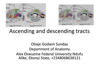
- 1. Ascending and descending tracts Obaje Godwin Sunday Department of Anatomy Alex Ekwueme Federal University Ndufu Alike, Ebonyi State, +2348068638121
- 2. Characteristics of ascending tract • Made up of 3 neurons (1st, 2nd, and 3rd order) • The posterior column is called spinothalamic tract • Receptors are exteroceptive or proprioceptive from the origin
- 3. Characteristics of ascending tract • Terminates at the Brodmann areas(3,1,2), which is the primary sensory area in the cerebral areas • 1st order neuron located outside the CNS, somewhere in the dorsal root ganglion of spinal cord or in the sensory ganglia of cranial nerves • Except; trigeminal nerve whose 1st order neuron in the midbrain
- 4. What Brodman areas look like
- 5. Characteristics of ascending tract • 2nd order neurons located in the spinal cord or medulla oblongata • 2nd order neurons crossed to the opposite (contralateral) in the cerebral cortex • 3rd order neurons located in the ventral posterior side of thalamus
- 6. Characteristics of ascending tract • Information is carried from the environment and to the brain for interpretation (cerebral cortex) • Spinocerebellar Tract (SCT) is the ascending tract when it reaches the cerebellum • It has two neurons to reach the cerebellum
- 7. Characteristics of ascending tract • 1st order neuron of SCT is located outside CNS, probably in the dorsal root ganglion of spinal cord or ganglia of cranial nerves • Originated from the proprioceptive receptors • Terminated in the cerebellum
- 8. Characteristics of ascending tract • 2nd order neuron of SCT is located outside CNS, in the ganglia of spinal cord or medulla oblongata • Representation is ipsilateral in the cerebellum • Carry postural and equilibrium movements
- 9. Characteristics • Light touch and pressure, tickle, and itch ant. Spinothalamic tract • Pain and temperature by lat. Spinothalamic tract • Consciousness, fine touch, and two point discrimination by fasciculi gracilis and cuneatus • Unconsciousness proprioception by ant and post spinocerebellar tracts
- 13. Descending tract
- 14. Characteristic features • Involve 2 neurons ( upper and lower motor neurons) • The motor neurons located in the anterior horn of spinal cord and motor nuclei of cranial nerves are referred to as lower motor neurons
- 15. Characteristic features • The neurons present in the various parts of the brain which influence the activity of lower motor neurons are referred to as upper motor neurons • The control is usually contralateral.
- 16. Characteristic features • Descending tracts arising from motor and premotor area of cerebral cortex constitute pyramidal tracts (lateral and anterior corticospinal tracts)
- 17. Characteristic features • All other descending tracts pathways having their origin in subcortical areas are referred to as extrapyramidal tracts (tectospinal, rubrospinal, reticulospinal, olivospinal and vestibulospinal)
- 18. Sensations • Skilled voluntary movement by corticospinal tract (pyramidal), ant and post corticospnal • Extrapyramidal • Tectospinal tract for spinovisual reflexes like head and neck movements while eyes closed
- 19. Sensations • Rubrospinal srrves as facilitators for flexors and inhibitors for extensors • Vestibulospinal tract for postures and balancing
- 20. Sensations • Reticulospinal tract (medial) serves as facilitatory to extensor muscles • Reticulospinal tract (lat) serves as facilitatory to flexor muscles
- 22. Differences between UMN and LMN Features UMN LMN Paralysis Type Spastic Flaccid Muscle Tone Increased Decreased Muscle atrophy Absent Present Tendon reflexes Exaggerated Diminished Babinski’s sign Present Absent
- 23. What happens in CST injuries/paralysis? A solution has come from Prof Liu in Hong Kong
- 24. Babinski’s sign Dorsiflexion of great toe with fanning of lateral toes on scratching lateral aspect of plantar surface of foot In healthy/normal adults the toes are planter flexed
- 25. As soon as your baby’s born, you’ll notice their primitive reflexes — although you might not know them by name. Palmar grasp reflex Plantar reflex Sucking reflex Rooting reflex Stepping reflex
- 27. Characteristics • Is the lowermost part of the brainstem (other parts are pons and midbrain) • Continuous below with the spinal cord and above with pons.
- 28. Characteristics • Located in the posterior cranial fossa and is related to clivus anteriorly and cerebellum posteriorly. • Separated from the cerebellum by the cavity of fourth ventricle
- 30. Medulla oblongata • Function: Innervation to the viscera of the head, thorax and abdomen, heart rate. Involuntary actions like vomiting and sneezing • Nuclei: Cranial nerve nuclei (IX-XII): inferior salivatory nucleus, spinal nucleus of trig
- 31. Medulla oblongata • Tracts: Corticospinal (pyramidal) tract, cuneate fascicle, gracile fascicle, medial lemnisci
- 32. Medulla oblongata • Cone shaped neuronal mass, broad in the upper part and narrow in the lower part. • It’s myelencephalon • Approximately 3 cm in length
- 33. Medulla oblongata • Divided into two part: –Lower closed part: It has a central canal –Upper open part : In this part the central canal widens and opens dorsally to form the lower half of the floor of fourth ventricle
- 35. Medulla oblongata • The ventral aspect of medulla oblongata shows the following features from medial to lateral: • Anterior median fissure/sulcus in the median plane (divides ventral aspect into two symmetrical halves).
- 36. Medulla oblongata • Pyramids: Elongated elevation produced by underlying the corticospinal tract • Anterolateral sulcus: Hypoglossal nerve rootlets emerge along this sulcus
- 37. Medulla oblongata • Olive: Oval elevation produced by underlying Inferior olivary nucleus • Posterolateral sulcus: Rootlets of glossopharyngeal, vagus and cranial part of accessory nerves emerge along this sulcus from above downward
- 39. Medulla oblongata • Features of the dorsal surface of lower closed part of medulla oblongata from medial to lateral are: • Posterior median sulcus: In the median plane
- 40. Medulla oblongata • Fasciculus gracilis (lowerpart) and gracile tubercle (in the upper part): gracile tubercle is produces by the underlying nucleus gracilis, where the axons comprising fasciculus gracilis terminate
- 41. Medulla oblongata • Fasciculus cuneatus (lowerpart) and cuneate tubercle (in the upper part): cuneate tubercle is produces by the underlying nucleus cuneatus, where the axons comprising fasciculus cuneatus terminate. • Inferior cerebellar peduncle: Connects medulla to cerebellum.
- 43. Dorsal surface of upper open part of medulla oblongata • Dorsal surface of upper part of medulla forms the lower half of the floor of fourth ventricle • The transversely running stria medullaris fibers separate the dorsal surface of medulla from that of pons
- 44. Dorsal surface of upper open part of medulla oblongata • These fibers originate from arcuate nucleus ( dislodged pontine nuclei) and reach the cerebellum via inferior cerebellar peduncle
- 45. Dorsal surface of upper open part of medulla oblongata from medial to lateral are: • Median sulcus: in the median plane. • Medial eminence: shows two triangular areas, hypoglossal triangle above and vagal triangle below, which overlie the nucleus of hypoglossal nerve and dorsal motor nucleus of vagus respectively
- 46. Dorsal surface of upper open part of medulla oblongata from medial to lateral are: • Below the vagal triangle lies area postrema ( chemoreceptor trigger zone (CTZ) which trigger vomiting in response to presence of emetic substances in the blood). Area postrema is devoid of blood brain barrier
- 47. Dorsal surface of upper open part of medulla oblongata from medial to lateral are: • Sulcus limitans: separates medial eminence from vestibular area. • Vestibular area: inferior and medial verstibular nuclei lie deep to this area
- 48. Transverse sections of the medulla oblongata at the level of pyramidal decussation
- 49. Transverse sections of the medulla oblongata at the level of sensory decussation
- 50. Transverse sections of the medulla oblongata at the level of olive (open part of medulla)
- 51. The medulla oblongata is supplied by the following arteries: • Vertebral arteries. • Anterior and posterior spinal arteries. • Anterior and posterior inferior cerebellar arteries. • Basilar artery.
- 52. Applied Aspects • Medial medullary syndrome • It occurs due to injury to the branches of anterior spinal artery supplying the paramedian region of medulla
- 53. Applied Aspects • Following structures are affected: • Hypoglossal nucleus • Medial lemniscus • Corticospinal tract (pyramid)
- 55. The symptoms are as follows • Contralateral hemiplegia (UMN Paralysis • Ipsilateral paralysis of tonque muscles (LMN Paralysis) • Contralateral loss of conscious proprioception of position and vibration
- 56. Lateral medullary syndrome It occurs due to thrombosis of posterior inferior cerebellar artery, which supplies a wedge-shaped area on the dorsolateral aspect of the medulla The following structures are affected: • Vestibular nucleus • Inferior cerebellar peduncle
- 57. Lateral medullary syndrome • Spinal nucleus and tract of trigeminal • Nucleus ambiguus • Reticular formation
- 59. For some of you who may think that I have wasted their time, please forgive me. If you don’t like my face help me watch this video to the end https://www.clutchprep.com/physiology/spinal-tracts-introduction-to-ascending- and-descending-pathways