Major histocompatility complex (Antigen Presentation to T cells, Autoimmunity, Transplantation)
•Télécharger en tant que PPTX, PDF•
10 j'aime•1,508 vues

MHC, HLA, Antigen Presentation, Autoimmunity, Tolerance, Tissue Transplantation

Recommandé
Contenu connexe
Tendances
Tendances (20)
Similaire à Major histocompatility complex (Antigen Presentation to T cells, Autoimmunity, Transplantation)
Similaire à Major histocompatility complex (Antigen Presentation to T cells, Autoimmunity, Transplantation) (20)
Plus de Pradeep Singh Narwat
Plus de Pradeep Singh Narwat (20)
Dernier
Dernier (20)
Major histocompatility complex (Antigen Presentation to T cells, Autoimmunity, Transplantation)
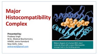
- 1. Major Histocompatibility Complex Presented by: Pradeep Singh M.Sc. Medical Biochemistry HIMSR, Jamia Hamdard New Delhi, India prdnarwat@gmail.com
- 2. Contents • Introduction of MHC molecules • History of MHC molecules • Structure of MHC molecules • General organization and inheritance of MHC • Functions of MHC
- 3. Introduction
- 5. • Cells of the innate immunity includes basophils, dendritic cells, eosinophils, Langerhans cells, mast cells, monocytes, macrophages, neutrophils and NK cells. • Cells of Adaptive immunity includes the B-cells and T-cells (TH cells and TC cells). Ques: How innate and adaptive immunity collaborate to resolve an infection? Ans: B-cells have the tendency to directly bind with the antigen while T-cells requires these MHC molecules for antigen presentation. First, pathogen is recognized by various receptors present on the innate immune cells. Second, this pathogen (antigen) is then presented to cells of adaptive immune system by cell-surface molecules called MHC for further action.
- 6. (Phagolysosome) (Phagosome) into small peptides Peptides expressed on cell surface as MHC-peptide complex
- 7. • MHC proteins are present on all nucleated cells and are unique to each individual. • MHC proteins are encoded by MHC genes. • In human, MHC locus is found on Chromosome 6 . (In human, MHC complex is known as human leukocyte antigen (HLA) complex because it was first identified on leukocytes) • In mice, MHC locus is found on chromosome 17. (In mice, MHC complex is known as Histocompatibility system 2 or just H-2)
- 8. History
- 9. HistoryGorer (1930s): • In mid 1930s, Peter Gorer identified four groups of genes that encode blood-cell antigens using inbred strains of mice. He designated these as I, II, III and IV. • He concluded that the rejection of foreign tissue transplanted between individuals in a species was the result of an immune response mounted against these cell surface molecules. Gorer and Snell (1940s & 1950s): • Antigens encoded by the genes in the group II took part in the rejection of transplanted tumors and other tissues. • Snell called these genes “Histocompatibility genes” (H-2 in mice). Dausset and Snell helped to characterize the functions controlled by the MHC, resulting in 1980s Nobel Prize in Medicine and Physiology. Follow-up studies by Rolf Zinkernagel and Peter Doherty illustrated that the proteins encoded by these genes play a seminal role in adaptive immunity.
- 11. Structure of MHC Molecules • There are two main classes of MHC molecules: class I and class II. • These are membrane bound glycoproteins that are closely related in both structure (quaternary) and function. • Each MHC molecules has three domains: i. Extracellular domain ii. Transmembrane domain iii. Cytoplasmic domain • Antigen binding cleft is present on the extracellular domain.
- 12. The α1 and α2 domains form the antigen binding cleft The α1 and β2 domains form the antigen binding cleft
- 13. MHC Class I Proteins • MHC class I proteins are present on the surface of all nucleated cells [Also present on platelets]. • Foreign antigens combined with MHC class I proteins are recognized by cytotoxic T cells. • Made up of a large α-subunit and a smaller β-subunit. (β-subunit is coded by genes outside the MHC locus) • The α-subunit has three extra-cellular domains (α1,α2 and α3), a trans membrane region and a cytoplasmic tail. • The α1 and α2 domains form the peptide binding cleft. • MHC I proteins bind fragments of proteins degraded by cytosolic pathway (Proteasomal / Ubiquitin Pathway ). • α3-domain & β2 macroglobulin domain have structural and amino acid sequence homologous with Ig constant domain of Ig Gene Superfamily. MHC Class II Proteins • MHC class II proteins are present on the surface of Antigen Presenting Cells (macrophages, B-cells, dendritic cells etc). • Foreign antigens combined with MHC class II proteins are recognized by helper T cells. • MHC class II proteins are made up of almost equal sized α-subunit and β-subunit. Each subunit has two extracellular domains, a trans membrane region and a cytoplasmic tail. • The α1 and β1 domains form the peptide binding cleft. • MHC class II proteins bind fragments of proteins degraded by lysosomal or endocytic pathway. • α2-domain & β2 macroglobulin domain have structural and amino acid sequence homologous with Ig constant domain of Ig Gene Superfamily.
- 15. General Organization & Inheritance of the MHC
- 16. General Organization & Inheritance of the MHC • MHC molecules are encoded by a cluster of genes collectively called MHC locus. MHC genes can be divided into: MHC class I genes MHC class II genes MHC class III genes
- 17. The MHC locus consist of three class of genes which encodes three major classes of molecules: • Class I MHC genes encode glycoproteins expressed on the surface of nearly all nucleated cells; the major function of class I gene products is presentation of endogenous peptide antigens to CD8+ T cells. [Membrane Proteins] • Class II MHC genes encode glycoproteins expressed predominantly on APCs (macrophages, dendritic cells, and B cells), where they primarily present exogenous antigenic peptides to CD4+ T cells. [Membrane Proteins] • Class III MHC genes encode several different proteins, some with immune functions, including components of the complement system (C4, C2 and factor B) and molecules involved in inflammation (TNF-α and Lymphotoxin- α [TNF-β]). [Not Membrane Proteins]
- 18. Fig: Comparison of the organization of the MHC in mouse and human. • Unlike in the human, class I MHC genes region in mouse is non-continuous, interrupted by the class II and class III regions. • Class I MHC molecules: α-chain molecules are encoded by the K and D regions in mice with an additional L region found in some strains, and by the A, B, and C loci in humans. (Collectively, these are referred to as classical class I molecules) [β2-microglobulin is encoded by genes outside the MHC locus] • Class II MHC molecules: encoded by IA and IE regions in mice and by the DP, DQ and DR regions in humans.
- 19. Classical Vs Non-classical class I MHC molecules • Classical class I molecules: Molecules encoded by K and D region in mice and A, B, C region in humans are referred to as classical class I molecules, all posses the functional capability of presenting protein fragments of antigen to T cells. • Non-classical class I molecules: Additional genes or groups of genes within the class I region of both mouse and human encode non-classical class I molecules that are expressed only in specific cell types. • Example of non-classical class I molecule: HLA-G molecule in humans. These are present on fetal cells at the maternal-fetal interface and are credited with inhibiting rejection by maternal CD8+ T cells by protecting the fetus from identification as foreign, which may occur when paternally derived antigens begin to appear on the developing fetus.
- 20. Classical Vs Non-classical class II MHC molecules • Classical class II molecules: Molecules encoded by IA and IE region in mice and DP, DQ and DR regions in humans are referred to as classical class II molecules, all posses the functional capability of presenting protein fragments of antigen to T cells. • Non-classical class II molecules: Additional genes or groups of genes within the class II region of both mouse and human encode non- classical class II molecules that are expressed only in specific cell types. • Example of non-classical class II molecule: Human non-classical class II genes are DM and DO. The DM genes encode a class II–like molecule (HLA-DM) that facilitates the loading of antigenic peptides into class II MHC molecules. Class II-like HLA-DO molecules, which are expressed only in the thymus and on mature B cells, have been shown to serve as regulators of class II antigen processing.
- 21. The Exon/Intron Arrangements of Class I and II Genes Reflects Their Domain Structure DNA mRNA Proteins synthesized on ER membrane folding into quaternary structure in ER Golgi Vesicles Cell Membrane Fig: Schematic diagram of (a) class I and (b) class II MHC genes, mRNA transcripts, and protein molecules.
- 22. Allelic Forms of MHC Genes Are Inherited In Linked Groups Called Haplotypes • The genes that reside within the MHC region are highly polymorphic. • Haplotypes: The individual genes of MHC loci (class I, II and III) lie so close together that their inheritance is linked. This set of linked alleles is referred to as a haplotype. • An individual inherits one haplotype from the mother and one haplotype from the father, two set of alleles i.e total 6 set of alleles each of class I and class II • MHC follows: 1. Polymorphism 2. Polygenism 3. Linkage disequilibrium 4. Co-Dominance
- 23. MHC Molecules Are Co-Dominantly Expressed • The genes within the MHC locus exhibit a co-dominant form of expression, meaning that both maternal and paternal gene products (from both haplotypes) are expressed at the same time and in the same cells.
- 24. Recombination • Genetic recombination can generate new allelic combinations, or haplotypes. • As a result of recombination and other mechanisms for generating mutations, it is rare for any two unrelated individuals to have identical sets of HLA genes. • This makes transplantation between individuals who are not identical twins quite challenging!
- 25. • As compared to MHC class I molecules, MHC class II molecules have even greater potential for diversity. • Each of the classical class II MHC molecules is composed of two different polypeptide chains (α & β) encoded by different loci. • A heterozygous individual can express - combinations that originate from the same chromosome (maternal only or paternal only) as well as class II molecules arising from unique chain pairing derived from separate chromosomes (new maternal-paternal - combinations). • The diversity generated by these new MHC molecules likely increases the number of different antigenic peptides that can be presented and is therefore advantageous to the organism in fighting infection.
- 26. Functions of MHC
- 27. The Role of the MHC and Expression Patterns Why an MHC molecule on the surface of a cell is important. In general, these include the following: • To display self class I to demonstrate that the cell is healthy • To display foreign peptide in class I to show that the cell is infected and to engage with TC cells • To display a self-peptide in class I and II to test developing T cells for autoreactivity (primary lymphoid organs) • To display a self-peptide in class I and II to maintain tolerance to self- proteins (secondary lymphoid organs) • To display a foreign peptide in class II to show the body is infected and activate TH cells Ques: Ans:
- 28. Function of MHC molecules 1. Antigen presentation 2. Autoimmune reactions 3. Transplant rejection
- 30. 1. Antigen Presentation • Types of antigens: i. Intracellular pathogen Presented by MHC class I molecules ii. Extracellular pathogen Presented by MHC class II molecules
- 32. Steps: • Degradation of proteins into smaller peptides by ubiquitin mediated pathway (proteasomal pathway) into the cytosol • Transport of small peptides inside the lumen of RER • Synthesis, folding of MHC molecules into their functional form in ER. • Loading of small peptides into the groove of MHC class I molecules in ER. • Transport of MHC-peptide complex from ER to Golgi apparatus in vesicle form. • Transport of vesicle containing MHC-peptide complex from Golgi to plasma membrane. • Fusion of vesicles with the plasma membrane. The Endogenous Pathway of Antigen Processing and Presentation (MHC class I Pathway)
- 33. • Intracellular proteins are degraded into short peptides by a cytosolic proteolytic system present in all cells, called the proteasome. • The immune system also utilizes this general pathway of protein degradation to produce small peptides for presentation by class I MHC molecules. • Small peptides are transported from the cytosol to the RER. Degradation of proteins into smaller peptides:
- 34. • There are transporter proteins designated TAP (for transporter associated with antigen processing) in the membrane of the RER that helps in the uptake of small peptides inside the RER lumen from cytosol. • TAP is a membrane-spanning heterodimer consisting of two proteins: TAP1 and TAP2. • Both TAP1 and TAP2 belong to the family of ATP-binding cassette proteins. • Both TAP1 and TAP2 proteins each have a domain projecting into the lumen of RER and an ATP-binding domain that projects into the cytosol.
- 35. Loading of peptides on MHC molecules • Like all other proteins destined for plasma membrane, the α-chain and β- chain of MHC class I and class II molecules are synthesized on ribosomes on RER. • Molecular chaperons facilitates the folding of these polypeptides into functional MHC molecules. • Tapasin (TAP-associated protein) brings the TAP transporter into proximity with the class I molecule and allows it to acquire an antigenic peptide. • Exoproteases in the ER will act on peptides not associated with class I MHC molecules.
- 36. • When β2-microglobulin binds to the chain, calnexin is released and the class I molecule associates with the chaperone calreticulin and with tapasin. • Tapasin (TAP-associated protein) brings the TAP transporter into proximity with the class I molecule and allows it to acquire an antigenic peptide. • On peptide binding, the class I molecules displays increased stability can dissociate from the complex with calreticulin, tapasin and ERp57. • The first molecular chaperone involved in class I MHC assembly is calnexin, a resident membrane protein of the ER. • ERp57, a protein with enzymatic activity, and calnexin associate with the free class I chain and promote its folding.
- 37. Clinical Focus: Bare Lymphocyte Syndrome • Deficiencies in TAP can lead to Bare Lymphocyte Syndrome. • Lymphocytes in individuals with TAP deficiency express low levels of class I molecules than those of normal controls. • Low levels of class I molecules leads to increased numbers of NK and γδT cells and decreased levels of CD8+ T cells. • In early life, the TAP-deficient individual suffers frequent bacterial infections of the upper respiratory tract and in the second decade begins to experience chronic infection of the lungs.
- 38. MIIC: MHC class II containing compartment The Exogenous Pathway of Antigen Processing and Presentation (MHC class II pathway) Steps: • Endocytosis of the antigen by APCs (Phagosome) • Fusion of phagosome with lysosomes leads to the formation of phagolysosme. • Degradation of antigen into smaller peptides inside phagolysosome by endocytic processing pathway. • Fusion of the late endosomes containing MHC class II molecules (from ER Golgi) with phagolysosomes. • Loading of small peptides into the groove MHC class II molecules in the phagolysosomes. • Transport of MHC-peptide complex from phagolysosomes to plasma membrane. • Fusion of vesicles with the plasma membrane.
- 39. Since APCs express both class I and class II MHC molecules. Some mechanism must exist to prevent the binding of peptide generated by proteasomal pathway to class II molecules in the ER lumen. Think About It!!!
- 40. Invariant Chain Guides Transport of Class II MHC Molecules to Endocytic Vesicles • When MHC class II molecules are synthesized in the RER, these class II chains associate with a protein called the invariant chain (Ii, CD74). • Ii (non-MHC encoded protein) interacts with the class II peptide-binding groove preventing any endogenously derived peptides from binding while the class II molecule is within the RER • Ii chain also appears to be involved in the folding of the class II α and β chains. • Ii contain contains sorting signals in its cytoplasmic tail that direct the transport of the class II MHC complex from the trans-Golgi network to the endocytic compartments. • Transfection experiments revealed that, in the absence of Ii, class II molecules remain primarily in the ER and do not transit past the cis-Golgi.
- 41. Assembly of Class II MHC Molecules • Class II MHC-invariant chain complexes are transported from the RER through the Golgi complex to MIIC late endosomes compartment. The proteolytic activity increases in each successive compartment, the invariant chain is gradually degraded. • A short fragment of the invariant chain termed CLIP (for class II–associated invariant chain peptide) remains bound to the class II molecule. • Like antigenic peptide, CLIP physically occupies the peptide-binding groove of the class II MHC molecule, preventing any premature binding of antigen-derived peptide. • A nonclassical class II MHC molecule called HLA-DM is required to catalyze the exchange of CLIP with antigenic peptides. • The nonclassical class II molecule HLA-DO may act as a negative regulator of class II antigen processing by binding to HLA-DM and inhibiting its role in the dissociation of CLIP from class II molecules.
- 42. Cross-Presentation of Exogenous Antigens
- 43. Cross-Presentation of Exogenous Antigens • First reported by Michael Bevan and later described in detail by Peter Cresswell and colleagues. What we know till now ? Intracellular pathogen Endogenous pathway MHC class I molecules CD8+ T cells Extracellular pathogen Exogenous pathway (APC) MHC class II molecules CD4+ T cells Ques: If a pAPC (peripheral APC) not harbouring an intracellular infection, such as viruses that have been engulfed from extracellular sources in ways that will activate the needed CTL (cytotoxic T lymphocytes) responses (usually mediated by endogenous pathway)? The answer to this dilemma is a process called cross-presentation. The phenomenon of cross-presentation requires that internalized antigens that would normally be handled by the exogenous pathway leading to class II MHC presentation somehow become redirected to a class I peptide loading pathway. [Extracellular Pathogen MHC class II molecules TH cells MHC class I molecules CD8+ T cells]
- 44. FIGURE: Activation of naïve Tc cells by exogenous antigen requires DC licensing and cross-presentation. (a) Dendritic cells (DCs) first internalize and process antigen through the exogenous pathway, presenting to CD4+ TH cells via MHC class II molecules and activating these cells through, among other things, CD40-CD40L engagement. (b) These activated TH cells can then serve as a bridge to help activate CTL responses; they provide local IL-2 and they in turn license the DC to cross-present internalized antigen in MHC class I, up-regulate costimulatory molecules, and down-regulate their inhibitory counterparts. DC licensing creates an ideal situation for the stimulation of antigen- specific CD8+ T cell responses. When the TLRs on these DCs are engaged, this further activates these cells, providing added encouragement for cross-presentation. Dashed arrows indicate antigen directed for cross-presentation. [Adapted from Kurts et al., 2010, Nature Reviews Immunology 10:403.]
- 45. Presentation of Non-peptide Antigens • It is well known that some non-protein antigens are also recognized by T cells, and in the 1980s T-cell proliferation was detected in the presence of non-protein antigens derived from infectious agents. • Various types of T cells (expressing as well as T–cell receptors) can react against lipid antigens, such as mycolic acid, derived from well- known pathogens, such as Mycobacterium tuberculosis. • These antigens are presented by members of the CD1 family of non- classical class I molecules.
- 46. What Are Superantigens ? • Superantigens are viral or bacterial proteins that bind simultaneously to specific V regions of T-cell receptors and to the chain of class II MHC molecules.
- 47. Figure: Endogenous (green) and Exogenous (red) pathways of antigen presentation.
- 48. Structure of T cell receptor
- 49. Structure of TC cells receptor Structure of TH cells receptor
- 50. T-cell Activation • CD4+ and CD8+ T cells leave the thymus and enter the circulation as resting cells in the G0 stage of the cell cycle. • Each naïve T cell recirculates from blood through lymph nodes and back again every 12 to 24 hours until it encounters and MHC-foreign peptide complex on phagocytic cells. • Activation of T-cells and elimination of the pathogen take place.
- 51. Two Signal Hypothesis • In 1987, Helen Quill and Ron Schwartz recognized that, in the absence of functional APCs, isolated high affinity TCR-MHC interactions actually led to T-cell non- responsiveness rather than activation—a phenomenon called T-cell anergy. • Their studies led to the simple but powerful notion that not one but two signals were required for full T-cell activation: Signal 1 is provided by antigen-specific TCR engagement (which can be enhanced by co-receptors and adhesion molecules) Signal 2 is provided by contact with a costimulatory ligand, which can only be expressed by a functional APC.
- 52. cSMAC: central supramolecular activating complex pSMAC: peripheral supramolecular activating complex
- 53. Costimulatory molecules can be: i. Negative costimulatory molecules: Inhibit TCR signaling ii. Positive costimulatory molecules: Activate TCR signaling • Role of negative costimulatory molecules: (1) maintaining peripheral T- cell tolerance and (2) reducing inflammation both after the natural course of an infection and during responses to chronic infection. E.g: CTLA-4 • Role of positive costimulatory molecules: Activate TCR signaling eg. CD28 binds to two distinct ligands of the B7 family of proteins., CD80 (B7-1) and CD86 (B7-2) of APCs. Cytokines provide the third signal
- 55. • MHC molecules can present both intracellular and extracellular pathogens. • Normally, our cells present self-peptides in the groove of MHC class I molecules. • The expression of self-MHC class I (with self peptides) signals that a cell is healthy; absence of self-MHC class I (as can occur in virus-infected and tumor cells) targets that cell for killing by NK cells. • When foreign proteins are present in the cytosol and begin to appear in the groove of MHC class I on the cell surface, this alerts CD8+ T cells to the presence of this unwelcome visitor, targeting the cell for destruction. • MHC class II molecules primarily display peptides that have come from the extracellular spaces (Opsonization).
- 57. 2. Autoimmune Reactions • Autoimmunity results from some failure of the host’s immune system to distinguish self from non-self, causing destruction of self cells and organs. • The mechanisms that protect an individual from anti-self immune attack are collectively termed as tolerance or self-tolerance. • In adults, most encounters with foreign antigen leads to an immune response aimed at eradication. This is not true in the foetus, where due to immature state of the immune system, exposure to antigen frequently results in tolerance. • These mechanisms that maintain self-tolerance also cause rejection of any transplanted tissues or cells that carry new proteins, as occurs whenever the donor is not genetically identical to the recipient.
- 58. • T cells without appropriate co-stimualtion results in a form of tolerance known as anergy (unresponsiveness to foreign antigen) whereas the same antigen presented with co-stimulatory molecules can become a potent immunogen. • Tolerance – Treg play an important role in maintain tolerance. Two types: i) Central tolerance tolerance in primary lymphoid organs ii) Peripheral tolerance tolerance in secondary lymphoid organs • Human individuals have been shown to posses mature, recirculating, self reactive lymphocytes. • Mechanism that maintain tolerance include the induction of cell death or cell anergy in lymphocytes and limitation on the activity of self- reactive cells by regulatory processes. • Peripheral tolerance regulate autoreactive cells in the circulation.
- 59. • Data showed that interaction between CD28 on T-cell and CD80/86 (B7) on the APC provided the co-stimulatory signal required for T-cell activation. • Further examination of these co-stimulatory molecules revealed the existence of other molecules that could bind to CD80/86 and the discovery of a related molecule called CTLA-4. This molecule inhibits rather than stimulation of T cell activation upon binding CD80/86. • Treg cells: i. nTreg cells (natural) ii. iTreg cells (inducible)
- 60. Autoimmunity • Autoimmune disease is caused by failure of the tolerance processes described earlier to protect the host from the action of self-reactive lymphocytes. • Production of auto-antibodies are the main offenders in majority of autoimmune diseases. • Autoimmune diseases: two types i. Organ specific autoimmune diseases ii. Systemic autoimmune diseases
- 62. Clinical Focus: Ankylosing Spondylitis (AS) • The word is from Greek, ankylose meaning “to unite or grow together”, spondylos meaning “vertebra” and itis meaning “inflammation”. • Initial symptoms are usually a chronic dull pain in the lower back or gluteal region combined with stiffness of the lower back. • More than 90% of the cases in the UK have a specific human leukocyte antigen, HLA-B27. • Individuals with the HLA-B27 variants are at a higher risk than the general population of developing the disorder. • Autoantibodies specific for AS have not been identified. • No cure for AS.
- 64. 3. Transplant Rejection • The first successful organ (kidney) transplant, performed in 1954 by Joseph Murray between identical twins, followed 3 years later by the first transplant between non-identical individuals. • Graft rejection occurs based on immunologic principles Autograft: self tissue transferred from one body site to another Isograft: tissue transferred between identical twins Allograft: tissue transferred between genetically different members of same species. Xenograft: tissue transferred between different species Role of blood group and MHC antigens in graft tolerance: • The most intense graft rejection reactions are due to differences between donor and recipient in ABO blood-group and MHC antigens. • First, donor and recipient to be screened for ABO compatibility. • Next, the MHC compatibility between potential donors and a recipient is determined.
- 65. • Tissues that share sufficient antigenic similarity, allowing transfer without immunologic rejection, are said to be histocompatible. • Tissues that display signifi cant antigenic differences are histoincompatible and typically induce an immune response that leads to tissue rejection. • Autografts and isografts are usually accepted, owing to the genetic identity between donor and recipient. • Skin grafts are generally rejected faster than other tissues, such as kidney or heart. • As the reaction develops, vascularized transplant becomes infiltrated with inflammatory cells. There is decreased vascularization of the transplanted tissue followed by visible necrosis and complete rejection.
- 66. • Rejection is an adaptive immune response via cellular immunity (mediated by killer T cells inducing apoptosis of target cells) as well as humoral immunity (mediated by activated B cells secreting antibody molecules). • Immunologic mechanisms of rejection i. Immunization: Dendritic cells, which are the primary antigen-presenting cells (APCs), of the donor tissue migrate to the recipient's peripheral lymphoid tissue (lymphoid follicles and lymph nodes), and present the donor's self peptides to the recipient's lymphocytes. The recipient's TH cells coordinate specific immunity directed at the donor's self peptides or at the donor's MHC molecules, or at both. ii. Immune memory iii. Cellular immunity iv. Humoral immunity v. Complement cascade All the above events finally leads to necrosis of the transplanted tissue.
- 68. Targets of Immunosuppressive therapy • MHC-Peptide complex • MHC-Peptide-T cell complex • Co-stimulatory ligands • Signalling cascade
- 69. Questions ?
- 70. Thank You !!!
Notes de l'éditeur
- Concept of Intracellular and extracellular pathogens.
- Important Point: Concept of Passive Immunity It is only the B-cells or antibodies that can be transferred to another individual not the T-cells. If a T cell is transferred from one person to another, it will not recognize the antigen.
- The MHC got its name from the fact that the genes this region encode proteins that determine whether a tissue transplanted between two individuals will be accepted or rejected. These main components of the adaptive immune system are missing not only in invertebrates but also in primitive 'jawless' vertebrates
- Inbred strains are individuals of a particular species which are nearly identical to each other in genotype due to long inbreeding. They may have similar or different MHC molecules.
- Concept behind *
- MHC molec
- Concept of classical vs non-classical molecules
- Polymorphic means many alternative forms of each gene or alleles, exist within the population. Most polymorphism are point mutations. MHC differs from individual to individual and from species to species. Linkage disequilibrium means all the linked genes are not inherited together.
- Small 19S regulatory subunit Large 20S subunit forms the base
- Transfection is the process of introducing naked or purified nucleic acids into eukaryotic cells
- To this point, the discussion of the presentation of antigens has been limited to protein antigens and their presentation by classical class I and II MHC molecules.
- Tolerance: The processs and mechanisms protect an individual from self-immune attack is called tolerance. Any defect in the process of tolerance leads to autoimmunity. Cytokines provide signal three. B7 is a type of peripheral membrane protein found on activated antigen-presenting cells
- Central tolerance is not perfect and some self-reactive lymphocytes find their way into the periphery and finally into secondary lymphoid tissue. These backup precautions include peripheral tolerance.
- An animal's exposure to the antigens of a different member of the same or similar species is allostimulation, and the tissue is allogenic. Transplanted organs are often acquired from a cadaver (usually a host who had succumbed to trauma), whose tissues had already sustained ischemia or inflammation.
- DTH – Delayed Type Hypersensitivity