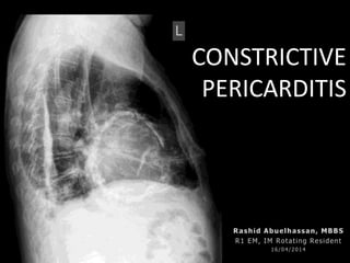
Constrictive Pericarditis
•
25 j'aime•7,392 vues

Constrictive Pericarditis, restrictive pericarditis

Recommandé
Contenu connexe
Tendances
Tendances (20)
Similaire à Constrictive Pericarditis
Similaire à Constrictive Pericarditis (20)
Plus de Rashid Abuelhassan
Plus de Rashid Abuelhassan (20)
Dernier
Dernier (20)
Constrictive Pericarditis
- 3. EFFUSION SEQUEL • Cardiac tamponade: acute or subacute(Variants include low pressure (occult) and regional tamponade. • Constrictive pericarditis: chronic, subacute, transient, and occult constriction. • Effusive-constrictive pericarditis – a mixed hemodynamic picture with features of both constriction and tamponade.
- 4. CAUSES • Idiopathic or viral 42 - 49 % • Post-cardiac surgery 11 to 37 % • Post-radiation therapy 9 to 31 %, (Hodgkin or breast ca.) • Connective tissue disorder 3 to 7 % • Postinfectious (tuberculous or purulent) 3 to 6 % • Miscellaneous causes 1 to 10 % – (malignancy, trauma, drug-induced, asbestosis, sarcoidosis, uremic pericarditis)
- 5. PREVELANCE • Idiopathic/viral – 0.76 cases per 1000 person/yr • Connective tissue/pericardial injury syndrome – 4.40 cases per 1000 person/yr • Neoplastic– 6.33 cases per 1000 person/yr • Tuberculous– 31.65 cases per 1000 person/yr • Purulent– 52.75 cases per 1000 person/yr
- 6. PRESENTATION • Symptoms related to fluid overload, ranging from peripheral edema to anasarca • Symptoms related to diminished cardiac output in response to exertion, such as fatigability and dyspnea on exertion
- 7. Physical examination • Elevated JVP has been reported in 93% • Pulsus paradoxus 20% • Kussmaul's sign 13%-21% • A pericardial knock 47% • Profound cachexia, peripheral edema, ascites, pulsatile hepatomegaly and pleural effusion are common findings .
- 8. EVALUATION • Electrocardiography (no pathognomonic findings.) • Chest radiograph • Echocardiography – M Mode – 2 dimensional ECHO – Doppler • CT scan (>4 mm) in 72 % • MRI * • Pro BNP (less than in other types of cardiomyopathy)
- 9. CONSTRICTIVE VS. RESTRICTIVE constrictive pericarditis restrictive cardiomyopathy Doppler ECHO Respiratory variation in ventricular inflow velocities increase in respiratory variation of the ventricular inflow velocities in patients with compared to a in patients normal pattern Hepatic venous flow reversal usually reverses during expiration in constrictive pericarditis reverses during inspiration in restrictive cardiomyopathy. Ventricular end-diastolic pressures (RVEDP and LVEDP) are equal or nearly equal LVEDP is usually higher than RVEDP in restrictive cardiomyopathy.
- 10. OCCULT CONSTRICTIVE PERICARDITIS Chest Pain, Dyspnea Fatigue reported in 1977 described 19 patients with a syndrome called occult constrictive pericardial disease
- 11. • history of acute pericarditis in 12 patients (which was recurrent in five), pericardial calcification in 2 patients, and nonspecific repolarization changes in 16 patients. A plausible cause for pericardial disease was present in 10 • Saline infusion caused an elevation and equalization of ventricular filling pressures, and development of pressure waveforms in diastole characteristic of constrictive pericarditisin the patients presumed to have occult constriction. • Ventricular filling pressures and diastolic waveform were unaltered in the subjects free from heart disease. • The patients with myocardial disease developed elevated ventricular filling pressures, but unequally on the two sides. • Eleven of the symptomatic patients underwent pericardiectomy with dramatic improvement. All 11 cases had mild gross or histologic evidence of pericardial disease. The fluid challenge was repeated postoperatively in five of the patients, with normal hemodynamic findings. OCCULT CONSTRICTIVE PERICARDITIS
- 12. Treatment • newly diagnosed hemodynamically stable + No chronic constriction may be given a trial of conservative management for 2-3(Grade 2C). • No chronic constriction (ie, no evidence of cachexia, weight loss, reduced cardiac output at rest, or hypoalbuminemia due to protein losing enteropathy and/or impaired hepatic function due to chronic congestion or cardiogenic cirrhosis • Treatment had included NSAIDs , steroids, antibiotics, chemotherapy, and ACEi plus diuretics.
- 13. • Transient > review of 212 patients with echocardiographic findings of constrictive pericarditis, 17 percent had follow-up studies showing resolution at an interval ranging from two months to two years • Chronic symptyomatic Pericardiectomy Treatment
Notes de l'éditeur
- Such patients may be mistakenly thought to have only cardiac tamponade; however, elevation of the right atrial and pulmonary wedge pressures after drainage of the pericardial fluid points to the underlying constrictive process.
- Knock (accentuated heart sound occurring slightly earlier than an S3)The " a " wave corresponds to right Atrial contraction and ends synchronously with the carotid artery pulse. The peak of the 'a' wave demarcates the end of atrial systole.The " c " wave corresponds to right ventricular Contraction causing the triCuspid valve to bulge towards the right atrium.The " x " descent follows the 'a' wave and corresponds to atrialrelaXation and rapid atrial filling due to low pressure.The " x' " (x prime) descent follows the 'c' wave and occurs as a result of the right ventricle pulling the tricuspid valve downward during ventricular systole. (As stroke volume is ejected, the ventricle takes up less space in pericardium, allowing relaXed atrium to enlarge). The x' (x prime) descent can be used as a measure of right ventricle contractility.The " v " wave corresponds to Venous filling when the tricuspid valve is closed and venous pressure increases from venous return - this occurs during and following the carotid pulse.The " y " descent corresponds to the rapid emptYing of the atrium into the ventricle following the opening of the tricuspid valve.
- Common findings on imaging studies include pericardial thickening with or without calcification, dilatation of the inferior vena cava and hepatic veins (plethora) with absent or diminished inspiratory collapse, abnormal passive filling of the ventricles during early diastole, and pronounced respiratory variation in ventricular filling.
- To test this hypothesis, they measured hemodynamics invasively before and after infusing a liter or warm saline over a period of six to eight minutes to determine if occult constriction would then become overt. Six patients known not to have heart disease and 12 patients with myocardial disease served as controls.
- Recommendation grades1. Strong recommendation: Benefits clearly outweigh the risks and burdens (or vice versa) for most, if not all, patients2. Weak recommendation: Benefits and risks closely balanced and/or uncertainEvidence gradesA. High-quality evidence: Consistent evidence from randomized trials, or overwhelming evidence of some other formB. Moderate-quality evidence: Evidence from randomized trials with important limitations, or very strong evidence of some other formC. Low-quality evidence: Evidence from observational studies, unsystematic clinical observations, or from randomized trials with serious flaws