Electrocardiogram (ECG) / ECG interpretation
•
8 j'aime•860 vues

This document provides information about electrocardiography (ECG) including its history, components, interpretation, and procedure. It discusses that ECG was invented in 1901 by Enthovan to record electrical impulses of the heart. It describes the normal conduction system, waves (P, Q, R, S, T), segments, intervals of ECG and placement of 12 leads. The document outlines the procedure for performing an ECG including preparing the patient, connecting the leads, and interpreting the results. It emphasizes the importance of properly performing and interpreting ECG to assess cardiac function and diagnose cardiac conditions.

Recommandé
Contenu connexe
Tendances
Tendances (20)
En vedette
En vedette (20)
Similaire à Electrocardiogram (ECG) / ECG interpretation
Similaire à Electrocardiogram (ECG) / ECG interpretation (20)
Dernier
Dernier (20)
Electrocardiogram (ECG) / ECG interpretation
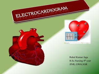
- 1. Rahul Kumar Jaga B.Sc.Nursing 4th year JINR, GWALIOR
- 2. INTRODUCTION Electrocardiogram (ECG) is invented by Enthovan in 1901. He is also known as father of ECG.
- 6. NORMAL ECG
- 7. DEFINITION It is a tracing made of the various phase of the heart’s action by means of an ECG. To understand the ECG, recall the events that takes place in the cardiac cycle.
- 8. IMPULSE CONDUCTION & THE ECG Sinoatrial node AV node Bundle of His Bundle Branches Purkinje fibers
- 9. purpose To assess the cardiac functions, e.g. rate , rhythm, and conduction. To diagnose the cardiac rhythm disorder, e.g. Arrhythmia. To diagnose cardiac disease, e.g. myocardial infarction. Electrolyte disturbances, especially in calcium and potassium levels. To evaluate effects of treatment, e.g. administration of cardiac drug.
- 10. Basic principles of ECG An ECG is a permanent record of the electrical impulses generated in the heart by the depolarization and repolarization (contraction and relaxation) of the myocardium. These impulses are transmitted to the surface of body where they are detected and picked up by the electrodes and measured by a galvanometer (electrocardiography).
- 11. Components of ECG ECG consist of :- Waves (P, Q, R, S, T, U) complex (QRS) segments (P-Q, S-T, T-P) Intervals(P-R, Q-T, R-R)
- 12. WAVES OR COMPLEX It is deflexion that can be positive, negative, or both.
- 13. THE “PQRST” P wave - Atrial depolarization • T wave - Ventricular repolarization • QRS - Ventricular depolarization
- 14. SEGMENTS A segments is the period of time between a wave or complex and another wave or complex; normally, it is a straight line (isotonic line).
- 15. INTERVALS An intervals is the period of time between two points on the ECG that includes a wave, a complex or both.
- 17. GRAPHICAL REPRESENTATION OF ECG 1.P WAVE :- P wave is the first positive deflection (appearing above the base line) seen on the ECG. It is smoothly rounded. It appears just before the QRS complex. Presence of P wave indicates that the stimulus began in the S.A.node (pacemaker) and subsequently spread through both atria causing atrial contraction. It represents the name taken by the atria to empty the blood in to the ventricles through the open A.V.valves. Normally, P wave has a duration of less than 0.11 second and a height of less than 2.5mV.(3 small squares width and 3 small squares height)
- 18. NORMAL ECG
- 19. Cont… ABNORMALITIES:- P wave may be hidden, absent, invented, or there may be more than one P wave. An abnormal P wave indicates that the excitation impulses is in a pacemaker other than S.A.node (ectopic focus) or atrial depolarization is normal. When there are more P waves then QRS complexes, it indicates atrio-ventricular conduction block. P wave is considered enlarge, if it is more than 3 small squares tall and / or 3 small squares wide, indicatives or atrial enlargement.
- 21. Cont… 2.P-R INTERVALS:- P-R intervals is measured from the beginning of P wave to the beginning of QRS complex. It represent the time duration which the wave of contraction passes through the entire conduction system from the A,V,node to the purkinje fibres in the myocardium. During the P-R interval, the filling of the ventricles is completed. Normally it measured less than 0.20 second (5 small squares in width).
- 22. NORMAL INTERVALS
- 23. THE PR INTERVAL Atrial depolarization + delay in AV junction (AV node/Bundle of His) (delay allows time for the atria to contract before the ventricles contract)
- 24. NORMAL PR INTERVAL 0.12 to 0.20 s (3 - 5 small squares). Short PR – Wolff-Parkinson-White syndrome. Long PR – 1st Degree AV block
- 25. Cont… ABNORMALITIES:- P-R intervals more than 0.20 second and less than 0.12 second are considered abnormal. If prolonged beyond 0.20 second, it indicates first degree atrio-ventricular block; P-R interval less than 0.12 second indicates a pacemaker in the atria than S.A.node or in the A.V.node.
- 27. Cont… 3.QRS COMPLEX :- The QRS complex consist of : A. Q wave – initial downward deflection. B. R wave – a large upward deflection. C. S wave – a second downward deflection. It represent the depolarization of ventricles. It represent the time required for the impulses to spread through the ventricles to complete the ventricular contraction. Normal QRS complex is narrow with a sharply pointed wave. It measures 0.08 to 0.11 second or less than 3 small squares in width.
- 28. Cont… ABNORMALITIES:- QRS complex more than 0.11 second in width and often with a bizarre appearance unusual indicate that depolarization is proceeding in abnormal sequence and direction. Widened QRS complex indicates of bundle branch block.
- 30. Cont… 4.S-T SEGMENTS:- S-T segments began at the end of the S wave of terminates at the beginning of the T wave. It correlates with the period between the ventricles depolarization and repolarization (reflactory period); the cardiac muscle cannot respond to stimuli during this period. Normally S-T segments remains on the isoelectric line.
- 31. Cont… ABNORMALITIES:- If the S-T segments is more than 1mm above or below the base line, it indicates possible myocardial ischaemia, or infarction. This may be associate with reciprocal depression or elevation in leads uninjured open.
- 33. Cont… 5.T WAVE:- T wave represent the electrical recovery of ventricles (repolarization). This is the most vulnerable period of the cardiac cycle. Normally it appears upright and measures less than 0.20 second in width and not more than 5mm in height.
- 34. Cont… ABNORMALITIES:- Flat T wave indicates myocardial ischaemia; inverted T wave indicates myocardial infarction, usually tall T wave indicate elevated serum potassium.
- 36. Cont… 6.U WAVE:- U wave is a small upright of low voltage, sometimes seen following T wave. Its significant is not known.
- 38. 12 LEADS OF ECG ECG consist of records from 12 leads. 3 Standard bipolar Leads (I, II, III) 3 unipolar Limb Leads(aVR, aVL, aVF) 6 Precordial/unipolar chest Leads (V1-V6)
- 39. LEAD PLACEMENT 3 STANDARD BIPOLAR LEADS LEAD I:- RIGHT ARM – LEFT ARM (recorded the difference of electrical potential between the right arm and left arm.) LEAD II:- RIGHT ARM – LEFT LEG (recorded the difference of electrical potential between the right arm and left leg.) LEAD III:- LEFT ARM TO LEFT LEG (recorded the difference of electrical potential between the left arm and left leg.)
- 41. 3 UNIPOLAR LIMB LEAD aVR (AUGMENTED VOLTAGE RIGHT):- Recorded the difference of electrical potential between the right limb and the central terminal. The latter is also called the indifferent electrode and is constructed within the ECG machine by connecting all three extremities. It has practically zero potential. aVL (AUGMENTED VOLTAGE LEFT):- Recorded the difference of electrical potential between left arm and central terminal. aVF ( AUGMENTED VOLTAGE FOOT):- Recorded the difference of electrical potential between the left leg and central terminal. NOTE :- one electrode is placed on the right leg and it serve as the grounded electrode.
- 43. 6 Precordial/unipolar chest Lead V1: Lead V1 is to be placed 4th RICS of the sternum. V2 : Lead V2 is to placed directly across from V1 in the 4th LICS, on the left side of the sternum. V4 : Lead V4 is placed in the 5th LICS, midclavicular. V3 : Lead V3 is placed directly horizontally & equidistant in between leads V2 and V4. V6 : Lead V6 is placed in the 5th LICS, midaxillary horizontally level with V4 . V5 : Lead V5 is placed horizontally & equidistant in between leads V4 and V6 (anterior axillary line )
- 46. Standard Chest Lead Electrode Placement The Right-Sided 12-Lead ECG The 15-Lead ECG
- 47. LEAD “VIEWS”
- 48. PROCEDURE 1.PREPRATION OF PATIENT:- There is no specific preparation for the investigation. Explain the procedure to the patient and relatives that the seemingly complex apparatus will do no harm but gill give information on the action of heart. There should not be any ornaments during ECG on the body or the leads should not come in contact with the ornaments. Apply jelly to the skin where electrode is to be attached to have a good contact between the skin and the electrode. Give flat and relaxed position to the patient because any movements or twitching recorded by the machine may alter the tracings. Clean the jelly off the electrodes site before leaving the patient. There are specific position for the placement of the chest. The improper placement of the chest leads can discomfort the tracing and alter the diagnosis.
- 49. CONT… 2.PREPROCEDUAL CARE:- Hand wash Check and arrange the ECG machine, cables and electrodes and needed articles ready in advance. Explain the procedure clearly to the patient and family. Check the doctor’s order for the ECG. Identify the patient name, age, Id No. and diagnosis. Inform the patient not to move during procedure time.
- 51. Cont…
- 52. Cont…
- 53. Cont…
- 54. CONT… 3.PROCEDURE:- Position the patient relaxed and flat. Inform the bystanders to keep away to prevent them touching the patient during procedure. Enter the identification data of the patient. Expose the needed area for connecting electrode. Stay with the patient till it get over.
- 55. CONT… 4.AFTER CARE:- Clean the patient electrodes site with gauze or tissue paper. Ambulate and transfer the patient send the ECG record to the doctor for interpretation. Replace the ECG machine and articles to proper place. Record and report in nurse’s sheet.
- 56. ANY QUESTIONS
- 57. Thank you for your continuous efforts to participate, be part of the team and be ready to do an ECG when needed. Perform an ECG, Make a difference, Save a life.