
Recommandé
Contenu connexe
Similaire à seminar 2.pptx
Similaire à seminar 2.pptx (20)
Plus de Roop
Plus de Roop (12)
Dernier
Dernier (20)
seminar 2.pptx
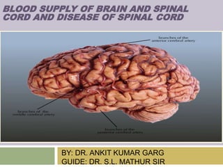
- 1. BY: DR. ANKIT KUMAR GARG GUIDE: DR. S.L. MATHUR SIR BLOOD SUPPLY OF BRAIN AND SPINAL CORD AND DISEASE OF SPINAL CORD
- 2. CONTENT BLOOD SUPPLY OF BRAIN CIRCLE OF WILLIS CAROTID SYSTEM ACA & MCA SUPPLY & STROKE VERTEBRAL SYSTEM BRAIN STEM STROKES BLOOD SUPPLY OF SPINAL CORD DISEASE OF SPINAL CORD
- 3. INTRODUCTION The continuous blood supply to brain is utmost important because of its high metabolic demands of oxygen and glucose Highly sensitive to hypoxia and hypoglycemia Consciousness is lost within 10 seconds of cessation of blood flow, irreversible damage starts to occur at about 4 minutes and is completed within 10 mins Although brain constitute 2%(1/50th) of total body weight , it receives 20%(1/5th) of total cardiac output. Adequate Normal cerebral blood flow 50-55ml/100gm/min. Electrical failure occurs when blood flow <18ml/100gm/min cell damage <10ml/100gm/min.
- 4. Blood supply of brain Carotid system vertebral system (internal carotid artery) (vertebrobasilar system) 80% 20%
- 5. Internal Carotid Artery : branch of Common Carotid artery Right Common Carotid arises from brachio-cephalic trunk(right subclavian & right common carotid) Left arises directly from arch of aorta Vertebral Artery : branch of postero-superior portion of Subclavian artery
- 9. CIRCLE OF WILLIS SITE: at the base of the brain FUNCTION: important anastomoses between two internal carotid arteries in front and vertebro- basilar system behind. The importance of this wide anatomoses is to maintain the blood circulation in brain even if any one or 2 component is compromised. ARTERIES forming it: Right and left internal carotid arteries Rt. and lt. ACA. Rt. and lt. PCA. Posterior communicating arteries .
- 11. BRANCHES OF ICA MEMONICS TO REMEMBER- OPAAM O- Opthalmic artery(1st branch) p- posterior communicating artery A-Anterior choriodal artery (post. Choriodal is a branch of PCA) A- Anterior cerebral artery M- Middle cerebral artery
- 12. BRANCHES OF ACA Medial striate artery(recurrent artery of Heubner) (supplies the lower part of the anterior limb and genu of the internal capsule) Orbitofrontal branches Frontopolar artery Callosomarginal artery Pericallosal artery
- 14. CORTICAL STROKE: Anterior cerebral artery stroke • 3 important structures supplied by anterior cerebral artery (ACA) are: a) Prefrontal cortex(executive functions)- responsible for personality, complex behaviour and decision making. b) Supplementary motor area- changing between motor behaviours, micturating inhibitory area. c) Paracentral lobule- have sensory and motor fibres of contralateral lower limb.
- 15. ACA BLOCKAGE Capsular branch occlusion(Heubner artery): Facio-brachial monoplegia Frontal branches occlusion 1.Severe behavior disturbance (apathy or abulia,akinetic) 2. Perseveration 3.Grasp reflexes, and diffuse rigidity –gegenhelton
- 17. MIDDLE CEREBRAL ARTERY (MCA) Passes through the sylvian fissure to appear superficially on lateral part of the brain giving various branches. In the sylvian fissure, it gives off lenticulostrial branches which supplies much of the basal ganglia and upper part of internal capsule. When superficial, its gives off a superior superficial branch(which runs superficially on lateral surface) and an inferior superficial branch which runs deep in the sylvian fissure. Superior branch supplies motor and the sensory strips/ areas. In the dominant hemisphere(commonly left hemisphere), it supplies the Brocas and the
- 19. 1. Superior division(frontoparietal area) A) Prefrontal B) Precentral (prerolandic) C) Central (rolandic) 2. Inferior division(temporoparietal area) A) Middle temporal B) Posterior temporal C) Angular D) Parietal
- 21. MCA STEM BLOCKAGE Contralateral hemiplegia, hemianesthesia Homonymous hemianopia with macula sparing Gaze preference towards side of lesion (in cerebral hemisphere) Dysarthria (due to facial weakness) Global aphasia---dominant hemisphere involvement Constructional apraxia, neglect, anosogonosia---non dominant hemisphere involvement
- 22. MCA: STROKE ARTERY AFFECTED AREA INVOLVED SYMPTOM LENTICULOSTRIAL ARTERY INTERNAL CAPSULE CONTRALATERAL MOTOR WEAKNESS +/- SESNSORY INVOLVEMENT+/- HOMONYMOUS HEMIANOPIA SUPERFICIAL ARTERY LATERAL SURFACE OF SAME HEMISPHERE – precentral /postcentral area WEAKNESS (MOTOR+/- SENSORY)OF CONTRALTERAL FACE AND UPPER LIMB *sparing lower limb as it is supplied by ACA a) if DOMINANT LOBE (Broca’s & Aphasia (MOTOR/SENSORY/GLO BAL)
- 23. POSTERIOR CEREBRAL ARTERY(PCA) • Early in its course it gives deep branches to thalamus and a part of midbrain. • Superficial branches to – 1) Occipital lobe 2) Inferior aspect of temporal lobe and part of parietal lobe. 3)Splenium of corpus callosum (carries fibres between two occipital lobes). • Structural variation is quite common in PCA. • In occipital lobe, we have our primary and associated visual area, important in perception and interpretation of visual information supplied by PCA.
- 24. PCA course and its branches
- 25. P1 Syndrome Midbrain, sub-thalamic and thalamic sign due to occlusion of P1 segment PCA or its penetrating branches. Infarction usually occurs in the ipsilateral sub-thalamus and medial thalamus and in the ipsilateral cerebral peduncle and mid brain. Claude’s syndrome: 3rd nerve palsy with contralateral ataxia Weber syndrome: 3rd nerve palsy with contralateral hemiplegia Thalami Dejerine Roussy syndrome: it is thalamic stroke. Bcoz involvement of spinothalamic tract, contralateral hemisensory loss followed by agonizing,searing or burning pain in affected areas
- 26. P2 SYNDROME Occlusion of the distal PCA causes infarction of the medial temporal and occipital lobes. Contralateral homonymous hemianopia without macular sparing Memory disturbance due to hippocampal involvement. Alexia without agraphia due to involvement of splenium of corpus callosum.
- 27. INTERNAL CAPSULE : blood supply
- 28. STROKE – Internal Capsule Can present as : A) Pure motor stroke caused by infarct in internal capsule is the most common form of lacunar stroke syndrome. B) Mixed sensorimotor weakness is also seen when posterior limb of internal capsule is affected. C) Upper motor neuron signs seen with internal capsule stroke are hyperreflexia, spasticity, babinski positive, clonus.
- 29. LACUNAR STROKE SYNDROME: 5 classic syndrome 1) Pure motor stroke- most common syndrome, usually occurs with infarction of posterior limb of internal capsule or basilar surface of pons. Affect face arm or leg with equal proportion. 2) Ataxic hemiparesis - 2nd most common. Seen with lacunar infarct of internal capsule, corona radiata,or basilar surface of pons. It is characterised by combination of cerebellar and motor signs. Affects foot/ leg more than hand/arm. 3) Pure sensory deficit – seen with infarction of VPL nucleus of thalamus 4) Dysarthria-clumsy hand syndrome- seen with infarction of Genu or basilar surface of pons. 5) Mixed sensorimotor – seen with infarction thalamus
- 30. WATERSHED AREAS &ITS STROKE • Defined as an ischemic or blood flow blockage that is localised to the border zone between the territorial of 2 major or arteries in brain. • 10 % of all ischemic strokes area watershed strokes. • 2 primary region in brain are cortical/external and internal/subcortical. Cortical (external )watershed are localised between the watershed territories of ACA/MCA/PCA. Internal watershed are in the white matter along and slightly along the lateral ventricle between superficial and deep arterial system of MCA or between superficial system of MCA & ACA.
- 31. WATERSHED AREAS
- 33. Branches of 4th part of Vertebral Artery Anterior spinal artery Posterior spinal A Posterior inferior cerebellar artery (PICA) Medullary artery Meningeal artery
- 34. Branches of Basilar Artery Anterior inferior cerebellar A Superior cerebellar A PCA Pontine artery Labyrinthine artery
- 35. BLOOD SUPPLY OF MIDBRAIN Posterior cerebral artery (PCA) Superior cerebellar artery Posterior communicating artery Anterior choroidal artery
- 36. BLOOD SUPPLY OF PONS Basilar artery(paramedian area) Superior cerebellar artery Pontine branches(upper lat. Area) Anterior inferior cerebellar artery(lower lat. Area)
- 37. BLOOD SUPPLY OF MEDULLA Vertebral artery PICA Anterior spinal artery Posterior spinal artery
- 38. BLOOD SUPPLY OF CEREBELLUM Superior cerebellar A(sup. /upper part) Anterior inferior cerebellar artery (lateral part) Posterior inferior cerebellar Artery(lower part)
- 39. BRAIN STEM STROKE SYNDROMES
- 40. PONTINE SYNDRMES
- 41. LATERAL MEDULLARY (WALLENBERG) SYNDROME Occlusion of any of five vessel may be responsible- vertebral, PICA, superior, middle or inferior lateral medullary arteries. ON THE SIDE OF LESION: 1. Pain, numbness, impaired sensation over one half of face: descending tract and nucleus of 5th nerve. 2. Ataxia of limb, falling to the side of lesion: restiform body, cerebellar hemisphere, cerebellar fiber, spinocerebellar tracts.
- 42. 4. Nystagmus, nausea, vomiting, vertigo: vestibular nucleus(8th) 5. Horner’s syndrome: descending sympathetic tract. 6. Dysphagia, hoarseness, paralysis of palate and vocal cord: issuing fiber of 9th and 10th nerve. 7. Numbness of ipsilateral arm, trunk or leg: cuneate and gracile nuclei. ON SIDE OPPOSITE TO LESION: 1. Impaired pain and temperature over half the body: spinothalamic tract
- 43. MEDIAL MEDULLARY SYNDROME (dejerine anterior bulbar syndrome) • Occurring due to involvement of basilar artery or occlusion of vertebral artery. AFFECTED STRUCTURE SYMPTOMS 12TH NERVE NUCLEI Tongue deviation to same side MEDIAL LEMINISCUS Loss of joint position sense and vibration on opposite side PYRAMIDS Weakness of opposite side limbs
- 44. VENOUS DRAINAGE OF BRAIN • Venous drainage of brain is by two type of vessels – sinuses and veins. Sinuses are vessels between meningeal layers and periostium. They are not true blood vessels and site outside dura. • Veins are true blood vessels. Separated into two 2 system : 1) Superficial cerebral vein- around superficial surface of hemisphere under Dura in subarachanoid space draining the hemisphere. • 2) Deep vein – drain subcortical matter and white matter around lateral ventricle.
- 46. • Principle sinus is the Superior Saggital Sinus (SSS) in the falx cerebri. Superficial cerebral vein drain into SSS via venous lacunae. • Superior middle cerebral vein travel along lateral sulcus and then drain into cavernous sinus which then drain into sigmoid sinus via inferior petrusal sinus . SUP. SAGGITAL SINUS CONFLUENCE OF SINUSES RIGHT AND LEFT LATERAL SINUSES SIGMOID SINUS
- 47. DEEP VENOUS SYSTEM • Inferior saggital sinus along superior surface of corpus callosum running parallel to superior saggital sinus. • Internal cerebral vein run along the top of the third ventricle to form great cerebral vein ( great vein Galen ) . • Inferior sagital sinus STRAIGHT SINUS LATERAL SINUS SIGMOID SINUS INTERNAL JUGULAR VEIN Anterior cerebral vein (travel along ACA ) drains medial and inferior part of hemisphere which the drain into deep middle cerebral vein to form BASILAR vein which drains into great vein Galen via internal cerebral vein .
- 49. Spinal cord anatomy It is 46 cm in length. Extends from lower medulla to lower border of L1. whereas dura or arachnoid matter upto S2. piamatter attach to coccyx. Conus- it is lower most portion of spinal cord. (S3,4,5,cx) Epiconus-just above the conus. (L4,5,S1,2) Cauda euina-roots below L2.
- 51. Blood supply of spinal cord Anterior spinal artery- branch of vertebral artery. Single in number. Supplies anterior 2/3rd of spinal cord. Posterior spinal artery- branch of vertebral/PICA. 2 in numbers. Supllies posterior 1/3rd of spinal cord. Radicular artery/radiculomedullary artery. Artera radicularis magna(artery of adamkiewicz) arises from aorta unilateral,usually from left side. In the lower thoracic or the upper lumbar levels(T9 and L2) supplies the lumbar enlargement.
- 56. Brown sequard syndrome Most common in extramedullary lesion. BELOW LEVEL- C/L loss of pain and temp.(lat spinothalamic tract) I/L proprioceptive I/L UMN AT LEVEL- I/L loss of pain and temp. I/L proprioceptive LMN
- 57. Central cord syndrome Causes- hyperextension injury(men in barrel type) central mass syringomyelia Arm weakness out of proportion to leg weakness dissociated sensory loss(loss of pain and temp. sensations over the shoulders,lower neck, and upper trunk(cape distribution)
- 58. THANK YOU!! Have a nice day ...