Patrick updown
•Télécharger en tant que PPT, PDF•
0 j'aime•181 vues

SHAPE Society

Recommandé
Recommandé
Contenu connexe
Tendances
Tendances (16)
En vedette
En vedette (18)
Similaire à Patrick updown
Similaire à Patrick updown (20)
Plus de Society for Heart Attack Prevention and Eradication
Plus de Society for Heart Attack Prevention and Eradication (20)
Dernier
Dernier (20)
Patrick updown
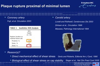
- 1. Plaque rupture proximal of minimal lumen Coronary artery Fujii, et al. Circulation 2003 Carotid artery Lovett and Rothwell, Cerebrovasc Dis 2003 Dirksen et al., Circulation 1998 Masawa, Pathology International 1994 Reason(s)? Direct mechanical effect of shear stress Gertz and Roberts, Editorial Am J Card, 1990 Biological effect of shear stress on cap stability Hemodynamics Laboratory Thoraxcenter Rotterdam Slager et al., Nat Clin Pract Card 2005
- 2. Biological observations related to flow direction Dirksen et al., Circ. 1998 Flow High density Low density High densityLow density Tricot et al., Circ. 2000 Macrophages Flow Smooth muscle cells 2.7% 18.8 % Endothelial cells in apoptosis Flow Hemodynamics Laboratory Thoraxcenter Rotterdam
- 3. Shear stress distribution over advanced plaque High shear stress Cap Blood flow Lipid core Low shear stress Hemodynamics Laboratory Thoraxcenter Rotterdam
- 4. Spatially restricted endothelial anti-inflammatory signaling Slager et al., Nat Clin Pract Card 2005 Hemodynamics Laboratory Thoraxcenter Rotterdam
- 6. Cap weakening due to High Shear Stress
- 7. Cap weakening due to High Shear Stress
Notes de l'éditeur
- Finding in literature, both for coronary and carotid arteries show that plaque rupture occurs predominately at the proximal (upstream region) part of the plaque.
- Dirksen et al. showed in longitudinal cross-sections of plaques a different distribution for both MF and SMCs upstream versus downstream. This indicates a higher matrix degradation by MFs and lower possible synthesis by SMCs at the upstream region compared with the downstream region. But why is not understood yet. Maybe there is a link with the Ecs. Tricot et al. showed in longitudinal cross-sections a higher cell apoptosis downstream compared with upstream. This could indicate that the endothelial cells at the upstream region have regained their functionality and response inflammatory to the high shear stress.
- Zooming in at the advanced plaque, we notice a high shear stress region at the upstream part and a low shear stress region at the downstream part of the plaque, both previously located at the inner curve and thus at a low shear stress region.