Seal of Good Local Governance (SGLG) 2024Final.pptx
Tissues
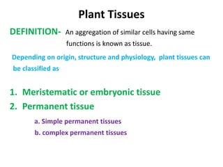
1. Plant Tissues
DEFINITION- An aggregation of similar cells having same
functions is known as tissue.
Depending on origin, structure and physiology, plant tissues can
be classified as
1. Meristematic or embryonic tissue
2. Permanent tissue
a. Simple permanent tissues
b. complex permanent tissues
4. EPIDERMIS
Outermost cellular layer
Covers the whole plant structure
Covers roots, stem, leaves, flowers and fruit.
Living cells.
Closely packed, without intercellular spaces or
chloroplasts.
Outer wall thickened, covered by a waxy,
waterproof cuticle which are made up of cutin.
Stomata found in the epidermis of leaves and stem.
root hairs (trichomes) found in the epidermis of
leaves and stems.
5. 3
A stoma is an opening (pore)
Which is bounded by two bean shaped cells
called guard cells .
Guard cells are covered by various numbers of
subsidiary cells.
Stomata 5 types
1. Paracytic
2. Diacytic
3. Anisocytic
4. Anomocytic
5. Actinocytic
structure
8. Functions
Protect the underlying cells.
Cuticle prevents the loss of moisture from
leaves and stems.
The transparent epidermal cells allow sunlight (for
photosynthesis) to pass through to the chloroplasts in
the mesophyll tissue.
The stomata of leaves and stems allow gaseous
exchange to take place which is necessary for
photosynthesis and respiration.
Help in transpiration, water vapor may be given off
through the stomata.
Trichomes excrete water and volatile oils etc.
9. Parenchyma
Composed of living cells
Thin-walled, made with cellulose.
Found in all organs of higher plants.
Iso-diametric
Intercellular space is present.
Contain vacuolated protoplast.
Main function is manufacturing and
storage of food.
10. 7
The secondary thickening may give rise to various
types of parenchyma, such as
1. simple parenchyma.
2. Parenchyma with intercellular space.
3. lignified parenchyma
4. Reticulate parenchyma
11. Collenchyma
Supporting tissue
found underneath of epidermis of stem. leaf
Thick, soft, plastic and non-lignified cell wall.
Walls have lots of pectin, which holds water.
Also contains lots of cellulose in the cell wall.
Generally longer than parenchyma cells.
Cells have protoplast, chloroplasts are normally
absent.
Thick walled cells give mechanical support to
plant.
12. 9
Type of collenchyma
1. Angular - Walls thickened where cells meet in the
corners.
2. Lamellar or plate – thickened on tangential walls. Cells
appear to line up in rows just below epidermis.
3. Lacunar, i.e. with a hole - thickening around the spaces
between the cells. Hole may be closed later by pectin
substances.
13. Sclerenchyma
• Mature sclerenchyma cells are dead.
• Have secondary cell walls thickened with cellulose and
usually impregnated with lignin.
• The cell cavity or lumen is very small or it may
disappear completely.
• It is found in stems, roots and leaves.
Functions:
• sclerenchyma is an important supporting tissue in
plants, provide mechanical support plant.
• sclereids are responsible for the hardness of seeds and
the shell of walnut.
• fibres probably play a role in the transport of water in
the plant,
14. 11
There are two types of sclerenchyma cells, namely
sclereids and fibres.
Sclereids:
The cells are irregular in shape.
The cell walls are thick, hard and lignified which makes
the lumen very small.
Simple pits (canals) are found in the thickened cell
walls and link adjacent cells.
Sclereids are commonly found in fruit and seeds.
They are found in groups or single.
15. 12
Fibres:
The cells are needle-shaped with pointed tips, thick walls and
rather small lumen.
Secondary cell walls, impregnated with, are formed.
Simple pits are also present.
Fibres are abundant in the vascular tissue of angiosperms,
i.e. flowering plants.
They are also called as internal hairs.
They gradually loose protoplast and become dead.
They are pericyclic fibres, xylem fibres, phloem fibres etc.
16. CORK
Cork is a tissue found in many vascular plants as part of the
periderm.
The cork is a lateral meristem
A mature cork cell is non-living.
Cell walls are composed of a waxy substance that is highly
impermeable to gases and water called suberin.
cork cell may be filled with air or may contain traces of lignin,
tannins, or fatty acids.
May vary in thickness from one to the next.
Packed closely together.
17. 14
Cells are generally arranged in radial rows.
Separation among the cells is achieved by structures
arising from the cork cambium called lenticels.
These pore-like structures allow gases to be exchanged
between the plant stem and the outside environment.
The cork cambium provides the internal cells of the
plants with extra insulation and protection.
18. Xylem
• Xylem is one of the transport tissue in vascular
plants.
• The basic function of xylem is to transport
water, but it also transports some nutrients.
• Give mechanical support to the plant.
• Composed of tracheids, tracheae or vessel,
xylem parenchyma and xylem fibres.
• Tracheids are elongated cells with large cavities
and tapering end.
• tracheids are dead cells.
19. 15
• Cell wall is hard, lignified with bordered pits.
Vessel/trachea:
• They are long tube like bodies. Trachae is formed
from a row of cylindrical cells.
• The cell wall is hard & lignified with annular to
pitted.
• It should be noted that trachied is a single cell, but
a trachea or vessel is a tube like body formed from
a row of cells.
20. 20
• The initially formed xylem vessels are smaller
cavities & annular with spiral thickening called as
Protoxylem vessel.
• But lately formed vessels are reticulate with
pitted thickening called metaxylem vessel.
23. PHLOEM
• Phloem is the living tissue .
• The phloem is the innermost layer of the bark.
• The phloem is concerned mainly with the
transport of soluble organic material made
during photosynthesis.
• Phloem composed of Sieve tube, Companion
cell, phloem parenchyma also phloem fibres.
26. • Seive tube is living, having lining cytoplasm with a large
central vacuole.
• The nucleus disintegrates with the maturity of the tube.
• Cell wall is thin & made up of cellulose.
• Vacuole is rich in nitrogenous matter.
• A carbohydrate called callose is deposited on the seive
plate in the form of a pad called callous pad.
• Companion cells are closely associated with sieve tubes
& are connected with them by pores.