The presentation includes an overview of hypersensitivity and type 1 hypersensitivity with certain pictures elaborating the mechanism. The presentation also talks about asthma very briefly as an example of type 1 hypersensitivity.
2. Immunity
• Immunity is the ability of the body to protect against all types of foreign
body like bacteria , Virus , Toxic substances etc. which enter the body .
• Immunity is provided by immune system which is a complex network of
lymphoid organs such as bone marrow, thymus, spleen etc.
3. Immune system
• The immune system is made up of a network of cells, tissues, and organs
that work together to protect the body. One of the important cells
involved are white blood cells, also called leukocytes, which come in two
basic types that combine to seek out and destroy disease-causing
organisms or substances.
• It can be classified as:
Innate Immunity (Natural)
- Early reactions
Acquired Immunity (Adaptive)
- Late Reactions - Self /Non Self recognition
- High specificity - Antigenic Specificity
4. The Immune System In Health &
Disease
The immune system enables us to
recognize self from non-self and
thereby confer protection against
disease.
It is made up of complex network
of
Lymphocytes
in tonsils, lymph nodes,
thymus, spleen and bone marrow,
Monocytes and macrophages
Plasma proteins
Cytokines (Chemical messengers)
The most common clinical
problems are:
Over activity of the immune
response leading to allergic and
autoimmune disease.
Under activity resulting in
immunodeficiency.
Inappropriate activity
of the immune system resulting in
autoimmune disorders.
5. Types of Cells involved in the Immune
response
Lymphoid Cells
T cells
B Cells
Natural Killer Cells
Mononuclear Phagocytes
Antigen-Presenting Cells
Polymorph nuclear Granulocytes and Platelets
Basophils and Mast Cells
Platelets
Antigens
Haptens
6. Self tolerance
• The antigens on own cells are known as self-antigens, while those that
do not originate in body or enter the body from outside are called non-
self antigens. Immune cells called lymphocytes recognize non-self
antigens and produce antibodies that bind specifically to each antigen.
• The ability to discriminate between self and nonself antigens is vital to
the functioning of the immune system as a specific defence against
invading microorganisms.
• Failure of the immune system to "tolerate" self tissues can result in
disorders of the immune system.
7. Disorders of the Immune System
The immune system is a remarkably effective structure that incorporates
specificity, inducibility and adaptation. Failures of host defence fall into
three broad categories:
Immunodeficiency
Autoimmunity
Hypersensitivity reactions
8. Hypersensitivity
• An immunological state in which the immune system “over-reacts” to
foreign antigen such that the immune response itself is more harmful
than the antigen.
• Hypersensitivity refers to undesirable reactions produced by the
normal immune system, including allergies and autoimmunity.
• A common cause of hypersensitivity disease is failure of self-tolerance
[property of the immune system that ensures individuals don not
respond to their own antigens (self antigens) ]
9. Hypersensitivity
ALLERGEN:
A non-parasitic antigen capable of stimulating hypersensitivity reactions.
Hypersensitivity reaction depends on:
1) chemical nature of allergen
2) route involved in sensitization i.e. inhalation, ingestion, injection
3) physiological state of individual / genetic potential
• Hypersensitivity can be :
IMMEDIATE
o The reactions occurs within the humoral branch of immunity mediated by Antibody or
Antibody or Antigen complexes .
o Symptoms manifest within Minutes or hours after a sensitized recipient encounters an
antigen .
DELAYED
• The reactions are caused by the formation of T Helper cells in the Cell mediated branch of
immunity .
• Symptoms are delayed until days after exposure of the sensitized individual.
10. Types of Hypersensitivity
The four types of hypersensitivity are:
TYPE I – IMMEDIATE, ATOPIC, ANAPHYLACTIC
: allergic reactions
: IgE mediated and very rapid (2-30 minutes)
TYPE II – ANTIBODY MEDIATED
: cytotoxic reactions
: cell damage due to complement activation via IgM or IgG
TYPE III – IMMUNE COMPLEX
:cell damage due to excess antibody/antigen complexes
TYPE IV – CELL MEDIATED / DELAYED
:cell damage involving T cells & macrophages
11. TYPE 1 HYPERSENSITIVITY
DEFINITION:
a hypersensitivity due to excessive production of the class of antibody known
as IgE. Reactions between allergens and IgE bound to mast cells and basophils
cause a greatly heightened inflammatory response.
SYMPTOMS:
redness, swelling, itching, mucus etc.
may vary from minor inconvenience to death.
RESPONSE TIME:
Usually take 10 to 30 mins to appear after exposure to antigen. Sometimes
delayed onset of reaction (10-12 hours).
BASIC ELEMENTS:
Mediator – histamine, leukotrienes and cytokines.
Primary cellular components- mast cell and basophils.
Amplifier- platelets, neutrophils and eosinophils.
12. TYPE 1 HYPERSENSITIVITY
• Allergic (anaphylactic) reactions involve the activation of mast cells or
basophils through the binding of antigen to IgE on the cell surface:
mast cells & basophils have IgE receptors that bind the constant region of
any IgE antibody
“cross-linking” of IgE molecules on the cell surface by binding to
antigen triggers the release of histamine, prostaglandins & leukotrienes that
causes the redness, swelling, itching, mucus, etc.
14. MECHANISM OF ACTION
Exposure of antigen to antigen presenting cell
Recognition by T-Helper cells
Activation of B- cells into plasma and memory cells
Secretion of antibody IgE
IgE binds to high affinity receptors on the surface of mast cells
Subsequent exposure of antigen
Antigen binds with IgE on the surface of mast cells
Release of primary inflammatory metabolites ( histamine, serotonin etc)
Activation of secondary metabolites
Inflammatory response
15.
16.
17. Many local type I hypersensitivity reactions have two well-defined phases:
•The immediate, or initial, response within 5 to 30 minutes is characterized
by:
vasodilatation,
vascular leakage, and
smooth muscle spasm or
glandular secretions.
Due to release of primary mediators.
The late-phase reaction sets in 2 to 24 hours later is characterized by
infiltration of tissues with eosinophils, neutrophils, basophils,
monocytes, and CD4+ T cells as well as
tissue destruction, typically in the form of mucosal epithelial cell
damage due to release of secondary mediators.
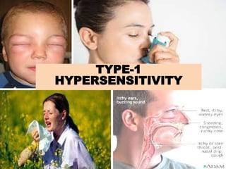
18. Most allergic reactions are local:
• itching, redness, hives in the skin, mucus, sneezing
• usually due to inhaled or ingested antigens
Systemic allergic reactions can be lethal:
severe loss of blood pressure, breathing difficulty (anaphylactic shock)
usually due to animal venoms or certain foods
epinephrine can “shut down” the allergic reaction
CLINICAL DISEASES WITH TYPE 1 HYPERSENSITIVITY: Anaphylaxis, Asthma, Allergic
Rhinitis (hay fever), and food allergy etc.
DIAGNOSIS:
Prick test – skin test
Transdermal test
ELISA- measures IgE level
TREATMENT:
Antihistamines (blocks histamine receptor)
Chromolyn sodium (inhibits mast cell degranulation)
Leukotriene receptor blockers
Use of IgG antibodies
Hyposensitization
19.
20. A chronic inflammatory airway disorder marked by airway hyper
responsiveness with recurrent episodes of wheezing, coughing, tightness of the
chest, and shortness of breath.
Affects approximately 300 million people around the world.
In children, males have a higher asthma risk; in adults, females have a higher
prevalence.
Asthma can affect the trachea, bronchi, and bronchioles.
Asthma lead to an increase in mucus-secreting cells with expansion of mucus-
secreting glands. The increased mucus secretion causes thick mucus plugs that
blocks the airway. Injury to the epithelium may cause epithelial peeling, which
may result in extreme airway impairment. Loss of the epithelium’s barrier
function allows allergens to penetrate, causing the airways to become
hyperresponsive—a major feature of asthma.
ASTHMA- TYPE 1 HYPERSENSITIVITY
21.
22. Asthma also causes loss of enzymes that normally break down inflammatory
mediators, with ensuing reflexive neural effects from sensory nerve exposure.
Without proper treatment and control, asthma may cause airway remodelling
leading to changes to cells and tissues in the lower respiratory tract; these
changes cause permanent fibrotic damage.