SlideShare une entreprise Scribd logo
1  sur  23
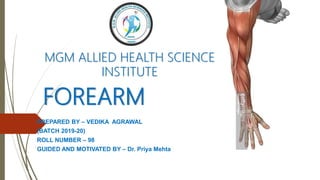
FOREARM
PREPARED BY – VEDIKA AGRAWAL
(BATCH 2019-20)
ROLL NUMBER – 98
GUIDED AND MOTIVATED BY – Dr. Priya Mehta
MGM ALLIED HEALTH SCIENCE
INSTITUTE
INTRODUCTION
 Forearm extends between elbow and wrist joints.
 Radius and ulna forms its skeleton.
 These two bones articulate at both their ends to form superior
and inferior radioulnar joints.
 In the upper part radius and ulna articulates with humerus to
form elbow joint.
 In lower part radius and ulna articulate with carpel bones to
form wrist joint.
 To understand forearm we study it in two sections:
1. Anterior compartment/ Front of forearm
2. Posterior compartment/ Back of forearm
FRONT OF FOREARM
SURFACE LANDMARKS
 Medial epicondyle of humerus is more prominent than the lateral.
 Ulnar nerve can be rolled on posterior surface of medial
epicondyle.
 Head of radius can be palpated on posterolateral aspect of
extended elbow, distal to lateral epicondyle.
 Styloid process of radius is 1cm below ulna and can be palpated
in the upper part of anatomical snuff box.
 Head of ulna forms surface elevation on posteromedial side when
hand is pronated.
 Pisiform bone can be felt at base of the hypothenar eminence
and can me palpated when wrist is fully extended.
 Tubercle of scaphoid lies beneath lateral part of distal transverse
crease of extended wrist.
MUSCLES
 It is divided into superficial and deep groups.
 superficial group has five muscles
 Deep group has three muscles.
 There are 2 arteries that and three nerves that accompany the
muscles in the front of forearm.
SUPERFICIAL GROUP
MUSCLES ORIGIN INSERSION NERVE
SUPPLY
ACTIONS
Pronator
Teres
Medial epicondyle of
humerus
Middle of lateral aspect
of shaft of radius
Median
nerve
Pronation of
forearm
Flexor Carpi
Radialis
Medial epicondyle of
humerus
Bases of second and
third metacarpal bones
Median
nerve
Flexes and abducts
hand at wrist joint
Palmaris
Longus
Medial epicondyle of
humerus
Flexor retinaculum and
palmar aponeurosis
Median
nerve
Flexes wrist joint
Flexor
digitorum
Superficialis
Medial epicondyle of
humerus; medial
border of coronoid
process of ulna
Anterior oblique line
of shaft of radius
Muscle divides into 4
tendons. Each tendon
divides into 2 slips which
are inserted on sides of
middle phalanx of 2nd to
5th digits
Median
nerve
Flexes middle
phalanx of fingers
and assists in
flexing proximal
phalanx and wrist
joint
Flexor Carpi
Ulnaris
Medial epicondyle of
humerus Medial
aspect of olecranon
process and
posterior border of
ulna
Pisiform bone; insertion
prolonged to hook of
the hamate and base of
fifth metacarpal bone
Median
nerve
Flexes and adducts
the hand at the
wrist ioint
DEEP GROUP
MUSCLES ORIGIN INSERSION NERVE
SUPPLY
ACTION
FIexor
digitorum
profundus
• Upper three-fourths of the
anterior and medial surface
of the shaft of ulna .
• Upper three-fourths of the
posterior border of ulna .
• Medial surface of the
olecranon and coronoid
processes of ulna.
• Adjoining part of the
anterior surface of the
interosseous membrane
• The muscle forms 4 tendons
for the medial 4 digits which
enter the palm by passing
deep to the flexor
retinaculum.
• Each tendon is inserted on
the palmar surface of the
base of the distal phalanx.
Median
half by
Ulnar
nerve
Lateral
half by
Anterior
interosse
ous nerve
• Flexor of distal
phalanges
• flexes the other
joints of the
digits, fingers, a
and the wrist.
• chief gripping
muscle
Flexor
pollicis
longus
• Upper three-fourths of the
anterior surface of the shaft
of radius,
• Adjoining part of the
interosseous membrane.
• The tendon enters the palm
by passing deep to the flexor
retinaculum
• It is inserted into the palmar
surface of the distal phalanx
of the thumb
Anterior
interosse
ous nerve
• Flexes the distal
phalanx of the
thumb
Pronator
quadratus
Oblique ridge on the lower one-
fourth of anterior surface of the
shaft of ulna, and the area
medial to it.
• Superficial fibres into the
lower one-fourth of the
anterior surface and the
anterior border of the radius.
• Deep fibres into the
triangular area above the
ulnar notch
Anterior
interosse
ous nerve
• Supeficial fibres
pronate the
forearm
• Deep fibres bind
the lower ends of
radius and ulna
RADIAL ARTERY
 Radial artery is the smaller terminal branch of the
brachial artery in the cubital fossa.
 As compared to the ulnar artery, it is quite superficial
throughout its whole course.
 Relations
1. Anteriorly – brachioradialis
2. Posteriorly – biceps brachii, flexor pollicis longus,
flexor digitorum superficialis and pronator
quadratus.
3. Medially – pronator teres, flexor carpi radialis
4. Laterally – brachioradialis, radial nerve
 Branches
1. Radial recurrent artery
2. Muscular branches
3. Palmar carpal branch
4. Dorsal carpal branch
5. Superficial palmar branch
ULNAR ARTERY
 Ulnar artery is the larger terminal branch of the brachial artery, and
begins in the cubital fossa.
 The artery runs obliquely downwards and medially in the upper one-
third of the forearm; but in the lower two-thirds of the forearm its
course is vertical.
 It enters the palm by passing superficial to the flexor retinaculum.
 Relations
1. Anteriorly; muscles arising from common flexor origin and
median nerve.
2. Posteriorly; flexor digitorum profundus.
3. Medially: ulnar nerve, flexor carpi ulnaris.
4. Laterally: flexor digitorum superficialis.
 Branches
1. Anterior and posterior ulnar recurrent arteries
2. Common interosseous artery
3. Muscular branches
4. Palmar and dorsal carpal branches
MEDIAN NERVE
Course
1. Median nerve lies medial to brachial artery and enters the
cubital fossa.
2. Then it enters the forearm to lie between flexor digitorum
superficialis and flexor digitorum profundus.
3. Then it reaches down the region of wrist where it lies deep and
lateral to palmaris longus tendon.
4. Lastly it passes deep to flexor retinaculum to enter the palm.
Branches
• Muscular branches
• Anterior interosseous branch
• Palmar cutaneous branch
• Articular branches
• Vascular branches
• Communicating branch
RADIAL NERVE
Course
• The radial nerve divides into its two terminal
branches in the cubital fossa just below the level of
the lateral epicondyle of the humerus.
Branches
• The deep terminal branch (posterior interosseous)
• The superficial terminal branch
ULNAR NERVE
Course
• Ulnar nerve is palpable as it lies behind medial
epicondyle of humerus and is nof a content of cubital
fossa
• It enters the forearm by passing between two heads
of flexor carpi ulnaris, to lie along the lateral border
of flexor carpi ulnaris.
• In the last phase, it courses superficial to the flexor
retinaculum, covered by its superficial slip or volar
carpal ligament to enter the region of palm.
Branches
• Muscular branches
• Palmar cutaneous branch
• Dorsal cutaneous branch
• Articular branches
CLINICAL ANATOMY
• The radial artery is used for feeling the (arterial) pulse at the wrist. The
pulsations can be felt well in this situation because of the presence of the flat
radius behind the artery.
• The ulnar nerve is commonly injured at the elbow, behind the medial
epicondyle or distal to elbow.
 Ulnar nerve injury at the elbow: Fexor carpi ulnaris and the medial half of
the flexor digitorum profundus are paralyzed.
 Due to this paralysis, the medial border of the forearm becomes flattened.
An attempt to produce flexion at the wrist result in abduction of the hand.
• The ulnar nerve controls fine movements of the fingers through its extensive
motor distribution to the short muscles of the hand.
• The median nerve controls coarse movements of the hand, as it supplies most
of the long muscles of the front of the forearm. It is, therefore, called the
laborer's nerve. It is also called "eye of the hand" as it is sensory to most of the
hand.
• When the median nerve is injured above the level of the elbow, as in supracondylar
fracture of the humerus:
 The flexor pollicis longus and lateral half of flexor digitorum profundus are paralyzed.
 The forearm is kept in a supine position due to paralysis of the pronators.
 The hand is adducted due to paralysis of the flexor carpi radialis.
 Flexion at the interphalangeal joints of the index and middle fingers is lost .
 Ape or monkey thumb deformity is present due to paralysis of the thenar muscles.
 Vasomotor and trophic changes: The skin on lateral three and a half-digit is warm, dry
and scaly. The nails get cracked easily .
BACK OF FOREARM
SURFACE LANDMARKS
• The olecranon process of the ulna is the most prominent bony point on back
of a flexed elbow.
• The head of the radius can be palpated in a depression on the posterolateral
aspect of an extended elbow just below the lateral epicondyle of the humerus.
• The posterior border of the ulna is subcutaneous. It can be felt in a
longitudinal groove on the back of the forearm when the elbow is flexed and
the hand is supinated.
• Tlte head of the ulna forms a surface elevation on the posteromedial. aspect
of the wrist in a pronated forearm.
• The dorsal tubercle of the radius (Lister's tubercle) can be palpated on the
dorsal surface of the lower end of the radius in line with the cleft between the
index and middle fingers.
• The heads of the metacarpals form the knuckles.
• The anatomical snuff box is a triangular depression on the lateral side of the
wrist. It is seen best when the thumb is extended.
EXTENSOR RETINACULUM
• The deep fascia on the back of the wrist is thickened to form the extensor
retinaculum which holds the extensor tendons in place
• ATTACHMENTS
 Laterally: Lower part of lhe anterior border of the radius.
 Medially: Styloid process of the ulna ,Triquetral ,Pisiform.
• COMPARTMENTS
 The retinaculum sends down septa which are attached to the longitudinal
ridges on the posterior surface of the lower end of radius.
 In this way, 6 Osseo fascial compartments are formed on the back of the wrist
MUSCLES
 It is divided into superficial and deep groups.
 superficial group has SEVEN muscles
 Deep group has FIVE muscles.
 There are 1 artery and one nerve that accompany
the muscles in the back of forearm.
SUPERFICIAL MUSCLES
ADDITIONAL POINTS
• The tendon to the index finger is joined on its medial side by the
tendon of the extensor indices, and the tendon to the little finger is
joined on its medial side by the two tendons of the extensor digiti
minimi.
• On the dorsum of the hand, adjacent tendons are variably connected
together by three intertendinous connections.
• The medial connection is strong; the lateral connection is weakest and
may be absent.
DORSAL DIGITAL EXPANSION
• It is a small triangular aponeurosis (related to each tendon of the extensor digitorum) covering the
dorsum of the proximal phalanx.
• The main tendon of the extensor digitorum occupies the central part of the extension, and is separated
from the Metacarpophalangeal joint by a bursa.
• The posterolateral corners of the extensor expansion are joined by tendons of the interossei and of
lumbrical muscles.
• The corners are attached to the deep transverse metacarpal ligament.
• Near the proximal interphalangeal joint the extensor tendon
divides into a central slip and two collateral slips.
• The central slip are inserted on the dorsum of the base of the
middle phalanx.
• Collateral slips join each other and are inserted on the dorsum of
the base of the distal phalanx.
• The retinacular ligaments (link ligaments) extend from the side of
the proximal phalanx, to the margins of the extensor expansion
to reach the base of the distal phalanx.
DEEP MUSCLES
Nerve Supply Action
Radial Nerve Supination of forearm when
elbow is extended
Radial nerve Abducts and extends thumb
Radial nerve Extends metacarpophalangeal joint
of thumb
Radial nerve Extends distal phalanx of thumb
Radial nerve Extends metacarpophalangeal joint
of index finger
PROSTERIOR INTROSSEOUS NERVE
• It is the chief nerve of the back of the forearrn. It is a branch of the radial nerve given
off in the cubital fossa.
• COURSE
 It begins in cubital fossa. Passes through supinator muscle to reach back of
forearm, where it descends downwards. It ends in a pseudoganglion in the 4th
compartment of extensor retinaculum.
• RELATIONS
 It leaves the cubital fossa and enters the back of the forearm by passing between
the two planes of fibers of the supinator. It emerges from the supinator on the
back of the forearm.
 At the lower border of the extensor pollicis brevis, it passes deep to the extensor
pollicis longus and runs up to the wrist where it enlarges into a pseudoganglion.
• BRANCHES
 Muscular Branches
 Articular Branches
 Sensory Branches
PROSTERIOR INTROSSEOUS ARTERY
 COURSE
 Posterior interosseous artery is the smaller terminal branch of the common
interosseous, given in the cubital fossa,
 It terminates by anastomosing with the anterior interosseous artery.
• RELATIONS
 It enters the back of the forearm by passing between the oblique cord and the
upper margin of the interosseous membrane.
 It appears on the back of the forearm in the interval between the supinator and
the abductor pollicis longus and thereafter accompanies the posterior
interosseous nerve.
• BRANCHES
 Interosseous recurrent branch which runs upwards and takes part in the
anastomosis on the back of the lateral epicondyle of the humerus
Any Doubts

Contenu connexe

Tendances

Anatomy of anterior compartment of forearm
Anatomy  of anterior compartment of forearmAnatomy  of anterior compartment of forearm
Anatomy of anterior compartment of forearmDr. Mohammad Mahmoud
 
Blood supply of upper limb (part2)
Blood supply of upper limb (part2)Blood supply of upper limb (part2)
Blood supply of upper limb (part2)Idris Siddiqui
 
Anatomy of posterior compartment of the arm
Anatomy of  posterior compartment of the armAnatomy of  posterior compartment of the arm
Anatomy of posterior compartment of the armDr. Mohammad Mahmoud
 
Anatomy of forearm
Anatomy of forearmAnatomy of forearm
Anatomy of forearmsongao
 
Anatomy of foot and arches of foot
Anatomy of foot and arches of footAnatomy of foot and arches of foot
Anatomy of foot and arches of footShiva Kandanulu
 
Pectoral region
Pectoral regionPectoral region
Pectoral regionHETA PATEL
 
Hand anatomy 2017 new microsoft power point presentation
Hand anatomy 2017 new microsoft power point presentationHand anatomy 2017 new microsoft power point presentation
Hand anatomy 2017 new microsoft power point presentationessameahady
 
Anterior compartment of thigh copy
Anterior compartment of thigh   copyAnterior compartment of thigh   copy
Anterior compartment of thigh copyAasmaMehak
 
Anatomy of flexor compartment of forearm
Anatomy of  flexor compartment of forearm Anatomy of  flexor compartment of forearm
Anatomy of flexor compartment of forearm Shaifaly madan rustagi
 
cubital fossa & elbow anastomosis.pptx
cubital fossa & elbow anastomosis.pptxcubital fossa & elbow anastomosis.pptx
cubital fossa & elbow anastomosis.pptxSrinivasagamRajaSank
 
Muscles of the forearm
Muscles of the forearmMuscles of the forearm
Muscles of the forearmMohaned Lehya
 

Tendances (20)

Anatomy of anterior compartment of forearm
Anatomy  of anterior compartment of forearmAnatomy  of anterior compartment of forearm
Anatomy of anterior compartment of forearm
 
The arm
The armThe arm
The arm
 
Slideshow: Lateral Leg
Slideshow: Lateral LegSlideshow: Lateral Leg
Slideshow: Lateral Leg
 
Blood supply of upper limb (part2)
Blood supply of upper limb (part2)Blood supply of upper limb (part2)
Blood supply of upper limb (part2)
 
Anatomy of posterior compartment of the arm
Anatomy of  posterior compartment of the armAnatomy of  posterior compartment of the arm
Anatomy of posterior compartment of the arm
 
Radial nerve-anatomy
Radial nerve-anatomyRadial nerve-anatomy
Radial nerve-anatomy
 
Anatomy of forearm
Anatomy of forearmAnatomy of forearm
Anatomy of forearm
 
Anatomy of foot and arches of foot
Anatomy of foot and arches of footAnatomy of foot and arches of foot
Anatomy of foot and arches of foot
 
Pectoral region
Pectoral regionPectoral region
Pectoral region
 
Hand anatomy 2017 new microsoft power point presentation
Hand anatomy 2017 new microsoft power point presentationHand anatomy 2017 new microsoft power point presentation
Hand anatomy 2017 new microsoft power point presentation
 
Anterior compartment of thigh copy
Anterior compartment of thigh   copyAnterior compartment of thigh   copy
Anterior compartment of thigh copy
 
Anatomy of flexor compartment of forearm
Anatomy of  flexor compartment of forearm Anatomy of  flexor compartment of forearm
Anatomy of flexor compartment of forearm
 
Ulnar nerve ppt
Ulnar nerve pptUlnar nerve ppt
Ulnar nerve ppt
 
Arm & Cubital Fossa.pptx
Arm & Cubital Fossa.pptxArm & Cubital Fossa.pptx
Arm & Cubital Fossa.pptx
 
cubital fossa & elbow anastomosis.pptx
cubital fossa & elbow anastomosis.pptxcubital fossa & elbow anastomosis.pptx
cubital fossa & elbow anastomosis.pptx
 
8. lateral and back of the leg
8. lateral and back of the leg8. lateral and back of the leg
8. lateral and back of the leg
 
Sole of foot
Sole of footSole of foot
Sole of foot
 
3.medial side of the thigh
3.medial side of the thigh3.medial side of the thigh
3.medial side of the thigh
 
1.front of the thigh
1.front of the thigh1.front of the thigh
1.front of the thigh
 
Muscles of the forearm
Muscles of the forearmMuscles of the forearm
Muscles of the forearm
 

Similaire à Forearm anatomy complete

disection of palm and sole.pptx
disection of palm and sole.pptxdisection of palm and sole.pptx
disection of palm and sole.pptxMeetVaghasiya20
 
vnd.ms-powerpoint&rendition=1-1.ppt
vnd.ms-powerpoint&rendition=1-1.pptvnd.ms-powerpoint&rendition=1-1.ppt
vnd.ms-powerpoint&rendition=1-1.pptDRJS74
 
5 anterior forearm.ppt
5  anterior forearm.ppt5  anterior forearm.ppt
5 anterior forearm.pptmehrdad asgari
 
UPPER LIMBS - WRIST AND HAND MAY 2022.pdf
UPPER LIMBS - WRIST AND HAND MAY 2022.pdfUPPER LIMBS - WRIST AND HAND MAY 2022.pdf
UPPER LIMBS - WRIST AND HAND MAY 2022.pdfManishaRao30
 
Front of forearm by vidya prashant
Front of forearm by vidya prashantFront of forearm by vidya prashant
Front of forearm by vidya prashantPrashant Bhavani
 
upper limb muscle anatomy.pdf
upper limb muscle anatomy.pdfupper limb muscle anatomy.pdf
upper limb muscle anatomy.pdfghhhh2
 
upper limb muscle anatomy.pdf
upper limb muscle anatomy.pdfupper limb muscle anatomy.pdf
upper limb muscle anatomy.pdfghhhh2
 
Structures of the Hand PPT
Structures of the Hand PPTStructures of the Hand PPT
Structures of the Hand PPTStacey Turner
 
Anatomy of radial,median &ulnar nerve
Anatomy of radial,median &ulnar nerveAnatomy of radial,median &ulnar nerve
Anatomy of radial,median &ulnar nerveAnkur Mittal
 
lecture 6aThe Fore am.pdf
lecture 6aThe Fore am.pdflecture 6aThe Fore am.pdf
lecture 6aThe Fore am.pdfNatungaRonald1
 
THE Intrinsic muscle OF THE HAND Prof.Ugo 02-10-23.pptx
THE Intrinsic muscle OF THE HAND Prof.Ugo 02-10-23.pptxTHE Intrinsic muscle OF THE HAND Prof.Ugo 02-10-23.pptx
THE Intrinsic muscle OF THE HAND Prof.Ugo 02-10-23.pptxKawukiIsah
 
Flexor compartment of forearm
Flexor compartment of forearmFlexor compartment of forearm
Flexor compartment of forearmedwardindla
 
Ul- flexor - Forearm.pptx
Ul- flexor  - Forearm.pptxUl- flexor  - Forearm.pptx
Ul- flexor - Forearm.pptxGUNASEKARANM20
 
Anatomy of upper limb rehan
Anatomy of upper limb rehanAnatomy of upper limb rehan
Anatomy of upper limb rehanRehan Asad
 

Similaire à Forearm anatomy complete (20)

disection of palm and sole.pptx
disection of palm and sole.pptxdisection of palm and sole.pptx
disection of palm and sole.pptx
 
vnd.ms-powerpoint&rendition=1-1.ppt
vnd.ms-powerpoint&rendition=1-1.pptvnd.ms-powerpoint&rendition=1-1.ppt
vnd.ms-powerpoint&rendition=1-1.ppt
 
5 anterior forearm.ppt
5  anterior forearm.ppt5  anterior forearm.ppt
5 anterior forearm.ppt
 
Lower limbs
Lower limbsLower limbs
Lower limbs
 
UPPER LIMBS - WRIST AND HAND MAY 2022.pdf
UPPER LIMBS - WRIST AND HAND MAY 2022.pdfUPPER LIMBS - WRIST AND HAND MAY 2022.pdf
UPPER LIMBS - WRIST AND HAND MAY 2022.pdf
 
Front of forearm by vidya prashant
Front of forearm by vidya prashantFront of forearm by vidya prashant
Front of forearm by vidya prashant
 
upper limb muscle anatomy.pdf
upper limb muscle anatomy.pdfupper limb muscle anatomy.pdf
upper limb muscle anatomy.pdf
 
upper limb muscle anatomy.pdf
upper limb muscle anatomy.pdfupper limb muscle anatomy.pdf
upper limb muscle anatomy.pdf
 
Ulnar Nerve.pptx
Ulnar Nerve.pptxUlnar Nerve.pptx
Ulnar Nerve.pptx
 
Structures of the Hand PPT
Structures of the Hand PPTStructures of the Hand PPT
Structures of the Hand PPT
 
Forearm muscles
Forearm musclesForearm muscles
Forearm muscles
 
Anatomy of radial,median &ulnar nerve
Anatomy of radial,median &ulnar nerveAnatomy of radial,median &ulnar nerve
Anatomy of radial,median &ulnar nerve
 
forearm.ppt
forearm.pptforearm.ppt
forearm.ppt
 
lecture 6aThe Fore am.pdf
lecture 6aThe Fore am.pdflecture 6aThe Fore am.pdf
lecture 6aThe Fore am.pdf
 
THE Intrinsic muscle OF THE HAND Prof.Ugo 02-10-23.pptx
THE Intrinsic muscle OF THE HAND Prof.Ugo 02-10-23.pptxTHE Intrinsic muscle OF THE HAND Prof.Ugo 02-10-23.pptx
THE Intrinsic muscle OF THE HAND Prof.Ugo 02-10-23.pptx
 
Wrist Joint & Hand
Wrist Joint & HandWrist Joint & Hand
Wrist Joint & Hand
 
Flexor compartment of forearm
Flexor compartment of forearmFlexor compartment of forearm
Flexor compartment of forearm
 
Ul- flexor - Forearm.pptx
Ul- flexor  - Forearm.pptxUl- flexor  - Forearm.pptx
Ul- flexor - Forearm.pptx
 
The wrist and hand
The wrist and handThe wrist and hand
The wrist and hand
 
Anatomy of upper limb rehan
Anatomy of upper limb rehanAnatomy of upper limb rehan
Anatomy of upper limb rehan
 

Dernier

1029 - Danh muc Sach Giao Khoa 10 . pdf
1029 -  Danh muc Sach Giao Khoa 10 . pdf1029 -  Danh muc Sach Giao Khoa 10 . pdf
1029 - Danh muc Sach Giao Khoa 10 . pdfQucHHunhnh
 
Seal of Good Local Governance (SGLG) 2024Final.pptx
Seal of Good Local Governance (SGLG) 2024Final.pptxSeal of Good Local Governance (SGLG) 2024Final.pptx
Seal of Good Local Governance (SGLG) 2024Final.pptxnegromaestrong
 
ComPTIA Overview | Comptia Security+ Book SY0-701
ComPTIA Overview | Comptia Security+ Book SY0-701ComPTIA Overview | Comptia Security+ Book SY0-701
ComPTIA Overview | Comptia Security+ Book SY0-701bronxfugly43
 
Web & Social Media Analytics Previous Year Question Paper.pdf
Web & Social Media Analytics Previous Year Question Paper.pdfWeb & Social Media Analytics Previous Year Question Paper.pdf
Web & Social Media Analytics Previous Year Question Paper.pdfJayanti Pande
 
Sociology 101 Demonstration of Learning Exhibit
Sociology 101 Demonstration of Learning ExhibitSociology 101 Demonstration of Learning Exhibit
Sociology 101 Demonstration of Learning Exhibitjbellavia9
 
Unit-IV; Professional Sales Representative (PSR).pptx
Unit-IV; Professional Sales Representative (PSR).pptxUnit-IV; Professional Sales Representative (PSR).pptx
Unit-IV; Professional Sales Representative (PSR).pptxVishalSingh1417
 
General Principles of Intellectual Property: Concepts of Intellectual Proper...
General Principles of Intellectual Property: Concepts of Intellectual  Proper...General Principles of Intellectual Property: Concepts of Intellectual  Proper...
General Principles of Intellectual Property: Concepts of Intellectual Proper...Poonam Aher Patil
 
Ecological Succession. ( ECOSYSTEM, B. Pharmacy, 1st Year, Sem-II, Environmen...
Ecological Succession. ( ECOSYSTEM, B. Pharmacy, 1st Year, Sem-II, Environmen...Ecological Succession. ( ECOSYSTEM, B. Pharmacy, 1st Year, Sem-II, Environmen...
Ecological Succession. ( ECOSYSTEM, B. Pharmacy, 1st Year, Sem-II, Environmen...Shubhangi Sonawane
 
microwave assisted reaction. General introduction
microwave assisted reaction. General introductionmicrowave assisted reaction. General introduction
microwave assisted reaction. General introductionMaksud Ahmed
 
Russian Escort Service in Delhi 11k Hotel Foreigner Russian Call Girls in Delhi
Russian Escort Service in Delhi 11k Hotel Foreigner Russian Call Girls in DelhiRussian Escort Service in Delhi 11k Hotel Foreigner Russian Call Girls in Delhi
Russian Escort Service in Delhi 11k Hotel Foreigner Russian Call Girls in Delhikauryashika82
 
TỔNG ÔN TẬP THI VÀO LỚP 10 MÔN TIẾNG ANH NĂM HỌC 2023 - 2024 CÓ ĐÁP ÁN (NGỮ Â...
TỔNG ÔN TẬP THI VÀO LỚP 10 MÔN TIẾNG ANH NĂM HỌC 2023 - 2024 CÓ ĐÁP ÁN (NGỮ Â...TỔNG ÔN TẬP THI VÀO LỚP 10 MÔN TIẾNG ANH NĂM HỌC 2023 - 2024 CÓ ĐÁP ÁN (NGỮ Â...
TỔNG ÔN TẬP THI VÀO LỚP 10 MÔN TIẾNG ANH NĂM HỌC 2023 - 2024 CÓ ĐÁP ÁN (NGỮ Â...Nguyen Thanh Tu Collection
 
Python Notes for mca i year students osmania university.docx
Python Notes for mca i year students osmania university.docxPython Notes for mca i year students osmania university.docx
Python Notes for mca i year students osmania university.docxRamakrishna Reddy Bijjam
 
Unit-V; Pricing (Pharma Marketing Management).pptx
Unit-V; Pricing (Pharma Marketing Management).pptxUnit-V; Pricing (Pharma Marketing Management).pptx
Unit-V; Pricing (Pharma Marketing Management).pptxVishalSingh1417
 
Mixin Classes in Odoo 17 How to Extend Models Using Mixin Classes
Mixin Classes in Odoo 17  How to Extend Models Using Mixin ClassesMixin Classes in Odoo 17  How to Extend Models Using Mixin Classes
Mixin Classes in Odoo 17 How to Extend Models Using Mixin ClassesCeline George
 
1029-Danh muc Sach Giao Khoa khoi 6.pdf
1029-Danh muc Sach Giao Khoa khoi  6.pdf1029-Danh muc Sach Giao Khoa khoi  6.pdf
1029-Danh muc Sach Giao Khoa khoi 6.pdfQucHHunhnh
 
Key note speaker Neum_Admir Softic_ENG.pdf
Key note speaker Neum_Admir Softic_ENG.pdfKey note speaker Neum_Admir Softic_ENG.pdf
Key note speaker Neum_Admir Softic_ENG.pdfAdmir Softic
 
Advanced Views - Calendar View in Odoo 17
Advanced Views - Calendar View in Odoo 17Advanced Views - Calendar View in Odoo 17
Advanced Views - Calendar View in Odoo 17Celine George
 
Introduction to Nonprofit Accounting: The Basics
Introduction to Nonprofit Accounting: The BasicsIntroduction to Nonprofit Accounting: The Basics
Introduction to Nonprofit Accounting: The BasicsTechSoup
 
Z Score,T Score, Percential Rank and Box Plot Graph
Z Score,T Score, Percential Rank and Box Plot GraphZ Score,T Score, Percential Rank and Box Plot Graph
Z Score,T Score, Percential Rank and Box Plot GraphThiyagu K
 

Dernier (20)

1029 - Danh muc Sach Giao Khoa 10 . pdf
1029 -  Danh muc Sach Giao Khoa 10 . pdf1029 -  Danh muc Sach Giao Khoa 10 . pdf
1029 - Danh muc Sach Giao Khoa 10 . pdf
 
Seal of Good Local Governance (SGLG) 2024Final.pptx
Seal of Good Local Governance (SGLG) 2024Final.pptxSeal of Good Local Governance (SGLG) 2024Final.pptx
Seal of Good Local Governance (SGLG) 2024Final.pptx
 
ComPTIA Overview | Comptia Security+ Book SY0-701
ComPTIA Overview | Comptia Security+ Book SY0-701ComPTIA Overview | Comptia Security+ Book SY0-701
ComPTIA Overview | Comptia Security+ Book SY0-701
 
Web & Social Media Analytics Previous Year Question Paper.pdf
Web & Social Media Analytics Previous Year Question Paper.pdfWeb & Social Media Analytics Previous Year Question Paper.pdf
Web & Social Media Analytics Previous Year Question Paper.pdf
 
Sociology 101 Demonstration of Learning Exhibit
Sociology 101 Demonstration of Learning ExhibitSociology 101 Demonstration of Learning Exhibit
Sociology 101 Demonstration of Learning Exhibit
 
Unit-IV; Professional Sales Representative (PSR).pptx
Unit-IV; Professional Sales Representative (PSR).pptxUnit-IV; Professional Sales Representative (PSR).pptx
Unit-IV; Professional Sales Representative (PSR).pptx
 
General Principles of Intellectual Property: Concepts of Intellectual Proper...
General Principles of Intellectual Property: Concepts of Intellectual  Proper...General Principles of Intellectual Property: Concepts of Intellectual  Proper...
General Principles of Intellectual Property: Concepts of Intellectual Proper...
 
Ecological Succession. ( ECOSYSTEM, B. Pharmacy, 1st Year, Sem-II, Environmen...
Ecological Succession. ( ECOSYSTEM, B. Pharmacy, 1st Year, Sem-II, Environmen...Ecological Succession. ( ECOSYSTEM, B. Pharmacy, 1st Year, Sem-II, Environmen...
Ecological Succession. ( ECOSYSTEM, B. Pharmacy, 1st Year, Sem-II, Environmen...
 
microwave assisted reaction. General introduction
microwave assisted reaction. General introductionmicrowave assisted reaction. General introduction
microwave assisted reaction. General introduction
 
Russian Escort Service in Delhi 11k Hotel Foreigner Russian Call Girls in Delhi
Russian Escort Service in Delhi 11k Hotel Foreigner Russian Call Girls in DelhiRussian Escort Service in Delhi 11k Hotel Foreigner Russian Call Girls in Delhi
Russian Escort Service in Delhi 11k Hotel Foreigner Russian Call Girls in Delhi
 
TỔNG ÔN TẬP THI VÀO LỚP 10 MÔN TIẾNG ANH NĂM HỌC 2023 - 2024 CÓ ĐÁP ÁN (NGỮ Â...
TỔNG ÔN TẬP THI VÀO LỚP 10 MÔN TIẾNG ANH NĂM HỌC 2023 - 2024 CÓ ĐÁP ÁN (NGỮ Â...TỔNG ÔN TẬP THI VÀO LỚP 10 MÔN TIẾNG ANH NĂM HỌC 2023 - 2024 CÓ ĐÁP ÁN (NGỮ Â...
TỔNG ÔN TẬP THI VÀO LỚP 10 MÔN TIẾNG ANH NĂM HỌC 2023 - 2024 CÓ ĐÁP ÁN (NGỮ Â...
 
Python Notes for mca i year students osmania university.docx
Python Notes for mca i year students osmania university.docxPython Notes for mca i year students osmania university.docx
Python Notes for mca i year students osmania university.docx
 
Unit-V; Pricing (Pharma Marketing Management).pptx
Unit-V; Pricing (Pharma Marketing Management).pptxUnit-V; Pricing (Pharma Marketing Management).pptx
Unit-V; Pricing (Pharma Marketing Management).pptx
 
Mixin Classes in Odoo 17 How to Extend Models Using Mixin Classes
Mixin Classes in Odoo 17  How to Extend Models Using Mixin ClassesMixin Classes in Odoo 17  How to Extend Models Using Mixin Classes
Mixin Classes in Odoo 17 How to Extend Models Using Mixin Classes
 
1029-Danh muc Sach Giao Khoa khoi 6.pdf
1029-Danh muc Sach Giao Khoa khoi  6.pdf1029-Danh muc Sach Giao Khoa khoi  6.pdf
1029-Danh muc Sach Giao Khoa khoi 6.pdf
 
Key note speaker Neum_Admir Softic_ENG.pdf
Key note speaker Neum_Admir Softic_ENG.pdfKey note speaker Neum_Admir Softic_ENG.pdf
Key note speaker Neum_Admir Softic_ENG.pdf
 
Advanced Views - Calendar View in Odoo 17
Advanced Views - Calendar View in Odoo 17Advanced Views - Calendar View in Odoo 17
Advanced Views - Calendar View in Odoo 17
 
Introduction to Nonprofit Accounting: The Basics
Introduction to Nonprofit Accounting: The BasicsIntroduction to Nonprofit Accounting: The Basics
Introduction to Nonprofit Accounting: The Basics
 
Asian American Pacific Islander Month DDSD 2024.pptx
Asian American Pacific Islander Month DDSD 2024.pptxAsian American Pacific Islander Month DDSD 2024.pptx
Asian American Pacific Islander Month DDSD 2024.pptx
 
Z Score,T Score, Percential Rank and Box Plot Graph
Z Score,T Score, Percential Rank and Box Plot GraphZ Score,T Score, Percential Rank and Box Plot Graph
Z Score,T Score, Percential Rank and Box Plot Graph
 

Forearm anatomy complete

  • 1. FOREARM PREPARED BY – VEDIKA AGRAWAL (BATCH 2019-20) ROLL NUMBER – 98 GUIDED AND MOTIVATED BY – Dr. Priya Mehta MGM ALLIED HEALTH SCIENCE INSTITUTE
  • 2. INTRODUCTION  Forearm extends between elbow and wrist joints.  Radius and ulna forms its skeleton.  These two bones articulate at both their ends to form superior and inferior radioulnar joints.  In the upper part radius and ulna articulates with humerus to form elbow joint.  In lower part radius and ulna articulate with carpel bones to form wrist joint.  To understand forearm we study it in two sections: 1. Anterior compartment/ Front of forearm 2. Posterior compartment/ Back of forearm
  • 3. FRONT OF FOREARM SURFACE LANDMARKS  Medial epicondyle of humerus is more prominent than the lateral.  Ulnar nerve can be rolled on posterior surface of medial epicondyle.  Head of radius can be palpated on posterolateral aspect of extended elbow, distal to lateral epicondyle.  Styloid process of radius is 1cm below ulna and can be palpated in the upper part of anatomical snuff box.  Head of ulna forms surface elevation on posteromedial side when hand is pronated.  Pisiform bone can be felt at base of the hypothenar eminence and can me palpated when wrist is fully extended.  Tubercle of scaphoid lies beneath lateral part of distal transverse crease of extended wrist.
  • 4. MUSCLES  It is divided into superficial and deep groups.  superficial group has five muscles  Deep group has three muscles.  There are 2 arteries that and three nerves that accompany the muscles in the front of forearm.
  • 5. SUPERFICIAL GROUP MUSCLES ORIGIN INSERSION NERVE SUPPLY ACTIONS Pronator Teres Medial epicondyle of humerus Middle of lateral aspect of shaft of radius Median nerve Pronation of forearm Flexor Carpi Radialis Medial epicondyle of humerus Bases of second and third metacarpal bones Median nerve Flexes and abducts hand at wrist joint Palmaris Longus Medial epicondyle of humerus Flexor retinaculum and palmar aponeurosis Median nerve Flexes wrist joint Flexor digitorum Superficialis Medial epicondyle of humerus; medial border of coronoid process of ulna Anterior oblique line of shaft of radius Muscle divides into 4 tendons. Each tendon divides into 2 slips which are inserted on sides of middle phalanx of 2nd to 5th digits Median nerve Flexes middle phalanx of fingers and assists in flexing proximal phalanx and wrist joint Flexor Carpi Ulnaris Medial epicondyle of humerus Medial aspect of olecranon process and posterior border of ulna Pisiform bone; insertion prolonged to hook of the hamate and base of fifth metacarpal bone Median nerve Flexes and adducts the hand at the wrist ioint
  • 6. DEEP GROUP MUSCLES ORIGIN INSERSION NERVE SUPPLY ACTION FIexor digitorum profundus • Upper three-fourths of the anterior and medial surface of the shaft of ulna . • Upper three-fourths of the posterior border of ulna . • Medial surface of the olecranon and coronoid processes of ulna. • Adjoining part of the anterior surface of the interosseous membrane • The muscle forms 4 tendons for the medial 4 digits which enter the palm by passing deep to the flexor retinaculum. • Each tendon is inserted on the palmar surface of the base of the distal phalanx. Median half by Ulnar nerve Lateral half by Anterior interosse ous nerve • Flexor of distal phalanges • flexes the other joints of the digits, fingers, a and the wrist. • chief gripping muscle Flexor pollicis longus • Upper three-fourths of the anterior surface of the shaft of radius, • Adjoining part of the interosseous membrane. • The tendon enters the palm by passing deep to the flexor retinaculum • It is inserted into the palmar surface of the distal phalanx of the thumb Anterior interosse ous nerve • Flexes the distal phalanx of the thumb Pronator quadratus Oblique ridge on the lower one- fourth of anterior surface of the shaft of ulna, and the area medial to it. • Superficial fibres into the lower one-fourth of the anterior surface and the anterior border of the radius. • Deep fibres into the triangular area above the ulnar notch Anterior interosse ous nerve • Supeficial fibres pronate the forearm • Deep fibres bind the lower ends of radius and ulna
  • 7. RADIAL ARTERY  Radial artery is the smaller terminal branch of the brachial artery in the cubital fossa.  As compared to the ulnar artery, it is quite superficial throughout its whole course.  Relations 1. Anteriorly – brachioradialis 2. Posteriorly – biceps brachii, flexor pollicis longus, flexor digitorum superficialis and pronator quadratus. 3. Medially – pronator teres, flexor carpi radialis 4. Laterally – brachioradialis, radial nerve  Branches 1. Radial recurrent artery 2. Muscular branches 3. Palmar carpal branch 4. Dorsal carpal branch 5. Superficial palmar branch
  • 8. ULNAR ARTERY  Ulnar artery is the larger terminal branch of the brachial artery, and begins in the cubital fossa.  The artery runs obliquely downwards and medially in the upper one- third of the forearm; but in the lower two-thirds of the forearm its course is vertical.  It enters the palm by passing superficial to the flexor retinaculum.  Relations 1. Anteriorly; muscles arising from common flexor origin and median nerve. 2. Posteriorly; flexor digitorum profundus. 3. Medially: ulnar nerve, flexor carpi ulnaris. 4. Laterally: flexor digitorum superficialis.  Branches 1. Anterior and posterior ulnar recurrent arteries 2. Common interosseous artery 3. Muscular branches 4. Palmar and dorsal carpal branches
  • 9. MEDIAN NERVE Course 1. Median nerve lies medial to brachial artery and enters the cubital fossa. 2. Then it enters the forearm to lie between flexor digitorum superficialis and flexor digitorum profundus. 3. Then it reaches down the region of wrist where it lies deep and lateral to palmaris longus tendon. 4. Lastly it passes deep to flexor retinaculum to enter the palm. Branches • Muscular branches • Anterior interosseous branch • Palmar cutaneous branch • Articular branches • Vascular branches • Communicating branch
  • 10. RADIAL NERVE Course • The radial nerve divides into its two terminal branches in the cubital fossa just below the level of the lateral epicondyle of the humerus. Branches • The deep terminal branch (posterior interosseous) • The superficial terminal branch
  • 11. ULNAR NERVE Course • Ulnar nerve is palpable as it lies behind medial epicondyle of humerus and is nof a content of cubital fossa • It enters the forearm by passing between two heads of flexor carpi ulnaris, to lie along the lateral border of flexor carpi ulnaris. • In the last phase, it courses superficial to the flexor retinaculum, covered by its superficial slip or volar carpal ligament to enter the region of palm. Branches • Muscular branches • Palmar cutaneous branch • Dorsal cutaneous branch • Articular branches
  • 12. CLINICAL ANATOMY • The radial artery is used for feeling the (arterial) pulse at the wrist. The pulsations can be felt well in this situation because of the presence of the flat radius behind the artery. • The ulnar nerve is commonly injured at the elbow, behind the medial epicondyle or distal to elbow.  Ulnar nerve injury at the elbow: Fexor carpi ulnaris and the medial half of the flexor digitorum profundus are paralyzed.  Due to this paralysis, the medial border of the forearm becomes flattened. An attempt to produce flexion at the wrist result in abduction of the hand. • The ulnar nerve controls fine movements of the fingers through its extensive motor distribution to the short muscles of the hand. • The median nerve controls coarse movements of the hand, as it supplies most of the long muscles of the front of the forearm. It is, therefore, called the laborer's nerve. It is also called "eye of the hand" as it is sensory to most of the hand.
  • 13. • When the median nerve is injured above the level of the elbow, as in supracondylar fracture of the humerus:  The flexor pollicis longus and lateral half of flexor digitorum profundus are paralyzed.  The forearm is kept in a supine position due to paralysis of the pronators.  The hand is adducted due to paralysis of the flexor carpi radialis.  Flexion at the interphalangeal joints of the index and middle fingers is lost .  Ape or monkey thumb deformity is present due to paralysis of the thenar muscles.  Vasomotor and trophic changes: The skin on lateral three and a half-digit is warm, dry and scaly. The nails get cracked easily .
  • 14. BACK OF FOREARM SURFACE LANDMARKS • The olecranon process of the ulna is the most prominent bony point on back of a flexed elbow. • The head of the radius can be palpated in a depression on the posterolateral aspect of an extended elbow just below the lateral epicondyle of the humerus. • The posterior border of the ulna is subcutaneous. It can be felt in a longitudinal groove on the back of the forearm when the elbow is flexed and the hand is supinated. • Tlte head of the ulna forms a surface elevation on the posteromedial. aspect of the wrist in a pronated forearm. • The dorsal tubercle of the radius (Lister's tubercle) can be palpated on the dorsal surface of the lower end of the radius in line with the cleft between the index and middle fingers. • The heads of the metacarpals form the knuckles. • The anatomical snuff box is a triangular depression on the lateral side of the wrist. It is seen best when the thumb is extended.
  • 15. EXTENSOR RETINACULUM • The deep fascia on the back of the wrist is thickened to form the extensor retinaculum which holds the extensor tendons in place • ATTACHMENTS  Laterally: Lower part of lhe anterior border of the radius.  Medially: Styloid process of the ulna ,Triquetral ,Pisiform. • COMPARTMENTS  The retinaculum sends down septa which are attached to the longitudinal ridges on the posterior surface of the lower end of radius.  In this way, 6 Osseo fascial compartments are formed on the back of the wrist
  • 16. MUSCLES  It is divided into superficial and deep groups.  superficial group has SEVEN muscles  Deep group has FIVE muscles.  There are 1 artery and one nerve that accompany the muscles in the back of forearm.
  • 18. ADDITIONAL POINTS • The tendon to the index finger is joined on its medial side by the tendon of the extensor indices, and the tendon to the little finger is joined on its medial side by the two tendons of the extensor digiti minimi. • On the dorsum of the hand, adjacent tendons are variably connected together by three intertendinous connections. • The medial connection is strong; the lateral connection is weakest and may be absent.
  • 19. DORSAL DIGITAL EXPANSION • It is a small triangular aponeurosis (related to each tendon of the extensor digitorum) covering the dorsum of the proximal phalanx. • The main tendon of the extensor digitorum occupies the central part of the extension, and is separated from the Metacarpophalangeal joint by a bursa. • The posterolateral corners of the extensor expansion are joined by tendons of the interossei and of lumbrical muscles. • The corners are attached to the deep transverse metacarpal ligament. • Near the proximal interphalangeal joint the extensor tendon divides into a central slip and two collateral slips. • The central slip are inserted on the dorsum of the base of the middle phalanx. • Collateral slips join each other and are inserted on the dorsum of the base of the distal phalanx. • The retinacular ligaments (link ligaments) extend from the side of the proximal phalanx, to the margins of the extensor expansion to reach the base of the distal phalanx.
  • 20. DEEP MUSCLES Nerve Supply Action Radial Nerve Supination of forearm when elbow is extended Radial nerve Abducts and extends thumb Radial nerve Extends metacarpophalangeal joint of thumb Radial nerve Extends distal phalanx of thumb Radial nerve Extends metacarpophalangeal joint of index finger
  • 21. PROSTERIOR INTROSSEOUS NERVE • It is the chief nerve of the back of the forearrn. It is a branch of the radial nerve given off in the cubital fossa. • COURSE  It begins in cubital fossa. Passes through supinator muscle to reach back of forearm, where it descends downwards. It ends in a pseudoganglion in the 4th compartment of extensor retinaculum. • RELATIONS  It leaves the cubital fossa and enters the back of the forearm by passing between the two planes of fibers of the supinator. It emerges from the supinator on the back of the forearm.  At the lower border of the extensor pollicis brevis, it passes deep to the extensor pollicis longus and runs up to the wrist where it enlarges into a pseudoganglion. • BRANCHES  Muscular Branches  Articular Branches  Sensory Branches
  • 22. PROSTERIOR INTROSSEOUS ARTERY  COURSE  Posterior interosseous artery is the smaller terminal branch of the common interosseous, given in the cubital fossa,  It terminates by anastomosing with the anterior interosseous artery. • RELATIONS  It enters the back of the forearm by passing between the oblique cord and the upper margin of the interosseous membrane.  It appears on the back of the forearm in the interval between the supinator and the abductor pollicis longus and thereafter accompanies the posterior interosseous nerve. • BRANCHES  Interosseous recurrent branch which runs upwards and takes part in the anastomosis on the back of the lateral epicondyle of the humerus