SlideShare une entreprise Scribd logo
1  sur  57
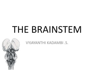
THE BRAINSTEM
VYJAYANTHI KADAMBI .S.
PONS
MIDBRAIN
MEDULLA
PARTS OF THE BRAIN STEM
DORSAL VIEW
VENTRAL VIEW
LATERAL VIEW
VENTRAL ASPECT
DORSAL ASPECT
OLIVES
DECUSSATION
OF PYRAMIDS
PYRAMIDS
MEDULLA
3 PARTS
• MID BRAIN
• PONS
• MEDULLA
• Present in the Posterior Cranial fossa.
• Connects narrow spinal cord with the
expanded Forebrain.
3 broad functions
1. It serves as a conduit for the ascending tracts
and descending tracts connecting the spinal
cord to the different parts of the higher
centers in the forebrain
2. Inportant reflex centers present - respiration,
cvs and control of consiousness
3. Nuclei for CN 3 to 12
THE FOURTH
VENTRICLE
FOURTH VENTRICLE
MEDULLA OBLONGATA
• Connects Pons with Spinal cord
• Conical in shape
• The central canal in lower half continues as
that of spinal cord while the upper half
expands as the cavity of fourth ventricle
• The region between the anterior median sulcus
and the antero lateral sulcus is occupied (on
either side of the midline) by an elevation called
the pyramid.
• The elevation is caused by a large bundle of fibres
that descend from the cerebral cortex to the spinal
Cord (corticospinal fibres)
• Some of these fibres cross from one side to
the other in the lower part of the medulla,
obliterating the anterior median fissure. These
crossing fibres constitute the decussation of the
pyramids.
• Some other fibres emerge from the anterior
median fissure, above the decussation, and
wind laterally over the surface of the medulla
to enter the cerebellum. These are the
anterior external arcuate fibres.
• OLIVES = INFERIOR OLIVARY NUCLEI
• In the groove between the pyramid and olive
emerge the rootlets of hypoglossal nerve.
Hypoglossal nerve
pyramid
olive Vagus
Glossopharyngeal nerve
Inferior cerebellar peduncle
Spinal accessory nerve
• The posterior surface of the superior half of
medulla forms the lower part of the floor of the
fourth ventricle
• The posterior surface of the inferior half of the
medulla is continuous with posterior aspect of the
spinal cord and has POSTERIOR MEDIAN SULCUS
• GRACILE TUBERCLE= GRACILE NUCLEUS(MEDIAL)
• CUNEATE TUBERCLE=CUNEATE NUCLEUS(LATERAL)
INTERNAL STRUCTURE OF THE
MEDULLA
• Considered at 4 levels
1. Level of decussation of Pyramids
2. Level of decussation of lemnisci
3. Level of the olives
4. Level just inferior to the Pons
Level of decussation of Pyramids
Sensory decussation/ level of lemnisci
• The region just behind the pyramids is occupied by a
prominent bundle of fibres, the medial lemniscus, on
either side of the midline.
• The medial lemniscus is formed by fibres arising in the
nucleus gracilis and the nucleus cuneatus. These fibres
cross the midline and turn upwards in the lemniscus of
the opposite side.
• The crossing fibres of the two sides constitute the
sensory decussation.
• The region lateral to the medial lemniscus contains
scattered neurons mixed with nerve fibres. This region
is the reticular formation.
• More laterally there is a mass of white matter
containing various tracts.
Level of the olives
• The pyramids, the medial lemniscus, the spinal nucleus and tract of the
trigeminal nerve, and the reticular formation are present in the same
relative position as at lower levels. The medial lemniscus is, however, much
more prominent and is somewhat expanded anteriorly.
• Lateral to the spinal nucleus (and tract) of the trigeminal nerve we see a
large compact bundle of fibres. This is the inferior cerebellar peduncle
which connects the medulla to the cerebellum.
• Posteriorly, the medulla forms the floor of the fourth ventricle. Here it is
lined by a layer of grey matter in which are located several important cranial
nerve nuclei
• The inferior olivary nucleus forms a prominent feature in the anterolateral
part of the medulla at this level. It is made up of a thin lamina of grey matter
that is folded on itself like a crumpled purse. The nucleus has a hilum that is
directed medially.
Olivary nuclear complex
• Largest = Inferior olivary nucleus
• Smaller dorsal and medial accessory olivary nuclei also are present
• The cells of the inferior olivary nucleus send fibres medially across the
midline to enter the cerebellum through inferior cerebellar peduncle.
• Afferent fibres reach from spinal cord via spino-olivary tracts and from the
cerebellum and cerebral cortex
• Function of olivary nuclei= voluntary muscle movement
PONS
• The Pons is anterior to the cerebellum and connects the medulla with the
midbrain.
• 1 inch
• Anterior surface has basilar groove which lodges the basilar artery.
• Connected to cerebellum via Middle cerebellar peduncle.
• On either side of the lower part of the Pons there is a region called the
cerebello-pontine angle. This region lies near the lateral aperture of the
fourth ventricle. The facial, vestibulocochlear and Glossopharyngeal
nerves, the nervus intermedius, and sometimes the labyrinthine arteries
lie in this region.
• The pons is divisible into a ventral part and a dorsal part
• The ventral (or basilar) part contains numerous transverse and vertical
fibres. Amongst the fibres are groups of cells that constitute the pontine
nuclei.
• When traced laterally the transverse fibres form the middle cerebellar
peduncle.
• The vertical fibres are of two types. Some of them descend from the
cerebral cortex to end in the pontine nuclei. Others are corticospinal fibres
that descend through the pons into the medulla where they form the
pyramids.
POSTERIOR SURFACE OF PONS
INTERNAL STRUCTURE OF PONS
• Divided into
• Studied at two levels :
1. At level of facial colliculus(caudal part)
2. At level of trigeminal nuclei(cranial part)
Trapezoid body (anterior)Tegmentum (posterior)
Transverse section through caudal part
Transverse section through cranial part
MIDBRAIN
• 0.8 inch in length
• Connects Pons and cerebellum with the forebrain
• The midbrain is traversed by NARROW CHANNEL, THE CEREBRAL
AQUEDUCT, filled with CSF
• Posterior surface- 4 colliculi- Corpora Quadrigemina
• Superior colliculi- Visual
• Inferior colliculi- auditory
• In the midline below the Inferior colliculus- trochlear nerves emerge
• When the midbrain is viewed from the anterior aspect, we see two large
bundles of fibres, one on each side of the middle line. These are the crura
of the midbrain
• The crura are separated by a deep fissure. Near the pons the fissure is narrow,
but broadens as the crura diverge to enter the corresponding cerebral
hemispheres. The parts of the crura just below the cerebrum form the
posterior boundary of a space called the interpeduncular fossa. The
oculomotor nerve emerges from the medial aspect of the crus of the same
side.
• Each colliculus is related laterally to a ridge called the brachium.
• The superior brachium (also called the superior quadrigeminal brachium, or
brachium of superior colliculus) connects the superior colliculus to the lateral
geniculate body.
• Similarly, the inferior brachium (also called the inferior quadrigeminal
brachium or brachium of inferior colliculus) connects the inferior colliculus to
the medial geniculate body.
• Just below the colliculi, there is the uppermost part of a membrane, the
superior medullary velum, which stretches between the two superior
cerebellar peduncles, and helps to form the roof of the fourth ventricle.
• For convenience of description, the midbrain may be divided as follows
A. The part lying behind a transverse line drawn through the cerebral
aqueduct is called the tectum. It consists of the superior and inferior
colliculi of the two sides.
B. The part lying in front of the transverse line is made up of right and left
halves called the cerebral peduncles.
• Each peduncle consists of three parts. From anterior to posterior side
these are the
1. crus cerebri (or basis pedunculi),
2. the substantia nigra and
3. the tegmentum.
The main features to be seen through a section of
upper part of the Midbrain
Section through lower part of midbrain
• The crus cerebri (or basis pedunculi) consists of fibres descending from
the cerebral cortex. Its medial one sixth is occupied by corticopontine
fibres descending from the frontal lobe; and the lateral one-sixth is
occupied by similar fibres from the temporal, occipital and parietal lobes.
The intermediate two-thirds of the crus cerebri are occupied by
corticospinal and corticonuclear fibres. The fibres for the leg are most
lateral and those for the head are most medial.
• The substantia nigra lies immediately behind and medial to the basis
pedunculi. It appears dark in unstained sections as neurons within it
contain pigment (neuromelanin).
• The substantia nigra is divisible into a dorsal part, the pars compacta; and
a ventral part, the pars reticularis.
• The pars compacta contains dopaminergic and cholinergic neurons. Most
of the neurons in the pars reticularis are GABAergic. Superiorly, the pars
reticularis becomes continuous with the globus pallidus. The substantia
nigra is closely connected, functionally, with the corpus striatum.
Section through upper part of midbrain
IMPORTANT POINTS
• Pretectal nucleus- light reflex
• The oculomotor nucleus lies in relation to the ventral part of the central grey
matter. The nuclei of the two sides lie close together forming a single complex. The
Edinger Westphal nucleus (which supplies the sphincter pupillae and ciliaris
muscle) forms part of the oculomotor complex. The oculomotor complex is related
ventrally to the medial longitudinal fasciculus.
• Connections of the red nucleus
The red nucleus consists of a cranial parvocellular part and a magnocellular part.
The magnocellular part is prominent in lower species, but in man it is much
reduced and is distinctly smaller than the parvicellular part.
• The red nucleus receives its main afferents from:
a) the cerebral cortex (directly, and as collaterals from the corticospinal tract); and
b) the cerebellum (dentate, emboliform and globose nuclei).
• The efferents of the nucleus are as follows.
The magnocellular part projects to the spinal cord through the rubrospinal tract;
and to motor cranial nerve nuclei (III, IV, V, VI, VII) through the rubrobulbar tract.
Some fibres reach the reticular formation. The parvicellular part of the nucleus
gives origin to fibres that descend through the central tegmental fasciculus to
reach the inferior olivary nucleus. Some fibres reach the reticular formation.
LEVEL CAVITY NUCLEI MOTOR TRACT SENSORY
TRACT
INFERIOR
COLLICULI
CEREBRAL
AQUEDUCT
•INFERIOR
COLLICULUS
•SUBS NIGRA
•4TH NUCLEUS
•MESENCEPHALIC
NUCLEUS OF CN 5
•CORTICONUCLE
AR +
CORTICOSPINAL
•TEMPOROPONT
INE
•FRONTOPONTIN
E
•MLF
•LATERAL
•TRIGEMINAL
•SPINAL
•MEDIAL
•DECUSSATION
OF SUPR
CEREBELLAR
PEDUNCLES
SUPERIOR
COLLICULI
CEREBRAL
AQUEDUCT
•SUPERIOR
COLLICULUS
•SUBS NIGRA
•3RD NUCLEUS
•EDINGER
WESTPHAL N.
•RED NUCLEUS
•MESENCEPHALIC
NUCLEUS OF CN 5
•CORTICONUCLE
AR +
CORTICOSPINAL
•TEMPOROPONT
INE
•FRONTOPONTIN
E
•MLF
•DECUSSATION
RUBROSPINAL
•TRIGEMINAL
•SPINAL
•MEDIAL
• The term reticular formation was originally used to designate areas of the central
nervous system which were not occupied by well defined nuclei or fibre bundles,
but consisted of a network of fibres within which scattered neurons were situated.
Such areas are to be found at all levels in the nervous system. In the spinal cord
there is an intermingling of grey and white matter on the lateral side of the neck of
the dorsal grey column. This area is sometimes referred to as the reticular
formation of the spinal cord. The reticular formation is, however, best defined in
the brainstem where it is now recognised as an area of considerable importance.
• The reticular formation extends throughout the length of the brainstem. In the
medulla it occupies the region dorsal to the inferior olivary nucleus. In the Pons it
lies in the dorsal part, while in the midbrain it lies in the tegmentum.
CEREBELLORETICULAR CONNECTIONS
• Ascending reticular activating system
It has been seen that many ascending tracts passing through the
brainstem are intimately related to the reticular formation. Many of
the fibres in these tracts give off collaterals to it. These come from
the spinothalamic tracts, from secondary trigeminal pathways and
from auditory pathways. These collaterals terminate predominantly
in the lateral part of the reticular formation. Fibres arising here
project to the intralaminar and reticular nuclei of the thalamus.
These nuclei in turn project to widespread areas of the cerebral
cortex. These pathways form part of the ascending reticular
activating system (ARAS) which is believed to be responsible for
maintaining a state of alertness
Summary of Functions of the Reticular Formation
A. Somatomotor control
Through its direct connections with the spinal cord; and
indirectly through the corpus striatum, the cerebral cortex
and the cerebellum, the reticular formation has an influence
on fine control of movements including those involved in
postural adjustments, locomotion, skilled use of the hands,
speech etc.
B. Somatosensory control
The reticular formation influences conduction through
Somatosensory pathways. Similar effects may also be
exerted on visual and auditory pathways.
.
C. Visceral control
Physiological studies have shown that stimulation of
certain areas in the reticular formation of the medulla has
great influence on respiratory and cardiovascular function.
The region influencing respiratory activity corresponds
approximately to the giganto-cellular nucleus and
parvocellular nucleus. Stimulation of the gigantocellular
nucleus and the upper part of the ventral reticular nucleus
causes depression of vasomotor activity while stimulation
of other areas has a pressor effect. These effects are
through connections between the reticular formation and
autonomic centres in the brainstem and spinal cord, but
the pathways concerned are not well defined.
D. Neuroendocrine control
Through its connections with the hypothalamus, the
reticular formation influences activity of the
adenohypophysis and of the neurohypophysis
CONTROL OF EYE MOVEMENTS
When the gaze is shifted from one object to another the eyes undergo sharp
movements called saccades. The controlling centres determine the velocity
and extent of a saccade. A different kind of control is required when the eyes
follow a moving object (pursuit movement).
A. The areas of cerebral cortex involved in programming and co-ordination of all
eye movements are:
(a) the frontal eye field (area 8);
(b) the supplemental eye field located on the medial surface of the hemisphere
(area 6).
(c) the prefrontal cortex (anterior to the frontal eye field); and
(d) the posterior eye field (in the parietal lobe).
• B. The areas of cerebral cortex listed above
project to the superior colliculus (some of
them passing via the basal nuclei). The
superior colliculus also receives fibres from
the retinae, from pretectal nuclei, and from
the cerebellum.
C. The superior colliculus projects to two important centres which are as follows.
(1) The vertical gaze centre lies in the midbrain. It is located in the rostral interstitial
nucleus (a collection of neurons within the medial longitudinal fasciculus), and in
the interstitial nucleus of Cajal. This centre controls vertical eye movements.
(2) The horizontal gaze centre lies in the pons, in the paramedian reticular formation.
The centre controls horizontal eye movements.
In addition to the afferents from the superior colliculus, these centres also
receive:
(a) some direct cortical fibres; and
(b) fibres from the cerebellum.
Efferents from the vertical and horizontal gaze centres project to the oculomotor,
trochlear and abducent nuclei.
D. The cranial nerve nuclei supplying muscles of the
eyeball receive the following afferents.
(a) Corticonuclear fibres.
(b) Fibres from the vertical and horizontal gaze centres
described above.
(c) Direct fibres from the superior colliculus and from
pretectal nuclei.
(d) Fibres from vestibular nuclei. These travel through
the medial longitudinal fasciculus. They provide
information about movements of the head and neck
(from the vestibular apparatus).
Through the vestibular nuclei, the oculomotor trochlear
and abducent nuclei are brought under cerebellar
control.
• E. The final control of vertical gaze is by fibres
passing from the oculomotor and trochlear nuclei
(to reach muscles producing upward and
downward movements of the eyeballs). The final
control of horizontal gaze is mainly by the
abducent nerve (lateral rectus), but is also
influenced by the oculomotor nerve (medial
rectus). The horizontal gaze centre is also
involved incontrol of eye movements when the
gaze follows a moving object. These are called
‘pursuit movements’. The control of these
movements is complex
CONTROL OF SACCADIC MOVEMENTS
CONTROL OF PURSUIT MOVEMENTS
The brainstem

Contenu connexe

Tendances (20)

Cerebrum
CerebrumCerebrum
Cerebrum
 
Anatomy of medulla oblongata
Anatomy of medulla oblongataAnatomy of medulla oblongata
Anatomy of medulla oblongata
 
Cerebellum-Connections and Functions
Cerebellum-Connections and FunctionsCerebellum-Connections and Functions
Cerebellum-Connections and Functions
 
Cerebellum
CerebellumCerebellum
Cerebellum
 
Anatomy of diencephalon
Anatomy of diencephalonAnatomy of diencephalon
Anatomy of diencephalon
 
CEREBRAL CORTEX
CEREBRAL CORTEXCEREBRAL CORTEX
CEREBRAL CORTEX
 
Cerebellum
CerebellumCerebellum
Cerebellum
 
Cerebellum Physiology
Cerebellum  PhysiologyCerebellum  Physiology
Cerebellum Physiology
 
NEUROANATOMY OF BRAINSTEM
NEUROANATOMY OF BRAINSTEMNEUROANATOMY OF BRAINSTEM
NEUROANATOMY OF BRAINSTEM
 
Cerebrum
CerebrumCerebrum
Cerebrum
 
Basal ganglia
Basal gangliaBasal ganglia
Basal ganglia
 
Spinal cord
Spinal cordSpinal cord
Spinal cord
 
Diencephalon 1
Diencephalon 1Diencephalon 1
Diencephalon 1
 
Thalamus
ThalamusThalamus
Thalamus
 
Anatomy and physiology of brain stem
Anatomy and physiology of brain stemAnatomy and physiology of brain stem
Anatomy and physiology of brain stem
 
Anatomy of meninges, ventricles, cerebrospinal fluid
Anatomy of meninges, ventricles, cerebrospinal fluidAnatomy of meninges, ventricles, cerebrospinal fluid
Anatomy of meninges, ventricles, cerebrospinal fluid
 
Medulla oblongata
Medulla oblongata Medulla oblongata
Medulla oblongata
 
MID BRAIN.pptx
MID BRAIN.pptxMID BRAIN.pptx
MID BRAIN.pptx
 
Anatomy of spinal cord
Anatomy of spinal cordAnatomy of spinal cord
Anatomy of spinal cord
 
MIDBRAIN basic anatomy and applied aspects.
MIDBRAIN basic anatomy and applied aspects.MIDBRAIN basic anatomy and applied aspects.
MIDBRAIN basic anatomy and applied aspects.
 

En vedette (20)

Anatomy of Midbrain and pons
Anatomy of Midbrain and ponsAnatomy of Midbrain and pons
Anatomy of Midbrain and pons
 
Brainstem demo neww
Brainstem demo newwBrainstem demo neww
Brainstem demo neww
 
Brain stem 2014
Brain stem 2014Brain stem 2014
Brain stem 2014
 
Anatomy of midbrain & pons
Anatomy of midbrain & ponsAnatomy of midbrain & pons
Anatomy of midbrain & pons
 
Brain stem
Brain stemBrain stem
Brain stem
 
2 brain stem akd
2 brain stem akd2 brain stem akd
2 brain stem akd
 
The Hindbrain And Midbrain
The Hindbrain And MidbrainThe Hindbrain And Midbrain
The Hindbrain And Midbrain
 
Sectional Anatomy of the Brain Stem
Sectional Anatomy of the Brain StemSectional Anatomy of the Brain Stem
Sectional Anatomy of the Brain Stem
 
Brain stem lessions in sensory symptoms
Brain stem lessions in sensory symptomsBrain stem lessions in sensory symptoms
Brain stem lessions in sensory symptoms
 
MRI SECTIONAL ANATOMY OF BRAIN
MRI SECTIONAL ANATOMY OF BRAIN MRI SECTIONAL ANATOMY OF BRAIN
MRI SECTIONAL ANATOMY OF BRAIN
 
Neuro clinics 31- The pupils -basic
Neuro clinics 31- The pupils -basicNeuro clinics 31- The pupils -basic
Neuro clinics 31- The pupils -basic
 
Tumeurs intracraniennes
Tumeurs intracraniennesTumeurs intracraniennes
Tumeurs intracraniennes
 
Temporal lobe
Temporal lobeTemporal lobe
Temporal lobe
 
Skull bones
Skull bonesSkull bones
Skull bones
 
Brainstem
BrainstemBrainstem
Brainstem
 
Lect -brainstem
Lect -brainstemLect -brainstem
Lect -brainstem
 
A and P Notes217 228
A and P Notes217 228A and P Notes217 228
A and P Notes217 228
 
Medulla
MedullaMedulla
Medulla
 
Midbrain And Cerebellum
Midbrain And CerebellumMidbrain And Cerebellum
Midbrain And Cerebellum
 
2/10 Brain stem
2/10 Brain stem2/10 Brain stem
2/10 Brain stem
 

Similaire à The brainstem

Anatomy of brain steam midbrain pons and medulla
Anatomy of brain steam midbrain pons and medullaAnatomy of brain steam midbrain pons and medulla
Anatomy of brain steam midbrain pons and medullaDr.Jitendra Patel
 
L5- BRAIN STEM.pptx
L5- BRAIN STEM.pptxL5- BRAIN STEM.pptx
L5- BRAIN STEM.pptxRawHady
 
INTRODUCTION TO THE BRAINSTEM.pptx
INTRODUCTION TO THE BRAINSTEM.pptxINTRODUCTION TO THE BRAINSTEM.pptx
INTRODUCTION TO THE BRAINSTEM.pptxSikander Sher
 
FINAL--PARTS OF BRAIN.pptx
FINAL--PARTS OF BRAIN.pptxFINAL--PARTS OF BRAIN.pptx
FINAL--PARTS OF BRAIN.pptxAsifBahadurAli
 
Detailed Anatomy of Medulla Oblongata ppt
Detailed Anatomy of Medulla Oblongata pptDetailed Anatomy of Medulla Oblongata ppt
Detailed Anatomy of Medulla Oblongata pptDr Geeta Dolli
 
The Rhombencephalon or Hind brain.ppt
The Rhombencephalon or Hind brain.pptThe Rhombencephalon or Hind brain.ppt
The Rhombencephalon or Hind brain.pptOkenyInnocent1
 
anatomy of central nervous system
anatomy of central nervous systemanatomy of central nervous system
anatomy of central nervous systemNeha Mathew
 
4th ventricle 21.12.2015.dk.
4th  ventricle 21.12.2015.dk.4th  ventricle 21.12.2015.dk.
4th ventricle 21.12.2015.dk.Deepak Khedekar
 
anatomyofbrain-180502045850.pdf
anatomyofbrain-180502045850.pdfanatomyofbrain-180502045850.pdf
anatomyofbrain-180502045850.pdfGUNASEKARANM20
 
Anatomy of brain
Anatomy of brainAnatomy of brain
Anatomy of brainThomas John
 
Lecture fourteen neuroanatomy, Embryology of the CNS.pdf
Lecture fourteen neuroanatomy, Embryology of the CNS.pdfLecture fourteen neuroanatomy, Embryology of the CNS.pdf
Lecture fourteen neuroanatomy, Embryology of the CNS.pdfNatungaRonald1
 
brainstem cerebellum cranial nerves forr
brainstem cerebellum cranial nerves forrbrainstem cerebellum cranial nerves forr
brainstem cerebellum cranial nerves forromarkhaled833960
 

Similaire à The brainstem (20)

BRAINSTEM-DETAILED.pptx
BRAINSTEM-DETAILED.pptxBRAINSTEM-DETAILED.pptx
BRAINSTEM-DETAILED.pptx
 
Anatomy of brain steam midbrain pons and medulla
Anatomy of brain steam midbrain pons and medullaAnatomy of brain steam midbrain pons and medulla
Anatomy of brain steam midbrain pons and medulla
 
L5- BRAIN STEM.pptx
L5- BRAIN STEM.pptxL5- BRAIN STEM.pptx
L5- BRAIN STEM.pptx
 
Brain stem.pdf
Brain stem.pdfBrain stem.pdf
Brain stem.pdf
 
INTRODUCTION TO THE BRAINSTEM.pptx
INTRODUCTION TO THE BRAINSTEM.pptxINTRODUCTION TO THE BRAINSTEM.pptx
INTRODUCTION TO THE BRAINSTEM.pptx
 
Brain stem.pptx
Brain stem.pptxBrain stem.pptx
Brain stem.pptx
 
FINAL--PARTS OF BRAIN.pptx
FINAL--PARTS OF BRAIN.pptxFINAL--PARTS OF BRAIN.pptx
FINAL--PARTS OF BRAIN.pptx
 
Brain Stem.pdf
Brain Stem.pdfBrain Stem.pdf
Brain Stem.pdf
 
Brain stem New.ppsx
Brain stem New.ppsxBrain stem New.ppsx
Brain stem New.ppsx
 
Detailed Anatomy of Medulla Oblongata ppt
Detailed Anatomy of Medulla Oblongata pptDetailed Anatomy of Medulla Oblongata ppt
Detailed Anatomy of Medulla Oblongata ppt
 
MIDBRAIN.DK.pptx
MIDBRAIN.DK.pptxMIDBRAIN.DK.pptx
MIDBRAIN.DK.pptx
 
The Rhombencephalon or Hind brain.ppt
The Rhombencephalon or Hind brain.pptThe Rhombencephalon or Hind brain.ppt
The Rhombencephalon or Hind brain.ppt
 
anatomy of central nervous system
anatomy of central nervous systemanatomy of central nervous system
anatomy of central nervous system
 
4th ventricle 21.12.2015.dk.
4th  ventricle 21.12.2015.dk.4th  ventricle 21.12.2015.dk.
4th ventricle 21.12.2015.dk.
 
anatomyofbrain-180502045850.pdf
anatomyofbrain-180502045850.pdfanatomyofbrain-180502045850.pdf
anatomyofbrain-180502045850.pdf
 
Anatomy of brain
Anatomy of brainAnatomy of brain
Anatomy of brain
 
Lecture fourteen neuroanatomy, Embryology of the CNS.pdf
Lecture fourteen neuroanatomy, Embryology of the CNS.pdfLecture fourteen neuroanatomy, Embryology of the CNS.pdf
Lecture fourteen neuroanatomy, Embryology of the CNS.pdf
 
brainstem cerebellum cranial nerves forr
brainstem cerebellum cranial nerves forrbrainstem cerebellum cranial nerves forr
brainstem cerebellum cranial nerves forr
 
Development of nervous system
Development of nervous systemDevelopment of nervous system
Development of nervous system
 
Spinal cord
Spinal cordSpinal cord
Spinal cord
 

Dernier

Premium Call Girls Nagpur {9xx000xx09} ❤️VVIP POOJA Call Girls in Nagpur Maha...
Premium Call Girls Nagpur {9xx000xx09} ❤️VVIP POOJA Call Girls in Nagpur Maha...Premium Call Girls Nagpur {9xx000xx09} ❤️VVIP POOJA Call Girls in Nagpur Maha...
Premium Call Girls Nagpur {9xx000xx09} ❤️VVIP POOJA Call Girls in Nagpur Maha...Sheetaleventcompany
 
Shazia Iqbal 2024 - Bioorganic Chemistry.pdf
Shazia Iqbal 2024 - Bioorganic Chemistry.pdfShazia Iqbal 2024 - Bioorganic Chemistry.pdf
Shazia Iqbal 2024 - Bioorganic Chemistry.pdfTrustlife
 
Premium Call Girls Dehradun {8854095900} ❤️VVIP ANJU Call Girls in Dehradun U...
Premium Call Girls Dehradun {8854095900} ❤️VVIP ANJU Call Girls in Dehradun U...Premium Call Girls Dehradun {8854095900} ❤️VVIP ANJU Call Girls in Dehradun U...
Premium Call Girls Dehradun {8854095900} ❤️VVIP ANJU Call Girls in Dehradun U...Sheetaleventcompany
 
🚺LEELA JOSHI WhatsApp Number +91-9930245274 ✔ Unsatisfied Bhabhi Call Girls T...
🚺LEELA JOSHI WhatsApp Number +91-9930245274 ✔ Unsatisfied Bhabhi Call Girls T...🚺LEELA JOSHI WhatsApp Number +91-9930245274 ✔ Unsatisfied Bhabhi Call Girls T...
🚺LEELA JOSHI WhatsApp Number +91-9930245274 ✔ Unsatisfied Bhabhi Call Girls T...soniya pandit
 
Whitefield { Call Girl in Bangalore ₹7.5k Pick Up & Drop With Cash Payment 63...
Whitefield { Call Girl in Bangalore ₹7.5k Pick Up & Drop With Cash Payment 63...Whitefield { Call Girl in Bangalore ₹7.5k Pick Up & Drop With Cash Payment 63...
Whitefield { Call Girl in Bangalore ₹7.5k Pick Up & Drop With Cash Payment 63...dishamehta3332
 
Electrocardiogram (ECG) physiological basis .pdf
Electrocardiogram (ECG) physiological basis .pdfElectrocardiogram (ECG) physiological basis .pdf
Electrocardiogram (ECG) physiological basis .pdfMedicoseAcademics
 
Genuine Call Girls Hyderabad 9630942363 Book High Profile Call Girl in Hydera...
Genuine Call Girls Hyderabad 9630942363 Book High Profile Call Girl in Hydera...Genuine Call Girls Hyderabad 9630942363 Book High Profile Call Girl in Hydera...
Genuine Call Girls Hyderabad 9630942363 Book High Profile Call Girl in Hydera...GENUINE ESCORT AGENCY
 
👉 Amritsar Call Girls 👉📞 8725944379 👉📞 Just📲 Call Ruhi Call Girl Near Me Amri...
👉 Amritsar Call Girls 👉📞 8725944379 👉📞 Just📲 Call Ruhi Call Girl Near Me Amri...👉 Amritsar Call Girls 👉📞 8725944379 👉📞 Just📲 Call Ruhi Call Girl Near Me Amri...
👉 Amritsar Call Girls 👉📞 8725944379 👉📞 Just📲 Call Ruhi Call Girl Near Me Amri...Sheetaleventcompany
 
Pune Call Girl Service 📞9xx000xx09📞Just Call Divya📲 Call Girl In Pune No💰Adva...
Pune Call Girl Service 📞9xx000xx09📞Just Call Divya📲 Call Girl In Pune No💰Adva...Pune Call Girl Service 📞9xx000xx09📞Just Call Divya📲 Call Girl In Pune No💰Adva...
Pune Call Girl Service 📞9xx000xx09📞Just Call Divya📲 Call Girl In Pune No💰Adva...Sheetaleventcompany
 
Goa Call Girl Service 📞9xx000xx09📞Just Call Divya📲 Call Girl In Goa No💰Advanc...
Goa Call Girl Service 📞9xx000xx09📞Just Call Divya📲 Call Girl In Goa No💰Advanc...Goa Call Girl Service 📞9xx000xx09📞Just Call Divya📲 Call Girl In Goa No💰Advanc...
Goa Call Girl Service 📞9xx000xx09📞Just Call Divya📲 Call Girl In Goa No💰Advanc...Sheetaleventcompany
 
Jaipur Call Girl Service 📞9xx000xx09📞Just Call Divya📲 Call Girl In Jaipur No💰...
Jaipur Call Girl Service 📞9xx000xx09📞Just Call Divya📲 Call Girl In Jaipur No💰...Jaipur Call Girl Service 📞9xx000xx09📞Just Call Divya📲 Call Girl In Jaipur No💰...
Jaipur Call Girl Service 📞9xx000xx09📞Just Call Divya📲 Call Girl In Jaipur No💰...Sheetaleventcompany
 
Cardiac Output, Venous Return, and Their Regulation
Cardiac Output, Venous Return, and Their RegulationCardiac Output, Venous Return, and Their Regulation
Cardiac Output, Venous Return, and Their RegulationMedicoseAcademics
 
❤️Call Girl Service In Chandigarh☎️9814379184☎️ Call Girl in Chandigarh☎️ Cha...
❤️Call Girl Service In Chandigarh☎️9814379184☎️ Call Girl in Chandigarh☎️ Cha...❤️Call Girl Service In Chandigarh☎️9814379184☎️ Call Girl in Chandigarh☎️ Cha...
❤️Call Girl Service In Chandigarh☎️9814379184☎️ Call Girl in Chandigarh☎️ Cha...Sheetaleventcompany
 
Jual Obat Aborsi Di Dubai UAE Wa 0838-4800-7379 Obat Penggugur Kandungan Cytotec
Jual Obat Aborsi Di Dubai UAE Wa 0838-4800-7379 Obat Penggugur Kandungan CytotecJual Obat Aborsi Di Dubai UAE Wa 0838-4800-7379 Obat Penggugur Kandungan Cytotec
Jual Obat Aborsi Di Dubai UAE Wa 0838-4800-7379 Obat Penggugur Kandungan Cytotecjualobat34
 
VIP Hyderabad Call Girls KPHB 7877925207 ₹5000 To 25K With AC Room 💚😋
VIP Hyderabad Call Girls KPHB 7877925207 ₹5000 To 25K With AC Room 💚😋VIP Hyderabad Call Girls KPHB 7877925207 ₹5000 To 25K With AC Room 💚😋
VIP Hyderabad Call Girls KPHB 7877925207 ₹5000 To 25K With AC Room 💚😋mahima pandey
 
Nagpur Call Girl Service 📞9xx000xx09📞Just Call Divya📲 Call Girl In Nagpur No💰...
Nagpur Call Girl Service 📞9xx000xx09📞Just Call Divya📲 Call Girl In Nagpur No💰...Nagpur Call Girl Service 📞9xx000xx09📞Just Call Divya📲 Call Girl In Nagpur No💰...
Nagpur Call Girl Service 📞9xx000xx09📞Just Call Divya📲 Call Girl In Nagpur No💰...Sheetaleventcompany
 
Chandigarh Call Girls Service ❤️🍑 9809698092 👄🫦Independent Escort Service Cha...
Chandigarh Call Girls Service ❤️🍑 9809698092 👄🫦Independent Escort Service Cha...Chandigarh Call Girls Service ❤️🍑 9809698092 👄🫦Independent Escort Service Cha...
Chandigarh Call Girls Service ❤️🍑 9809698092 👄🫦Independent Escort Service Cha...Sheetaleventcompany
 
❤️Chandigarh Escorts Service☎️9814379184☎️ Call Girl service in Chandigarh☎️ ...
❤️Chandigarh Escorts Service☎️9814379184☎️ Call Girl service in Chandigarh☎️ ...❤️Chandigarh Escorts Service☎️9814379184☎️ Call Girl service in Chandigarh☎️ ...
❤️Chandigarh Escorts Service☎️9814379184☎️ Call Girl service in Chandigarh☎️ ...Sheetaleventcompany
 
ANATOMY AND PHYSIOLOGY OF RESPIRATORY SYSTEM.pptx
ANATOMY AND PHYSIOLOGY OF RESPIRATORY SYSTEM.pptxANATOMY AND PHYSIOLOGY OF RESPIRATORY SYSTEM.pptx
ANATOMY AND PHYSIOLOGY OF RESPIRATORY SYSTEM.pptxSwetaba Besh
 
Dehradun Call Girls Service {8854095900} ❤️VVIP ROCKY Call Girl in Dehradun U...
Dehradun Call Girls Service {8854095900} ❤️VVIP ROCKY Call Girl in Dehradun U...Dehradun Call Girls Service {8854095900} ❤️VVIP ROCKY Call Girl in Dehradun U...
Dehradun Call Girls Service {8854095900} ❤️VVIP ROCKY Call Girl in Dehradun U...Sheetaleventcompany
 

Dernier (20)

Premium Call Girls Nagpur {9xx000xx09} ❤️VVIP POOJA Call Girls in Nagpur Maha...
Premium Call Girls Nagpur {9xx000xx09} ❤️VVIP POOJA Call Girls in Nagpur Maha...Premium Call Girls Nagpur {9xx000xx09} ❤️VVIP POOJA Call Girls in Nagpur Maha...
Premium Call Girls Nagpur {9xx000xx09} ❤️VVIP POOJA Call Girls in Nagpur Maha...
 
Shazia Iqbal 2024 - Bioorganic Chemistry.pdf
Shazia Iqbal 2024 - Bioorganic Chemistry.pdfShazia Iqbal 2024 - Bioorganic Chemistry.pdf
Shazia Iqbal 2024 - Bioorganic Chemistry.pdf
 
Premium Call Girls Dehradun {8854095900} ❤️VVIP ANJU Call Girls in Dehradun U...
Premium Call Girls Dehradun {8854095900} ❤️VVIP ANJU Call Girls in Dehradun U...Premium Call Girls Dehradun {8854095900} ❤️VVIP ANJU Call Girls in Dehradun U...
Premium Call Girls Dehradun {8854095900} ❤️VVIP ANJU Call Girls in Dehradun U...
 
🚺LEELA JOSHI WhatsApp Number +91-9930245274 ✔ Unsatisfied Bhabhi Call Girls T...
🚺LEELA JOSHI WhatsApp Number +91-9930245274 ✔ Unsatisfied Bhabhi Call Girls T...🚺LEELA JOSHI WhatsApp Number +91-9930245274 ✔ Unsatisfied Bhabhi Call Girls T...
🚺LEELA JOSHI WhatsApp Number +91-9930245274 ✔ Unsatisfied Bhabhi Call Girls T...
 
Whitefield { Call Girl in Bangalore ₹7.5k Pick Up & Drop With Cash Payment 63...
Whitefield { Call Girl in Bangalore ₹7.5k Pick Up & Drop With Cash Payment 63...Whitefield { Call Girl in Bangalore ₹7.5k Pick Up & Drop With Cash Payment 63...
Whitefield { Call Girl in Bangalore ₹7.5k Pick Up & Drop With Cash Payment 63...
 
Electrocardiogram (ECG) physiological basis .pdf
Electrocardiogram (ECG) physiological basis .pdfElectrocardiogram (ECG) physiological basis .pdf
Electrocardiogram (ECG) physiological basis .pdf
 
Genuine Call Girls Hyderabad 9630942363 Book High Profile Call Girl in Hydera...
Genuine Call Girls Hyderabad 9630942363 Book High Profile Call Girl in Hydera...Genuine Call Girls Hyderabad 9630942363 Book High Profile Call Girl in Hydera...
Genuine Call Girls Hyderabad 9630942363 Book High Profile Call Girl in Hydera...
 
👉 Amritsar Call Girls 👉📞 8725944379 👉📞 Just📲 Call Ruhi Call Girl Near Me Amri...
👉 Amritsar Call Girls 👉📞 8725944379 👉📞 Just📲 Call Ruhi Call Girl Near Me Amri...👉 Amritsar Call Girls 👉📞 8725944379 👉📞 Just📲 Call Ruhi Call Girl Near Me Amri...
👉 Amritsar Call Girls 👉📞 8725944379 👉📞 Just📲 Call Ruhi Call Girl Near Me Amri...
 
Pune Call Girl Service 📞9xx000xx09📞Just Call Divya📲 Call Girl In Pune No💰Adva...
Pune Call Girl Service 📞9xx000xx09📞Just Call Divya📲 Call Girl In Pune No💰Adva...Pune Call Girl Service 📞9xx000xx09📞Just Call Divya📲 Call Girl In Pune No💰Adva...
Pune Call Girl Service 📞9xx000xx09📞Just Call Divya📲 Call Girl In Pune No💰Adva...
 
Goa Call Girl Service 📞9xx000xx09📞Just Call Divya📲 Call Girl In Goa No💰Advanc...
Goa Call Girl Service 📞9xx000xx09📞Just Call Divya📲 Call Girl In Goa No💰Advanc...Goa Call Girl Service 📞9xx000xx09📞Just Call Divya📲 Call Girl In Goa No💰Advanc...
Goa Call Girl Service 📞9xx000xx09📞Just Call Divya📲 Call Girl In Goa No💰Advanc...
 
Jaipur Call Girl Service 📞9xx000xx09📞Just Call Divya📲 Call Girl In Jaipur No💰...
Jaipur Call Girl Service 📞9xx000xx09📞Just Call Divya📲 Call Girl In Jaipur No💰...Jaipur Call Girl Service 📞9xx000xx09📞Just Call Divya📲 Call Girl In Jaipur No💰...
Jaipur Call Girl Service 📞9xx000xx09📞Just Call Divya📲 Call Girl In Jaipur No💰...
 
Cardiac Output, Venous Return, and Their Regulation
Cardiac Output, Venous Return, and Their RegulationCardiac Output, Venous Return, and Their Regulation
Cardiac Output, Venous Return, and Their Regulation
 
❤️Call Girl Service In Chandigarh☎️9814379184☎️ Call Girl in Chandigarh☎️ Cha...
❤️Call Girl Service In Chandigarh☎️9814379184☎️ Call Girl in Chandigarh☎️ Cha...❤️Call Girl Service In Chandigarh☎️9814379184☎️ Call Girl in Chandigarh☎️ Cha...
❤️Call Girl Service In Chandigarh☎️9814379184☎️ Call Girl in Chandigarh☎️ Cha...
 
Jual Obat Aborsi Di Dubai UAE Wa 0838-4800-7379 Obat Penggugur Kandungan Cytotec
Jual Obat Aborsi Di Dubai UAE Wa 0838-4800-7379 Obat Penggugur Kandungan CytotecJual Obat Aborsi Di Dubai UAE Wa 0838-4800-7379 Obat Penggugur Kandungan Cytotec
Jual Obat Aborsi Di Dubai UAE Wa 0838-4800-7379 Obat Penggugur Kandungan Cytotec
 
VIP Hyderabad Call Girls KPHB 7877925207 ₹5000 To 25K With AC Room 💚😋
VIP Hyderabad Call Girls KPHB 7877925207 ₹5000 To 25K With AC Room 💚😋VIP Hyderabad Call Girls KPHB 7877925207 ₹5000 To 25K With AC Room 💚😋
VIP Hyderabad Call Girls KPHB 7877925207 ₹5000 To 25K With AC Room 💚😋
 
Nagpur Call Girl Service 📞9xx000xx09📞Just Call Divya📲 Call Girl In Nagpur No💰...
Nagpur Call Girl Service 📞9xx000xx09📞Just Call Divya📲 Call Girl In Nagpur No💰...Nagpur Call Girl Service 📞9xx000xx09📞Just Call Divya📲 Call Girl In Nagpur No💰...
Nagpur Call Girl Service 📞9xx000xx09📞Just Call Divya📲 Call Girl In Nagpur No💰...
 
Chandigarh Call Girls Service ❤️🍑 9809698092 👄🫦Independent Escort Service Cha...
Chandigarh Call Girls Service ❤️🍑 9809698092 👄🫦Independent Escort Service Cha...Chandigarh Call Girls Service ❤️🍑 9809698092 👄🫦Independent Escort Service Cha...
Chandigarh Call Girls Service ❤️🍑 9809698092 👄🫦Independent Escort Service Cha...
 
❤️Chandigarh Escorts Service☎️9814379184☎️ Call Girl service in Chandigarh☎️ ...
❤️Chandigarh Escorts Service☎️9814379184☎️ Call Girl service in Chandigarh☎️ ...❤️Chandigarh Escorts Service☎️9814379184☎️ Call Girl service in Chandigarh☎️ ...
❤️Chandigarh Escorts Service☎️9814379184☎️ Call Girl service in Chandigarh☎️ ...
 
ANATOMY AND PHYSIOLOGY OF RESPIRATORY SYSTEM.pptx
ANATOMY AND PHYSIOLOGY OF RESPIRATORY SYSTEM.pptxANATOMY AND PHYSIOLOGY OF RESPIRATORY SYSTEM.pptx
ANATOMY AND PHYSIOLOGY OF RESPIRATORY SYSTEM.pptx
 
Dehradun Call Girls Service {8854095900} ❤️VVIP ROCKY Call Girl in Dehradun U...
Dehradun Call Girls Service {8854095900} ❤️VVIP ROCKY Call Girl in Dehradun U...Dehradun Call Girls Service {8854095900} ❤️VVIP ROCKY Call Girl in Dehradun U...
Dehradun Call Girls Service {8854095900} ❤️VVIP ROCKY Call Girl in Dehradun U...
 

The brainstem

  • 7. 3 PARTS • MID BRAIN • PONS • MEDULLA • Present in the Posterior Cranial fossa. • Connects narrow spinal cord with the expanded Forebrain.
  • 8.
  • 9. 3 broad functions 1. It serves as a conduit for the ascending tracts and descending tracts connecting the spinal cord to the different parts of the higher centers in the forebrain 2. Inportant reflex centers present - respiration, cvs and control of consiousness 3. Nuclei for CN 3 to 12
  • 11. MEDULLA OBLONGATA • Connects Pons with Spinal cord • Conical in shape • The central canal in lower half continues as that of spinal cord while the upper half expands as the cavity of fourth ventricle
  • 12. • The region between the anterior median sulcus and the antero lateral sulcus is occupied (on either side of the midline) by an elevation called the pyramid. • The elevation is caused by a large bundle of fibres that descend from the cerebral cortex to the spinal Cord (corticospinal fibres) • Some of these fibres cross from one side to the other in the lower part of the medulla, obliterating the anterior median fissure. These crossing fibres constitute the decussation of the pyramids.
  • 13. • Some other fibres emerge from the anterior median fissure, above the decussation, and wind laterally over the surface of the medulla to enter the cerebellum. These are the anterior external arcuate fibres. • OLIVES = INFERIOR OLIVARY NUCLEI • In the groove between the pyramid and olive emerge the rootlets of hypoglossal nerve.
  • 14. Hypoglossal nerve pyramid olive Vagus Glossopharyngeal nerve Inferior cerebellar peduncle Spinal accessory nerve
  • 15. • The posterior surface of the superior half of medulla forms the lower part of the floor of the fourth ventricle • The posterior surface of the inferior half of the medulla is continuous with posterior aspect of the spinal cord and has POSTERIOR MEDIAN SULCUS • GRACILE TUBERCLE= GRACILE NUCLEUS(MEDIAL) • CUNEATE TUBERCLE=CUNEATE NUCLEUS(LATERAL)
  • 16.
  • 17.
  • 18.
  • 19. INTERNAL STRUCTURE OF THE MEDULLA • Considered at 4 levels 1. Level of decussation of Pyramids 2. Level of decussation of lemnisci 3. Level of the olives 4. Level just inferior to the Pons
  • 20. Level of decussation of Pyramids
  • 22. • The region just behind the pyramids is occupied by a prominent bundle of fibres, the medial lemniscus, on either side of the midline. • The medial lemniscus is formed by fibres arising in the nucleus gracilis and the nucleus cuneatus. These fibres cross the midline and turn upwards in the lemniscus of the opposite side. • The crossing fibres of the two sides constitute the sensory decussation. • The region lateral to the medial lemniscus contains scattered neurons mixed with nerve fibres. This region is the reticular formation. • More laterally there is a mass of white matter containing various tracts.
  • 23. Level of the olives
  • 24. • The pyramids, the medial lemniscus, the spinal nucleus and tract of the trigeminal nerve, and the reticular formation are present in the same relative position as at lower levels. The medial lemniscus is, however, much more prominent and is somewhat expanded anteriorly. • Lateral to the spinal nucleus (and tract) of the trigeminal nerve we see a large compact bundle of fibres. This is the inferior cerebellar peduncle which connects the medulla to the cerebellum. • Posteriorly, the medulla forms the floor of the fourth ventricle. Here it is lined by a layer of grey matter in which are located several important cranial nerve nuclei • The inferior olivary nucleus forms a prominent feature in the anterolateral part of the medulla at this level. It is made up of a thin lamina of grey matter that is folded on itself like a crumpled purse. The nucleus has a hilum that is directed medially.
  • 25. Olivary nuclear complex • Largest = Inferior olivary nucleus • Smaller dorsal and medial accessory olivary nuclei also are present • The cells of the inferior olivary nucleus send fibres medially across the midline to enter the cerebellum through inferior cerebellar peduncle. • Afferent fibres reach from spinal cord via spino-olivary tracts and from the cerebellum and cerebral cortex • Function of olivary nuclei= voluntary muscle movement
  • 26.
  • 27. PONS • The Pons is anterior to the cerebellum and connects the medulla with the midbrain. • 1 inch • Anterior surface has basilar groove which lodges the basilar artery. • Connected to cerebellum via Middle cerebellar peduncle.
  • 28. • On either side of the lower part of the Pons there is a region called the cerebello-pontine angle. This region lies near the lateral aperture of the fourth ventricle. The facial, vestibulocochlear and Glossopharyngeal nerves, the nervus intermedius, and sometimes the labyrinthine arteries lie in this region. • The pons is divisible into a ventral part and a dorsal part • The ventral (or basilar) part contains numerous transverse and vertical fibres. Amongst the fibres are groups of cells that constitute the pontine nuclei. • When traced laterally the transverse fibres form the middle cerebellar peduncle. • The vertical fibres are of two types. Some of them descend from the cerebral cortex to end in the pontine nuclei. Others are corticospinal fibres that descend through the pons into the medulla where they form the pyramids.
  • 30. INTERNAL STRUCTURE OF PONS • Divided into • Studied at two levels : 1. At level of facial colliculus(caudal part) 2. At level of trigeminal nuclei(cranial part) Trapezoid body (anterior)Tegmentum (posterior)
  • 33. MIDBRAIN • 0.8 inch in length • Connects Pons and cerebellum with the forebrain • The midbrain is traversed by NARROW CHANNEL, THE CEREBRAL AQUEDUCT, filled with CSF • Posterior surface- 4 colliculi- Corpora Quadrigemina • Superior colliculi- Visual • Inferior colliculi- auditory • In the midline below the Inferior colliculus- trochlear nerves emerge • When the midbrain is viewed from the anterior aspect, we see two large bundles of fibres, one on each side of the middle line. These are the crura of the midbrain
  • 34. • The crura are separated by a deep fissure. Near the pons the fissure is narrow, but broadens as the crura diverge to enter the corresponding cerebral hemispheres. The parts of the crura just below the cerebrum form the posterior boundary of a space called the interpeduncular fossa. The oculomotor nerve emerges from the medial aspect of the crus of the same side. • Each colliculus is related laterally to a ridge called the brachium. • The superior brachium (also called the superior quadrigeminal brachium, or brachium of superior colliculus) connects the superior colliculus to the lateral geniculate body. • Similarly, the inferior brachium (also called the inferior quadrigeminal brachium or brachium of inferior colliculus) connects the inferior colliculus to the medial geniculate body. • Just below the colliculi, there is the uppermost part of a membrane, the superior medullary velum, which stretches between the two superior cerebellar peduncles, and helps to form the roof of the fourth ventricle.
  • 35. • For convenience of description, the midbrain may be divided as follows A. The part lying behind a transverse line drawn through the cerebral aqueduct is called the tectum. It consists of the superior and inferior colliculi of the two sides. B. The part lying in front of the transverse line is made up of right and left halves called the cerebral peduncles. • Each peduncle consists of three parts. From anterior to posterior side these are the 1. crus cerebri (or basis pedunculi), 2. the substantia nigra and 3. the tegmentum.
  • 36.
  • 37. The main features to be seen through a section of upper part of the Midbrain
  • 38. Section through lower part of midbrain
  • 39. • The crus cerebri (or basis pedunculi) consists of fibres descending from the cerebral cortex. Its medial one sixth is occupied by corticopontine fibres descending from the frontal lobe; and the lateral one-sixth is occupied by similar fibres from the temporal, occipital and parietal lobes. The intermediate two-thirds of the crus cerebri are occupied by corticospinal and corticonuclear fibres. The fibres for the leg are most lateral and those for the head are most medial. • The substantia nigra lies immediately behind and medial to the basis pedunculi. It appears dark in unstained sections as neurons within it contain pigment (neuromelanin). • The substantia nigra is divisible into a dorsal part, the pars compacta; and a ventral part, the pars reticularis. • The pars compacta contains dopaminergic and cholinergic neurons. Most of the neurons in the pars reticularis are GABAergic. Superiorly, the pars reticularis becomes continuous with the globus pallidus. The substantia nigra is closely connected, functionally, with the corpus striatum.
  • 40. Section through upper part of midbrain
  • 41. IMPORTANT POINTS • Pretectal nucleus- light reflex • The oculomotor nucleus lies in relation to the ventral part of the central grey matter. The nuclei of the two sides lie close together forming a single complex. The Edinger Westphal nucleus (which supplies the sphincter pupillae and ciliaris muscle) forms part of the oculomotor complex. The oculomotor complex is related ventrally to the medial longitudinal fasciculus. • Connections of the red nucleus The red nucleus consists of a cranial parvocellular part and a magnocellular part. The magnocellular part is prominent in lower species, but in man it is much reduced and is distinctly smaller than the parvicellular part. • The red nucleus receives its main afferents from: a) the cerebral cortex (directly, and as collaterals from the corticospinal tract); and b) the cerebellum (dentate, emboliform and globose nuclei). • The efferents of the nucleus are as follows. The magnocellular part projects to the spinal cord through the rubrospinal tract; and to motor cranial nerve nuclei (III, IV, V, VI, VII) through the rubrobulbar tract. Some fibres reach the reticular formation. The parvicellular part of the nucleus gives origin to fibres that descend through the central tegmental fasciculus to reach the inferior olivary nucleus. Some fibres reach the reticular formation.
  • 42. LEVEL CAVITY NUCLEI MOTOR TRACT SENSORY TRACT INFERIOR COLLICULI CEREBRAL AQUEDUCT •INFERIOR COLLICULUS •SUBS NIGRA •4TH NUCLEUS •MESENCEPHALIC NUCLEUS OF CN 5 •CORTICONUCLE AR + CORTICOSPINAL •TEMPOROPONT INE •FRONTOPONTIN E •MLF •LATERAL •TRIGEMINAL •SPINAL •MEDIAL •DECUSSATION OF SUPR CEREBELLAR PEDUNCLES SUPERIOR COLLICULI CEREBRAL AQUEDUCT •SUPERIOR COLLICULUS •SUBS NIGRA •3RD NUCLEUS •EDINGER WESTPHAL N. •RED NUCLEUS •MESENCEPHALIC NUCLEUS OF CN 5 •CORTICONUCLE AR + CORTICOSPINAL •TEMPOROPONT INE •FRONTOPONTIN E •MLF •DECUSSATION RUBROSPINAL •TRIGEMINAL •SPINAL •MEDIAL
  • 43. • The term reticular formation was originally used to designate areas of the central nervous system which were not occupied by well defined nuclei or fibre bundles, but consisted of a network of fibres within which scattered neurons were situated. Such areas are to be found at all levels in the nervous system. In the spinal cord there is an intermingling of grey and white matter on the lateral side of the neck of the dorsal grey column. This area is sometimes referred to as the reticular formation of the spinal cord. The reticular formation is, however, best defined in the brainstem where it is now recognised as an area of considerable importance. • The reticular formation extends throughout the length of the brainstem. In the medulla it occupies the region dorsal to the inferior olivary nucleus. In the Pons it lies in the dorsal part, while in the midbrain it lies in the tegmentum.
  • 44.
  • 45.
  • 47. • Ascending reticular activating system It has been seen that many ascending tracts passing through the brainstem are intimately related to the reticular formation. Many of the fibres in these tracts give off collaterals to it. These come from the spinothalamic tracts, from secondary trigeminal pathways and from auditory pathways. These collaterals terminate predominantly in the lateral part of the reticular formation. Fibres arising here project to the intralaminar and reticular nuclei of the thalamus. These nuclei in turn project to widespread areas of the cerebral cortex. These pathways form part of the ascending reticular activating system (ARAS) which is believed to be responsible for maintaining a state of alertness
  • 48. Summary of Functions of the Reticular Formation A. Somatomotor control Through its direct connections with the spinal cord; and indirectly through the corpus striatum, the cerebral cortex and the cerebellum, the reticular formation has an influence on fine control of movements including those involved in postural adjustments, locomotion, skilled use of the hands, speech etc. B. Somatosensory control The reticular formation influences conduction through Somatosensory pathways. Similar effects may also be exerted on visual and auditory pathways. .
  • 49. C. Visceral control Physiological studies have shown that stimulation of certain areas in the reticular formation of the medulla has great influence on respiratory and cardiovascular function. The region influencing respiratory activity corresponds approximately to the giganto-cellular nucleus and parvocellular nucleus. Stimulation of the gigantocellular nucleus and the upper part of the ventral reticular nucleus causes depression of vasomotor activity while stimulation of other areas has a pressor effect. These effects are through connections between the reticular formation and autonomic centres in the brainstem and spinal cord, but the pathways concerned are not well defined. D. Neuroendocrine control Through its connections with the hypothalamus, the reticular formation influences activity of the adenohypophysis and of the neurohypophysis
  • 50. CONTROL OF EYE MOVEMENTS When the gaze is shifted from one object to another the eyes undergo sharp movements called saccades. The controlling centres determine the velocity and extent of a saccade. A different kind of control is required when the eyes follow a moving object (pursuit movement). A. The areas of cerebral cortex involved in programming and co-ordination of all eye movements are: (a) the frontal eye field (area 8); (b) the supplemental eye field located on the medial surface of the hemisphere (area 6). (c) the prefrontal cortex (anterior to the frontal eye field); and (d) the posterior eye field (in the parietal lobe).
  • 51. • B. The areas of cerebral cortex listed above project to the superior colliculus (some of them passing via the basal nuclei). The superior colliculus also receives fibres from the retinae, from pretectal nuclei, and from the cerebellum.
  • 52. C. The superior colliculus projects to two important centres which are as follows. (1) The vertical gaze centre lies in the midbrain. It is located in the rostral interstitial nucleus (a collection of neurons within the medial longitudinal fasciculus), and in the interstitial nucleus of Cajal. This centre controls vertical eye movements. (2) The horizontal gaze centre lies in the pons, in the paramedian reticular formation. The centre controls horizontal eye movements. In addition to the afferents from the superior colliculus, these centres also receive: (a) some direct cortical fibres; and (b) fibres from the cerebellum. Efferents from the vertical and horizontal gaze centres project to the oculomotor, trochlear and abducent nuclei.
  • 53. D. The cranial nerve nuclei supplying muscles of the eyeball receive the following afferents. (a) Corticonuclear fibres. (b) Fibres from the vertical and horizontal gaze centres described above. (c) Direct fibres from the superior colliculus and from pretectal nuclei. (d) Fibres from vestibular nuclei. These travel through the medial longitudinal fasciculus. They provide information about movements of the head and neck (from the vestibular apparatus). Through the vestibular nuclei, the oculomotor trochlear and abducent nuclei are brought under cerebellar control.
  • 54. • E. The final control of vertical gaze is by fibres passing from the oculomotor and trochlear nuclei (to reach muscles producing upward and downward movements of the eyeballs). The final control of horizontal gaze is mainly by the abducent nerve (lateral rectus), but is also influenced by the oculomotor nerve (medial rectus). The horizontal gaze centre is also involved incontrol of eye movements when the gaze follows a moving object. These are called ‘pursuit movements’. The control of these movements is complex
  • 55. CONTROL OF SACCADIC MOVEMENTS
  • 56. CONTROL OF PURSUIT MOVEMENTS