Coronary perforation
•Télécharger en tant que DOCX, PDF•
1 j'aime•49 vues

Coronary perforation, complication during PTCA

Recommandé
Contenu connexe
Tendances
Tendances (20)
Similaire à Coronary perforation
Similaire à Coronary perforation (20)
Plus de Ashish Golwara
Dernier
Dernier (20)
Coronary perforation
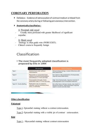
- 1. CORONARY PERFORATION Definition- Evidence of extravasation of contrastmedium or blood from the coronary artery during or following percutaneous intervention. Anatomically classifiedas:- 1) Proximal/ mid vessel - Usually more profound with greater likelihood of significant sequelae. - 2) Distal vessel - Etiology is often guide wire (WIRE EXIT). - Clinical courseis frequently benign Other classification Fukutomi Type 1- Epicardial staining without a contrast extravasation. Type 2- Epicardial staining with a visible jet of contrast extravasation. Kini Type 1 – Myocardial staining without contrast extravasation
- 2. Type 2- Contrast extravasation into pericardium, coronary sinus or cardiac chambers. Incidence 0.5% Complication rate:- ◦ POBA -0.1% ◦ Excimer laser -1.9% ◦ Rotational atherectomy – 1.3% Lesion prone for perforation CTO (27%) Calcification Tortuosity Eccentric plaque Bifurcation lesion AHA/ACC class B/C lesion Small calibre vessel (<2.5mm) Patient relatedrisk factors Older age Previous CABG Lower creatinine clearance Other risk factors Device-lumen mismatch Oversized compliant balloons (Balloon to artery ratio>1.2). High inflation pressure In case of perforation, GP 11b-111a inhibitors, is a/w - Higher incidence of tamponade - Greater requirement of emergency surgery.
- 3. - Guidewires Hydrophilic wire more prone for CAP More likely to cause distally, in the terminal sub-branches Less likely to cause frank rupture than a high pressure balloon barotrauma most of the guide wire mediated rupture is Ellis type1 or type 2 Prevention- create loop at the end of the wire. Prevention Keep ACT optimum IIIa-IIb use when indication is must UFH is preferable to Bivaluridin in complicated cases becauseeasy reversal with protamine Multiple views Dual injections Delayed watch
- 4. Start with workhorse wire or hydrophilic tipped/ stiff wires that are used to get through difficult lesions should be exchanged for workhorse wires with softer hydrophobic tips IVUS Confine wire to true lumen Do not dilate in side branch or collaterals Supportive measures:- I/V fluids Oxygen Analgesia Ionotropic support Atropine IABP Type 1 perforation treatment Usually respond to conservative measures. Close monitoring Serial echocardiography
- 5. Repeated injections of contrastmedia every 15-30 minutes No further action is required if degree of extravasation does not increase or diminishes. Increased extravasation is treated with reversal of anticoagulation and/or prolonged balloon inflation at or proximal to the perforated segment. Type 2/3 perforation Proximal/ mid vessel- Inflate balloon at the site of bleeding. Balloon inflation for upto 30 minutes usually at 2 atm If patient cannot tolerate ischemia (uncommon in CTO–PCIdue to presence of collaterals), then perfusion balloon can be used. Microcatheter over another guide wire is positioned distal to site of perforation and the patient's own arterial blood via microcatheter is injected (microcatheter distal perfusion technique). Anticoagulation- If the procedureis to be discontinued, reversalof heparin with protamine has shown to be effective. (But this should be deferred till balloons & wires are still in the artery. Antiplatelet GPI should be discontinued bcoz even trivial blush of extravasation may progress to severeproblem with this use. Abciximab bind irreversibly to platelet receptors, leading to platelet activity almost negligible for 24-36 hrs. Platelet transfusion may be required. However, in caseof tirofiban & eptifibatide, simply discontinuing the infusion is sufficient . Cardiac tamponade Urgent echo & pericardiocentesis. If there is no resolution of bleeding at 30 minutes, further action is required including surgery.
- 6. If the bleeding frompericardial tube is persistat a rate of 10mlper minute, despite mechanical & pharmacologicalaction, surgery is indicated. Measurein deploying covered stent- Deployed at high pressure(14-16 atm) with prolonged balloon inflation to allow optimal stent expansion – to ensure sealing of perforation & to reducethe risk of stent thrombosis 1) JOSTENTGraftMaster Stainless steel stent covered with polytetrafluoroethylene (PTFE) Wall thickness - 0.3mm Size - 3.0 to 5.0. Minimum Guiding catheter 6 to 7 F Bulkier than other covered stents 2) In situDirect stentstent-graft Stainless steel PTFE covered stent. Wall thickness 0.15mm Thinnest covered stent available (starting at 1.2mm.
- 7. Size – 2.5mmto 6.0mm Minimum Guiding catheter 6 to 7 F 3) Over & Under pericardiumcoveredstent Stainless steel stent covered with equine pericardium (105 um thickness). Highly flexible Size- 2.5 to 4.0mm More biocompatible, less risk of stent thrombosis. 4) PK Papyrus coveredcoronary stent Cobalt chromium stent covered with polyurethane(90um thickness) Highly flexible with low crossing profile. Size- 2.5 to 5.0mm Guiding catheter- 5 to 6Fr Low crossing profilereduces the stiffness of crimped stent graftby upto 58% Limitation of covered stents Bulkier than normal stents Reduced flexibility & trackability. Increserisk of stent thrombosis (5.7%)& restenosis (29%) Dual guiding catheter technique Aim– to reduce the time between deflation of sealing balloon & final delivery of the covered stent at the perforation site Steps- Sealing ballon is infalted Guide catheter is withdrawn slightly from coronary ostia Another guiding catheter (7Fr/8Fr) inserted from C/L femoral artery & engage the samecoronary ostia.
- 8. Covered stent graft(or coil in case of smaller & distal vessel) is advanced on a new wire via second guide catheter & placed just proximal to the sealing balloon. Sealing balloon is deflated & withdrawn proximally to allow passageof wire & covered stent which is to be deployed Sealing balloon, wire & guide catheter is removed only after gaining adequate seal of the lesion with covered stent