

Recommandé
Contenu connexe
Tendances
Tendances (20)
En vedette
Similaire à CAT
Similaire à CAT (20)
Dernier
Dernier (20)
CAT
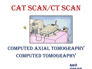
- 1. CAT Scan/CT Scan Computed Axial Tomography Computed Tomography Ravi
- 2. Basic principles • Mathematical principles of CT were first developed in 1917 by Radon • Proved that an image of an unknown object could be produced if one had an infinite number of projections through the object
- 3. X-Rays • photons produced by an electron beam • similar to visible light, but higher energy!
- 4. X-Rays - Physics • Associated with inner shell electrons • As the electrons decelerate in the target through interaction, they emit electromagnetic radiation in the form of x-rays. • patient between an x-ray source and a film -> radiograph cheap and relatively easy to use potentially damaging to biological tissue
- 5. Basic principles • Plain film imaging reduces the 3D patient anatomy to a 2D projection image • Density at a given point on an image represents the x-ray attenuation properties within the patient along a line between the x-ray focal spot and the point on the detector corresponding to the point on the image
- 6. Basic principles (cont.) • With a conventional radiograph, information with respect to the dimension parallel to the x-ray beam is lost • Limitation can be overcome, to some degree, by acquiring two images at an angle of 90 degrees to one another • For objects that can be identified in both images, the two films provide location information
- 7. CT - basics • CT's primary benefit is the ability to separate anatomical structures at different depths within the body. • A form of tomography can be performed by moving the X-ray source and detector during an exposure. • Anatomy at the target level remains sharp, while structures at different levels are blurred. • By varying the extent and path of motion, a variety of effects can be obtained, with variable depth of fieldand different degrees of blurring of 'out of plane' structures.
- 8. CT Basics
- 10. CT & CAT •CAT stands for Computerized Axial Tomography which was later changer to just CT or rather Computerized Tomography. •The scanners originally only did axial images (referring to the plane of the image) but later became able to do other planes such as coronal or saggital imaging and can now do anything with the block of information it collects.
- 11. Computed Axial Tomography • Godfrey Newbold Hounsfield introduced the system to the public in 1972 • Nobel Prize in 1979 • It looks like a donut – Made up of a series of X- ray sources and sensors – Tomogram
- 12. Process and Procedure • X-ray + Axial view + Computer Imaging = CAT Scan • X-ray + (digital) Computer Imaging = CT scan • Provides cross-sectional views and images of body organs and structures • Pixel, Radiodensity, Voxel, Windowing • Contrast Material
- 13. Computed Axial Tomography • Also called CAT scanning or “CT” • Image formed using a rotating thin beam(s) of ionizing radiation • Image “slices” reconstructed by computation • The image formed is related to the subjects density • Image display on computer or multiple films • New technology is multislice helical scanner
- 14. CT - principle • Because contemporary CT scanners offer isotropic, or near isotropic, resolution, display of images does not need to be restricted to the conventional axial images. • Instead, it is possible for a software program to build a volume by 'stacking' the individual slices one on top of the other. The program may then display the volume in an alternative manner.
- 15. Tomographic images • The tomographic image is a picture of a slab of the patient’s anatomy • The 2D CT image corresponds to a 3D section of the patient • CT slice thickness is very thin (1 to 10 mm) and is approximately uniform • The 2D array of pixels in the CT image corresponds to an equal number of 3D voxels (volume elements) in the patient • Each pixel on the CT image displays the average x-ray attenuation properties of the tissue in the corrsponding voxel
- 17. Tomographic acquisition • Single transmission measurement through the patient made by a single detector at a given moment in time is called a ray • A series of rays that pass through the patient at the same orientation is called a projection or view • Two projection geometries have been used in CT imaging: – Parallel beam geometry with all rays in a projection parallel to one another – Fan beam geometry, in which the rays at a given projection angle diverge
- 24. diagnostic Uses • The Head – Hemorrhaging, cerebrovascular accidents, trauma • The Abdomen, Pelvis, and Chest – Cancers, pneumonia, infection – Barium Sulfate • The Spine and Bones – Vertebrae, disc, spinal cord definition, and bone density – Indicator of Osteoporosis • Cardiac CT angiography.
- 25. CT - diagnostic use Cranial • diagnosis of cerebrovascular accidents and intracranial hemorrhage • CT generally does not exclude infarct in the acute stage of a stroke. For detection of tumors, CT scanning with IV contrast is occasionally used but is less sensitive than magnetic resonance imaging (MRI).
- 26. CT - diagnostic use Chest •CT is excellent for detecting both acute and chronic changes in the lung parenchyma. •For evaluation of chronic interstitial processes (emphysema, fibrosis, and so forth), thin sections with high spatial frequency reconstructions are used - often scans are performed both in inspiration and expiration. This special technique is called High resolution CT (HRCT). •For detection of airspace disease (such as pneumonia) or cancer, relatively thick sections and general Purpose image reconstruction techniques may be adequate.
- 27. CT - diagnostic use Abdominal and pelvic • CT is a sensitive method for diagnosis of abdominal diseases. It is used frequently to determine stage of cancer and to follow progress. It is also a useful test to investigate acute abdominal pain. • Renal/urinary stones, appendicitis, pancreatitis, diverticulitis, and bowel obstruction are conditions that are readily diagnosed and assessed with CT. • CT is also the first line for detecting solid organ injury after trauma.
- 28. CT – step by step
- 29. CT – step by step
- 30. CT – step by step
- 31. CT – step by step
- 32. Benefits: • Painless procedure…other than needing great deal of patience • Alternative to catheters and other guide camera options • A good option for those weary of enclosed spaces • Usually preferred over MRI, and costs less!!! • Improved technology has lead to Spiral and Helical Scans…faster processes
- 33. Spiral and Helical Scanners • Spiral • Circular Path • Constant gantry rotation and readings • Less exposure to radiation • Faster scan = better accuracy when patient must hold their breath
- 34. Potential Risks • Exposure to Radiation – Reduced with Spiral Scan • Reaction to contrast material – Diabetics – Isovue • Pregnant and nursing women should not be exposed
- 35. Investigating Emotionality and Psychopathology • Dementia • Anorexia Nervosa • Neurological Abnormalities • Schizophrenia
- 36. THANKS
- 37. CT - diagnostic use Cardiac • With the advent of subsecond rotation combined with multi- slice CT (up to 64-slice), high resolution and high speed can be obtained at the same time, allowing excellent imaging of the coronary arteries (cardiac CT angiography). • Images with an even higher temporal resolution can be formed using retrospective ECG gating. In this technique, each portion of the heart is imaged more than once while an ECG trace is recorded. The ECG is then used to correlate the CT data with their corresponding phases of cardiac contraction. Once this correlation is complete, all data that were recorded while the heart was in motion (systole) can be ignored and images can be made from the remaining data that happened to be acquired while the heart was at rest (diastole). In this way, individual frames in a cardiac CT investigation have a better temporal resolution than the shortest tube rotation time.