Airway management in the Emergency Department for Trainees
•Télécharger en tant que PPTX, PDF•
22 j'aime•10,871 vues

This is a power point presentation on Airway Management given by our deputy director in Emergency Medicine Training at the Wollongong Hospital, Paul Labana (consultant Emergency Physician) that presents a case illustrating difficulties in airway management and gives an overview of airway management in the emergency department. (Nb another video to do with airway management, and "airway exchange" can be found on this link http://youtu.be/6vaWNknIDQg) - thanks to Paul for sharing his educational material in the name of free open access meducation (#FOAMed)

Recommandé
Contenu connexe
Tendances
Tendances (20)
En vedette
En vedette (20)
Similaire à Airway management in the Emergency Department for Trainees
Similaire à Airway management in the Emergency Department for Trainees (20)
Plus de Bishan Rajapakse
Plus de Bishan Rajapakse (20)
Dernier
Dernier (20)
Airway management in the Emergency Department for Trainees
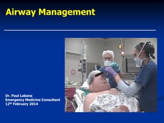
- 1. Airway Management Dr. Paul Labana Emergency Medicine Consultant 12th February 2014
- 2. Overview Case presentation Assessment Clinical features Decision making algorithms Equipment used
- 3. 35 yr male is t/f to TWH with 5% PT burns to face, neck, anterior chest after methylated spirits explodes from fire PMHx – Type I DM, hypercholesterolaemia, hypertension, smoker, occasional EtOH Coversyl +, Amlodipine, Atorvastatin, Protophane, Actrapid Allergic to penicillin On examination: A, B, C currently stable pain under control, covered in dressings with only nose showing, blisters/swelling around lips/oropharynx Starts to complain of SOB – vitals OK on high flow oxygen
- 4. What would you do next?
- 5. O/E facial swelling (but not in mouth), blisters on lips, nasal hair singed Decision for definitive airway Pre-ox, cricoid pressure, propofol, suxamethonium Initially grade 2 view with McCoy blade + stylet ETT dislodged on removing stylet (paraffin all over face/gloves making jaw thrust impossible) Bag & Mask ventilation Sux. wearing off, biting through guedel, desat 55% at lowest Further propofol, sux – Fastrach inserted #5 but big leak Replaced with classic – good ventilation Pt. became bradycardic – atropine 300 mcg Further attempts at ETT with swollen tongue, eventually intubated as confirmed by ETCO2
- 6. Progress Continued to improve over next 5 days Successfully extubated with nil stridor and good ABG’s D/C to burns unit Later d/c and was well
- 7. General Principles of Airway Management
- 8. Airway Management Aims of management Maintain oxygenation, prevent hypercarbia Minimise airway trauma (i.e. minimal instrumentation) 3 main scenarios Anticipated difficult airway e.g. burns Unanticipated difficult airway Facial/neck trauma
- 9. Methods of assessment Anaesthetic history (if at all available) History Previous difficult intubation/ventilation Congenital syndromes Down’s syndrome, large tongue, Atlanto-axial instability Nasal polyps, neck and TMJ problems, loose teeth Examination Facial hair Teeth: protruding or long upper incisors, prominent overbite Mouth opening (need at least 3-4 cm between incisors) High arched, narrow palate Mandibular protrusion (upper lip bite) Neck mobility and masses Thyromental distance (>6cm normal)
- 10. Examination General Habitus Ethnic differences: anterior larynx, small jaw, protruberant teeth Pregnant: Large breasts, mucosal hypervascularity, reduced oxygen reservoir with risk of desaturation Congenital syndromes: Downs’ syndrome, Goldenhaar, Pierre-Robin, Achondroplasia Orofacial trauma - (patient in C-spine collar?) Nose Nasal polyps, deviated septum, hypervascularity in pregnancy Mouth Small or restricted mouth opening, restricting laryngoscope passage Teeth Protruberant incisors, loose teeth (potential airway obstruction if knocked out) Tongue Macroglossia (Down’s Syndrome, Acromegaly) Jaw (incl. TMJ) Small or recessive jaw, previous TMJ surgery causing restriction may impede laryngoscopic view Neck Bullneck, previous surgery/tracheostomy Reduced ROM: Ankylosing Spondylitis, previous fusion, rheumatoid instability Environment Lack of equipment, skilled assistant
- 11. Relative Tongue/Pharyngeal size Modified Mallampati classification gives variable prediction of airway difficulty (rarely possible in emergency setting) I: Soft Palate, Fauces, Uvula, Ant + Post pillars II: Soft palate, Fauces, Uvula III: Soft palate, base of uvula IV: Hard palate only Performed with patient seated, head in neutral position, mouth open to widest extent and tongue maximally protruded without phonation Correlation with Cormack-Lehane grading: Class I: Grade I view 99-100% of the time Class IV: Grade III or IV view 100% of the time Class II and III are poor predictors
- 12. The Mallampati classification is a simple scoring system that relates the amount of mouth opening to the size of the tongue, and provides an estimate of space available for oral intubation by direct laryngoscopy. According to the Mallampati scale, class one is present when the soft palate, uvula, and pillars are visible, class two when the soft palate and uvula are visible, class three when the soft palate and only the base of the uvula are visible, and class four when only the hard palate is visible.
- 13. The Cormack-Lehane system for grading laryngoscopic view at intubation
- 14. Difficult ventilation Pregnant Increased breast tissue with reduced chest compliance Obese As above, plus increased pharyngeal tissue with increased upper airway resistance OSA / Snorers Beard Difficult to achieve seal with mask Edentulous Difficult to achieve seal with mask
- 15. Neck mobility and correct positioning 1. Bad : neutral position 2. Better : C6/C7 flexion 3. Best : Flexion at C6/C7 Extension at C1/C2 (~ 35º) “Sniffing the morning air…”
- 16. Complications of difficult airway access Hypoxaemia Soft tissue injury of the airway Increased gastric aspiration/regurgitation risk Haemodynamic stress of repeated laryngoscopic stress attempts Unnecessary tracheostomy Tooth damage
- 17. Facial/Neck Trauma & Burns Ensure senior anaesthetic/surgical help present Early consideration of intubation/surgical airway essential Childhood infections causing partial obstruction generally approached via gentle inhalational induction therefore call anaesthetics a.s.a.p. Can return to spontaneous breathing if laryngoscopic view poor, or offer option of FOB (bearing in mind it may occlude already narrow lumen) In cases of marked deformed anatomy, tracheostomy is generally the definitive management Open vs percutaneous dilational: No major difference in outcome, PDT associated with reduced risk of pneumomediastinum/bleeding May be performed under local anaesthetic (ketamine has also been described)
- 18. LEMON L ook (facies, anatomy, obesity etc.) E valuate 3-3-2 (see below) M allampati O bstruction (mass, infection, SOL) N eck mobility (“sniffing morning air”)
- 19. Preparation for intubation P repare patient & drugs & P ositioning E nd-tidal CO2 M ask (& bag connected to O2 AND ON) A djuncts (LMA/Guedel/Nasopharyngeal) I ntroducer L aryngoscope S uction (turned on at head of bed)
- 22. Laryngoscopes Macintosh (standard used on airway trolleys) McCoy Kessel Modified Macintosh blade with increased handle-blade angle (110˚): easier insertion in large-breasted (pregnant) women Miller Levered mobile tip allowing elevation of epiglottis Potentially reduces C-L grade by one Useful in cervical collar patients, anterior larynx Straight blade with curved tip for elevating epiglottis Thinner profile: easier insertion in small-mouth opening pts Huffman Macintosh blade with 30 or 80˚ refractive prism towards larynx: allows indirect laryngoscopy
- 23. Intubation Aides Gum elastic bougie (Eschmann tracheal tube introducer) 60cm, 15F (adults)/10F (children), Coude tip. Introduced in the 1970’s Portex reusable bougies most common. Disposable available but reported to have reduced success with intubation (?due to less malleable texture) Used in conjunction with laryngoscope to facilitate ETT passage where laryngeal inlet is incompletely visualised (or can be inserted blindly) Advantages are Longer compared with stylets Malleable, angled tip - ideally at 60˚ to capture tracheal “clicks” Flexible yet firm enough to have ETT railroaded over it Signs that bougie is endotracheal (therefore safe to railroad ETT) “Clicks” from tracheal rings Hold-up/Resistance at ~40cm (within bronchial tree) Coughing
- 24. Intubation Aides 2 Airway exchange catheters Functions as bougie, but with central lumen allowing ongoing oxygenation between intubation attempts Internal diameter varies between 3.7mm (Cook exchange catheters) to 4.7mm (Aintree airway catheters) NICE paper demonstrating use in conjunction with LMA and fibreoptic bronchoscope: Low-skill fibreoptic intubation: use of the Aintree catheter with the classic LMA. Anaesthesia 2005; 60: 915-920 Stylet Malleable, single use stylets used to shape ETT, allowing for easier passage into anterior larynxes BURP/2 person intubation
- 25. Laryngeal Mask Airway Most are disposable Once inserted, the mask is bounded by: Tip at level of inferior constrictor Sides abutting piriform fossae Top against base of tongue Provides airway support rather than definitive airway Valuable tool in rescue “can’t intubate, can’t mask ventilate” situations Serves as a conduit for endotracheal intubation Theoretically #4 can fit a 5.5 ETT, but clumsy fit at best Narrowest point is at LMA connector - ETT cuff may be torn here Length of LMA tube may lead to improper depth of ETT placement Very difficult to remove LMA once ETT is placed Gum-elastic bougie/Fibreoptic bronchoscope may be fed down lumen to facilitate endotracheal intubation and confirm position Bronchoscopic port connector allows simultaneous ventilation & FOB
- 26. Cook Kit (Seldinger technique) Seldinger technique safer than blind approach Aspirate air with the needle and syringe to check placement, cut with scalpel Remove syringe, insert wire into needle, remove needle Thread dilator with airway already loaded onto wire Remove wire
- 28. Needle cricothyroidotomy Oxygenation NOT ventilation Use 14 g cannula with syringe attached, once aspirating air, insert sheath and remove needle Connect a 3 way valve to sheath and to oxygen tubing 15 L/min oxygen for 1 sec followed by 4 secs expiration phase Approximately 45 mins to get definitive airway
- 29. Cricothyroid membrane between thyroid and cricoid cartilage Locate hyoid with thumb & index finger and middle finger will fall on correct area (1-1.5 cm below)
- 30. Standard Surgical Airway Equipment always available BUT need most senior help available Vertical incision in skin of neck Transverse incision of cricothyroid membrane Artery forceps/finger to open airway Insert cuffed ETT 6.0 into trachea
- 31. Surgical Airway
- 33. Questions ???