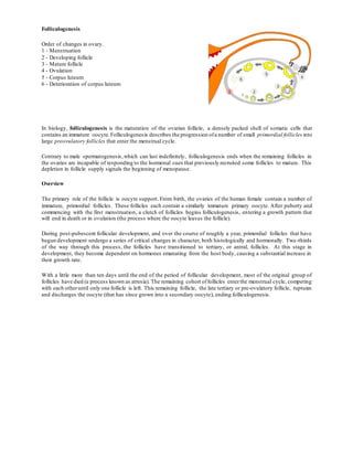
SlideShare une entreprise Scribd logo
1  sur  11
Folliculogenesis
Order of changes in ovary.
1 - Menstruation
2 - Developing follicle
3 - Mature follicle
4 - Ovulation
5 - Corpus luteum
6 - Deterioration of corpus luteum
In biology, folliculogenesis is the maturation of the ovarian follicle, a densely packed shell of somatic cells that
contains an immature oocyte.Folliculogenesis describes the progression ofa number of small primordial follicles into
large preovulatory follicles that enter the menstrual cycle.
Contrary to male spermatogenesis,which can last indefinitely, folliculogenesis ends when the remaining follicles in
the ovaries are incapable of responding to the hormonal cues that previously recruited some follicles to mature. This
depletion in follicle supply signals the beginning of menopause.
Overview
The primary role of the follicle is oocyte support. From birth, the ovaries of the human female contain a number of
immature, primordial follicles. These follicles each contain a similarly immature primary oocyte. After puberty and
commencing with the first menstruation, a clutch of follicles begins folliculogenesis, entering a growth pattern that
will end in death or in ovulation (the process where the oocyte leaves the follicle).
During post-pubescent follicular development, and over the course of roughly a year, primordial follicles that have
begun development undergo a series of critical changes in character, both histologically and hormonally. Two-thirds
of the way through this process, the follicles have transitioned to tertiary, or antral, follicles. At this stage in
development, they become dependent on hormones emanating from the host body, causing a substantial increase in
their growth rate.
With a little more than ten days until the end of the period of follicular development, most of the original group of
follicles have died (a process known as atresia). The remaining cohort offollicles enterthe menstrual cycle, competing
with each otheruntil only one follicle is left. This remaining follicle, the late tertiary or pre-ovulatory follicle, ruptures
and discharges the oocyte (that has since grown into a secondary oocyte), ending folliculogenesis.
Diagram of folliculogenesis, starting from pre-antral (late secondary), courtesy NCBI
Phases of development
Folliculogenesis lasts for approximately 375 days. It coincides with thirteen menstrual cycles. The process begins
continuously, meaning that at any time the ovary contains follicles in all stages of development, and ends when a
mature oocyte departs fromthe preovulatory follicle in a process called ovulation.
The growing follicle passes through the following distinct stages that are defined by certain structural characteristics
(unfamiliar terms will be defined in their respective sections):
In a larger perspective, the whole folliculogenesis, from primordial to preovulatory follicle, belongs to the stage of
ootidogenesis of oogenesis.
Stage Description Size
Primordial
Dormant, small, only one layer of
flat granulosa cells
Primordial follicles are about 0.03-0.05 mm in diameter.
Primary
Mitotic cells, cuboidal granulosa
cells
Almost 0.1 mm in diameter
Secondary
Presence of theca cells, multiple
layers of granulosa cells
The follicle is now 0.2 mm in diameter
Early
tertiary
The early tertiary follicle is arbitrarily divided into five classes.
Class 1 follicles are 0.2 mm in diameter, class 2 about 0.4 mm,
class 3 about 0.9 mm, class 4 about 2 mm, and class 5 about 5 mm.
Late tertiary
Fully formed antrum, no further
cytodifferentiation, no novel
progress
Class 6 follicles are about 10 mm in diameter, class 7 about 16 mm,
and class 8 about 20 mm. It is common for non-dominant follicles
to grow beyond class 5, but rarely is there more than one class 8
follicle.
Preovulatory
Building growth in estrogen
concentration, all other follicles
atretic or dead
In addition, follicles that have formed an antrum are called antral follicles or Graafian follicles. Definitions differ in
where this shift occurs in the staging given above,with some stating that it occurs when entering the secondary stage,[1]
and others stating that it occurs when entering the tertiary stage.[2]
Until the preovulatory stage, the follicle contains a primary oocyte that is arrested in prophase of meiosis I. During
the late preovulatory stage, the oocyte continues meiosis and becomes a secondary oocyte, arrested in metaphase II.
(a) The maturation of a follicle is shown in a clockwise direction proceeding from the primordial follicles. FSH
stimulates the growth of a tertiary follicle, and LH stimulates the production of estrogen by granulosa and theca cells.
Once the follicle is mature, it ruptures and releases the oocyte. Cells remaining in the follicle then develop into the
corpus luteum. (b) In this electron micrograph of a secondary follicle, the oocyte, theca cells (thecae folliculi), and
developing antrum are clearly visible. Electron microscopy images
Primordial
At 18–22 weeks post-conception, the cortex of the female ovary contains its peak number of follicles (about 4 to 5
million in the average case, but individual peak populations range from 6 to 7 million[3]). These primordial follicles
contain immature oocytes surrounded by flat, squamous granulosa cells (the support cells) that are segregated from
the oocyte's environment by the basal lamina. They are quiescent, showing little to no biological activity. Because
primordial follicles can be dormant for up to 50 years in the human, the length of the ovarian cycle does not include
this time.
The supply of follicles decreases slightly before birth, and to 180,000 by puberty for the average case (populations at
puberty range from 25,000 to 1.5 million).[3] By virtue of the "inefficient" nature of folliculogenesis (discussed later),
only 400 of these follicles will ever reach the preovulatory stage.At menopause, only 1,000 follicles remain. It seems
likely that early menopause occurs for women with low populations at birth, and late menopause occurs for women
with high populations at birth, but there is as yet no clinical evidence for this.[3]
The process by which primordial cells 'wake up' is known as initial recruitment. Research has shown that initial
recruitment is mediated by the counterbalance of various stimulatory and inhibitory hormones and locally produced
growth factors.[4]
Primary
The granulosa cells of these primordial follicles change from a flat to a cuboidal structure,marking the beginning of
the primary follicle. The oocyte genome is activated and genes become transcribed. Rudimentary paracrine signalling
pathways that are vital for communication between the follicle and oocyte are formed. Both the oocyte and the follicle
grow dramatically, increasing to almost 0.1 mm in diameter.
Primary follicles develop receptors to follicle stimulating hormone (FSH) at this time, but they are gonadotropin-
independent until the antral stage.Research has shown, however, that the presence of FSH accelerates follicle growth
in vitro.
A glycoprotein polymer capsule called the zona pellucida forms around the oocyte,separating it from the surrounding
granulosa cells. The zona pellucida, which remains with the oocyte after ovulation, contains enzymes that catalyze
with sperm to allow penetration.
Secondary
Stroma-like theca cells are recruited by oocyte-secreted signals.They surround the follicle's outermost layer, the basal
lamina, and undergo cytodifferentiation to become the theca externa and theca interna. An intricate network of
capillary vessels forms between these two thecal layers and begins to circulate blood to and fromthe follicle.
The late-term secondary follicle is marked histologically by a fully grown oocyte surrounded by a zona pellucida,
approximately nine layers of granulosa cells, a basal lamina, a theca interna, a capillary net, and a theca externa.
290 days have lapsed since recruitment.
Antrum formation
Further information: Antral follicle
The formation of a fluid-filled cavity adjacent to the oocyte called the antrum designates the follicle as an antral
follicle, in contrast to so a called preantral follicle that still lacks an antrum. An antral follicle is also called a Graafian
follicle.
Definitions differ in which stage this shift occurs, with some designating follicles in the secondary stage as antral,[1]
and others designating themas preantral.[2]
Early tertiary
In the tertiary follicle,the basic structure ofthe mature follicle has formed and no novelcells are detectable.Granulosa
and theca cells continue to undergo mitotis concomitant with an increase in antrum volume. Tertiary follicles can
attain a tremendous size that is hampered only by the availability of FSH, which it is now dependent on.
Under action of an oocyte-secreted morphogenic gradient, the granulosa cells of the tertiary follicle undergo
differentiation into four distinct subtypes: corona radiata,surrounding the zona pellucida; membrana, interior to the
basal lamina; periantral, adjacent to the antrum and cumulus oophorous, which connects the membrana and corona
radiata granulosa cells together. Each type of cell behaves differently in response to FSH.
Theca cells express receptors for luteinizing hormone (LH). LH induces the production of androgens by the theca
cells, most notably androstendione,which are aromatized by granulosa cells to produce estrogens, primarily estradiol.
Consequently, estrogen levels begin to rise.
Late tertiary and preovulatory (the follicular phase of the menstrual cycle)
At this point, the majority of the group of follicles that started growth 360 days ago have already died. This process
of follicle death is known as atresia, and it is characterized by radical apoptosis ofall constituent cells and the oocyte.
Although it is not known what causes atresia, the presence of high concentrations of FSH has been shown to prevent
it.
A rise in pituitary FSH caused by the disintegration of the corpus luteum at the conclusion of the twelfth menstrual
cycle precipitates the selection of five to seven class 5 follicles to participate in the thirteenth.These follicles enter the
end of the twelfth menstrual cycle and transition into the follicular phase of the thirteenth cycle. The selected follicles,
called antral follicles, compete with each other for growth-inducing FSH.
In response to the rise ofFSH, the antral follicles begin to secrete estrogen and inhibin,which have a negative feedback
effect on FSH.[5] Follicles that have fewer FSH-receptors will not be able to develop further; they will showretardation
of their growth rate and become atretic. Eventually, only one follicle will be viable. This remaining follicle, called the
dominant follicle,will grow quickly and dramatically—up to 20 mm in diameter—to become the preovulatory follicle.
Note: Many sources misrepresent the pace of follicle growth, some even suggesting that it takes only fourteen days
for a primordial follicle to become preovulatory. In all cases, the follicular phase of the menstrual cycle means the
time between selection of a tertiary follicle and its subsequent growth into a preovulatory follicle.
Ovulation and the corpus luteum
By the end of the follicular, or proliferative, phase of the thirteenth day of the menstrual cycle, the cumulus oophorus
layer of the preovulatory follicle will develop an opening, or stigma, and excrete the oocyte with a complement of
cumulus cells in a process called ovulation. The oocyte is now called the ovum and is competent to undergo
fertilization. The ovum will now travel down one of the fallopian tubes to eventually be discharged through
menstruation, if not fertilized by a sperm cell, or implanted in the uterus,if previously fertilized. The fully developed
oocyte (gamete) is now at the behest of the menstrual cycle.
The ruptured follicle will undergo a dramatic transformation into the corpus luteum, a steroidiogenic cluster of cells
that maintains the endometrium of the uterus by the secretion of large amounts of progesterone and minor amounts of
estrogen.
These two steps,while not part of folliculogenesis, are included for completeness.They are discussed in their entirety
by their respective articles, and placed into perspective by the menstrual cycle article. It is recommended that these
three topics be reviewed.
Hormone function
As with most things related to the reproductive system, folliculogenesis is controlled by the endocrine system. Five
hormones participate in an intricate process of positive and negative feedback to regulate folliculogenesis. They are:
 gonadotropin-releasing hormone (GnRH) secreted by the hypothalamus
 two gonadotropins:
o follicle-stimulating hormone (FSH)
o luteinizing hormone (LH)
 estrogen
 progesterone
GnRH stimulates the release of FSH and LH from the anterior pituitary gland that will later have a stimulatory effect
on follicle growth (not immediately, however, because only antral follicles are dependent on FSH and LH). When
theca cells form in the tertiary follicle the amount of estrogen increases sharply (theca-derived androgen is aromatized
into estrogen by the granulosa cells).
At low concentration,estrogen inhibits gonadotropins,but high concentration ofestrogen stimulates them.In addition,
as more estrogen is secreted, more LH receptors are made by the theca cells, inciting theca cells to create more
androgen that will become estrogen downstream. This positive feedback loop causes LH to spike sharply, and it is this
spike that causes ovulation.
Following ovulation, LH stimulates the formation of the corpus luteum. Estrogen has since dropped to negative
stimulatory levels after ovulation and therefore serves to maintain the concentration of FSH and LH. Inhibin, which
is also secreted by the corpus luteum, contributes to FSH inhibition.
The endocrine system coincides with the menstrual cycle and goes through thirteen cycles (and thus thirteen LH
spikes) during the course of normal folliculogenesis. However, coordinated enzyme signalling and the time-specific
expression of hormonal receptors ensures that follicle growth does not become disregulated during these premat ure
spikes.
Number of follicles
"Percentage of ovarian reserve related to increasing age. The curve describes the percentage of ovarian reserve
remaining at ages from birth to 55 years,based on the ADC model. 100% is taken to be the maximum ovarian reserve,
occurring at 18–22 weeks post-conception.The percentages apply to allwomen whose ovarian reserve declines in line
with our model (i.e. late and early menopause are associated with high and low peak NGF populations,respectively).
We estimate that for 95% of women by the age of 30 years only 12% of their maximum pre-birth NGF population is
present and by the age of 40 years only 3% remains. doi:10.1371/journal.pone.0008772.g005"[6]
Recently, two publications have challenged the idea that a finite number of follicles are set around the time of birth.[7][8]
Renewal of ovarian follicles from germline stem cells (originating from bone marrow and peripheral blood) was
reported in the postnatal mouse ovary. Studies attempting to replicate these results are underway, but a study of
populations in 325 human ovaries found no supporting evidence for follicular replenishment.[3]
In 2010, researchers at the University of Edinburgh determined that by the time women are 30 years old, only 10% of
their non-growing follicles (NGFs) remain.[6] At birth, women have all their follicles for folliculogenesis, and they
steadily decline until menopause.
Depletion of the ovarian reserve
As women (and mice) age, double-strand breaks accumulate in their primordial follicle reserve. These follicles contain
primary oocytes that are arrested in prophase of the first meiotic division. Meiosis, in eukaryotic organisms, is the
general process underlying germ cell formation, and it appears to be an adaptation for repairing DNA damages,
particularly double-strand breaks in germ line DNA.[9] (See Meiosis.) Double-strand breaks are accurately repaired
during meiosis by the particular process termed “homologous recombinational repair.” Titus et al.[10] (2013) found
that, as humans (and mice) age, expression of four key DNA repair genes necessary forhomologous recombinational
repair declines in oocytes. They hypothesized that DNA double-strand break repair is vital for the maintenance of
oocyte reserve, and that a decline in efficiency of repair with age plays a key role in the depletion of the ovarian
reserve (ovarian aging).
FOLLICULOGENESIS AND OVULATION
J.F. Guerin
Laboratory of Histology, Embryology and Reproductive Biology,
Faculty of Medicine Lyon Nord
8, Avenue Rockefeller - Lyon 8e, 69373 Lyon CEDEX 08
The ovaries, like the testicles, exert a double function, exocrine and endocrine, consisting ofthe production of
gametes, the oocytes, as well as sex hormones, estrogens and progesterone. Whereas in the testicles the two
functions are assured permanently from puberty onwards by two different structures, however, in the ovary
they are exerted cyclically, between puberty and the menopause, and result from the evolution of a same
morphological unit, the ovarian follicle, situated within the cortical stroma.
Histology of ovarian organelles
The gametogenic follicles
These follicles correspond to different stages of the evolution of primordial follicles up to the rupture of the mature
follicle (ovulation). Each one contains an oocyte and is the site ofoogenesis and ofthe production ofsteroid hormones.
The primordial follicle.
Around the 7th month of embryonic development, the ovarian cortex contains a definitive stock of several million
primordial follicles which progressively diminishes up to the menopause. Each follicle, within the cortical stroma, is
made up of a 1st order oocyte (oocyte 1) surrounded by a layer of flattened follicular cells, these cells being covered
by a basal membrane (membrane of Slavjanski). Oocyte 1 measures about 30 µm in diameter.
The primary follicle.
It is characterized by the transformation of the flattened follicular cells into cubic cells.
The secondary follicle.
This follicle is called secondary once the multiplication of the follicular cells constitutes a second layer around the
oocyte.The diameter of the follicle progressively increases up to about 180 µm. The follicular cells reach about 5000
in number and togetherconstitute the granulosa. Oocyte 1 begins its growth and its diameter increases from 40 up to
60 µm. At the last stage of its development, the secondary follicle appears surrounded by irregularly spaced islets of
differentiated epithelioid cells from stromal fibroblasts and in relation with the capillaries. Together the epithelioid
cells constitute the internal theca (theca interna) of the follicle. The secondary follicle, provided with its theca interna
is called a preantral follicle.
The tertiary follicle.
Also called cavitary follicle or antral follicle, it is characterized by the presence of a cavity (antrum) in the granulosa
and a theca externa, a fibrous layer around the theca interna. It increases considerably in volume because of the rapid
multiplication of the follicular cells which will reach about 50 million in number. At the end of its development, the
follicle (roughly 2 cm in diameter) will become a preovulatory or mature follicle.
In the clumps of the granulosa there appear small drops of liquid whose confluence forms the antrum which cont ains
the follicular fluid produced by the follicular cells. Around the oocyte, the granulosa projects into the follicular
cavity—the cumulus oophorus. The theca interna, separated from the granulosa by the membrane of Slavjanski, is
made up of numerous clusters ofepithelioid cells. Electron microscopy reveals that these cells have the characteristics
of steroidogenic cells, identical to those observed in Leydig cells. The theca externa is made up of a thick layer of
collagen fibres, traversed by numerous blood capillaries; it contains myofibroblasts differentiated from fibroblasts of
the stroma.
Up to the preovulatory stage offollicular evolution, the oocyte harboured in the cumulus is an oocyte 1 blocked at the
end of the prophase (diakinesis stage). Cytoplasmic growth continues and the oocyte attains around 120 µm in
diameter.
The preovulatory period and ovulation.
At the end of its growth, the mature follicle reacts to a discharge of gonadotropic hormones by great transformations
which end in follicular rupture (ovulation). The cumulus cells secrete large quantities of hyaluronic acid which
accumulates in the intercellular space and provokes the dissociation ofthe cumulus followed by its rupture: the oocyte
surrounded by certain quantity of follicular cells is released into the follicular fluid. The apical region, the ovarian
stroma, is the site of a vasoconstriction which results in an ischemia followed by necrosis,within a few hours,of the
stroma and the follicular wall. The gonadotropic discharge will give rise to a release of histamine and bradykinin,
leading to an edema of the theca. At the same time, the secretion of a plasminogen activator will also activate
collagenases which will dissociate the theca externa, this action being reinforced by the release of prostaglandins.
Lastly, the cells of the ovarian epithelium in the apical region, would appear to be subject to autolysis,leading to the
release of lysosomal hydrolases and thus to the dissociation of the apex (a mechanism which could be deficient in the
luteinized unruptured follicle [LUF] syndrome).
The oocyte completes its cytoplasmic and nuclear maturation in the cytoplasm, the cortical granules migrate to the
periphery and attach to the plasma membrane. Meiosis resumes but is again blocked in 2nd division metaphase
(metaphase II). Ovulation commences with the rupture of the necrosed tissues of the apex (stigma). The viscous
follicular fluid begins to flow. The decrease in pressure ofthe follicular liquid induces a series ofrhythmic contractions
of the myofibroblasts of the theca externa and of all the cortical stroma which lead to the expulsion of the follicular
fluid and oocyte II surrounded by cumulus cells.
The corpus luteum
After expulsion of the oocyte,the follicle presents a pleated aspect. It’s then called a dehiscent follicle. The membrane
of Slavjanski disappears completely and the blood capillaries of the theca rapidly invade the granulosa thereby
provoking the transformation of these cells (luteinization) by the constitution of the corpus luteum.
The blood vessels completely traverse the granulosa and open up in the follicular cavity, thereby causing a
circumscribed and rapidly coagulated hemorrhage (central coagulum). The granulosa cells are transformed into large
luteal cells, approximately 40 µm in diameter, whose ultrastructure is the same as that of steroidogenic cells. The cells
of the theca interna (hardly modified) constitute the small luteal or paraluteinic cells, situated at the periphery of the
corpus luteumand forming strings that penetrate more or less deeply into the layer of the large cells.
Follicular atresia and luteolysis
Between the 7th month of fetal life and the menopause, most of the gametogenic follicles undergo an involution
(involutive or atretic follicles). Only 300-400 follicles will reach the preovulatory stage. All the involutive follicles
which preserve for a certain time their theca interna are called thecogenic follicles. The theca cells of these follicles
as a whole constitute the interstitial gland of the ovary.
Involution of the corpus luteum, or luteolysis, occurs most often in the form of a fibrous or fibro-hyalin degeneration
with cell lysis and marked collagen fibre synthesis, which ends in the formation of a voluminous organelle called "
corpus albicans ". The process is relatively slow and spread over several weeks.
Dynamics of follicular growth
In the human being, the stock of primordial follicles, called " reserve follicles ", is about 1 million at birth, and at the
beginning of puberty a few hundred thousands.As already emphasized, practically all the follicles (over 99%) will be
affected by the phenomenon of atresia, but at variable stages in the course of development. The interregulation of
these two physiological phenomena—growth and atresia—is governed by complex mechanisms, which are now
beginning to be elucidated in the human female, through the works of Gougeon in particular.
It has been established that an average of85 days—i.e. corresponding to 3 ovarian cycles—separate the moment when
a follicle becomes preovulatory (stage 8 of Gougeon’s classification) and the moment when it has differentiated its
theca interna (i.e. is at stage 1 or " preantral "). This means that a preovulatory follicle enters the preantral stage 85
days earlier, in mid-cycle, at the time of the preovulatory discharge of the gonadotropic hormones follicle-stimulating
hormone (FSH) and luteinizing hormone (LH).
As it is also recognised that entry into the preantral stage occurs randomly at any moment during the cycle, it may be
deduced that all the follicles that differentiate their theca at a time that does not correspond to the preovulatory period
will evolve more or less rapidly to atresia. A hypothesis that has been advanced is that the concentration of plasma
FSH at the time of theca differentiation conditions the future quality of the theca, and more generally of the follicle to
which it belongs.
It is nevertheless recognized that, up to a diameter of 2-4 mm (stage 4-5), follicular growth requires only a minimal
concentration (basal) of FSH. Follicles up to 4 mm diameter may be found in impuberal girls or in women using
hormonal contraception. Further follicular growth requires stimulation by gonadotropic hormones, and more
especially by FSH. We can thus distinguish three steps:
1. Follicular recruitment, corresponding to entry into terminal growth of a group of follicles (stages 5 to 8).
2. Follicular selection, which will result in the emergence of the future ovulatory follicle.
3. Follicular dominance, exerted by the selected follicle and which will lead to the atretic evolution of the other
follicles.
In the human female, recruitment occurs during the first days of the cycle and affects a maximum of 5 follicles per
ovary, 3-5 mm in diameter (stage 5). It corresponds to an elevation in the plasma FSH level observed at the beginning
of the cycle. Selection becomes more obvious shortly after: it concerns the follicle with the highest mitotic index and,
generally, with the largest diameter. This follicle will continue its growth (stages 6-7) whereas the FSH level decreases
(under the action of a negative feedback due to the increase in estradiol), and signs of atresia appear in the other
follicles. It is of interest to note that if exogenous FSH is supplied, pure or associated with LH (human menopausal
gonadotropin [hMG]) these follicles can be " recuperated " and thereby avoid atresia. It is the principle of stimulatory
treatments of ovarian functions (hMG or pure FSH) which lead to multiple ovulations.
The dominance of the selected follicle is clearly evident in the second part of the follicular phase: growth continues
(stages 7-8) while the level of FSH continues to decrease:such a phenomenon may account fora betteruptake ofFSH,
but also for an amplified response to FSH, bringing into play an autocrine mechanism, corresponding to the production
of growth factors, as IGF-I, by the granulosa cells. In fact, for these large follicles, evolution to continued growth or
atresia is directly linked to the aromatization potentialities of the granulosa cell which will terminate in the
transformation into estrogens of androgens originating from the theca interna. The dominant follicle possesses,up to
the preovulatory gonadotropic discharge, a high aromatic activity. It might secrete a protein, called " regulatory ",
which could perhaps inhibit the aromatase activity of the other follicles through a paracrine mechanism.
Regulation of ovarian functions
Ovarian functions are under the control of cyclic pituitary gonadotropic hormones, which in turn are subjected to
stimulation by the hypothalamic peptide gonadotropin-releasing hormone (GnRH). Plasma FSH increases at the
beginning of a cycle, then decreases before a peak which reaches its summit about 24 hours before ovulation (i.e. D
13) and is thus synchronous with that of LH, constituting the preovulatory discharge of gonadotrophins.
Estradiol levels rise progressively during the follicular phase: estradiol is secreted by all the follicles recruited at the
beginning of the cycle, then, as atresia gradually affects the majority of these follicles, it is secreted by the dominant
follicle. It is accepted that estradiolexerts first a classicalnegative feedback on the pituitary gland which then becomes
positive as from a certain level, and then triggers the gonadotropic discharge in the 24 hours following the estradiol
peak. Progesterone then begins to be secreted by the mature preovulatory follicle, and can be detected in the follicular
fluid, but it is only once the corpus luteum is formed that it appears in large concentrations in the blood to reach a
maximum at the 21st day.
The important features may be summarized as follows: when the follicle reaches a diameter of approximately 5 mm
(stages 5-6), the mitotic indices of the theca and granulosa cells decrease,whereas their respective secretory functions
occurin a coordinated manner: stimulated by LH (only small quantities are needed),the theca cells produce increasing
quantities of androgens, which are transformed into estrogens by the granulosa cells exhibiting increased
aromatization capacities through FSH stimulation. FSH induces two important syntheses in these cells: the enzymatic
complex responsible for aromatization on the one hand, LH receptors on the other hand.
There occurs a reciprocal slowing-down in the synthesis ofprogesterone and aromatization, and therefore of estradiol
synthesis.Up to the gonadotropic surge, this balance is in favour of aromatization (inhibited progesterone synthesis).
In contrast,in the 24-48 hours before ovulation, the LH level rises whereas the number of its receptors increases,and
luteinization of the follicle begins, with slowing down of aromatization. In clinical practice it is known that
luteinization of a follicle that is still immature will perturb the ovarian functions and ovulation in particular.
After constitution of the corpus luteum, the granulosa luteal cells are mainly responsible for progesterone secretion,
whereas the theca luteal cells acquire the possibility to aromatize the androgens, and thus directly secrete estradiol.
The granulosa cell is subjected to a complex paracrine and autocrine regulation, whose general purpose is to control
aromatase activity. Among the positive effectors known, IGF-I is essentially important. Negative effectors are more
numerous: progesterone, inhibin (autocrine control), epidermal growth factor and 5a-dihydrotestosterone (paracrine
control).
Bibliography
1. Chappel, S.C., and Howles, C. (1991): Hum. Reprod., 6:1206-1212.
2. Driancourt, M.A., Gougeon, A., Royère, D., and Thibault, C. (1991): In: La reproduction chez les
mammifères et l’homme,edited by C.Thibault, and M.C. Levasseur, pp 273-298. Ellypses INRA, Edition
Marketing, Paris.
3. Gougeon,A. (1990): In: Establishing a successful human pregnancy, edited by R.G. Edwards, pp 49-62.
Raven Press, New York.
4. Thibault, C., and Levasseur, M.C. (1988): Hum. Reprod., 3:513-523.

Contenu connexe

Tendances

HORMONAL REGULATION OF OVULATION,PREGNANCY,PARTURITION
HORMONAL REGULATION OF OVULATION,PREGNANCY,PARTURITIONHORMONAL REGULATION OF OVULATION,PREGNANCY,PARTURITION
HORMONAL REGULATION OF OVULATION,PREGNANCY,PARTURITIONSudarshan Gokhale
 
SPERM FUNCTION TESTS
SPERM FUNCTION TESTSSPERM FUNCTION TESTS
SPERM FUNCTION TESTSRahul Sen
 
Gametogenesis
GametogenesisGametogenesis
Gametogenesisatul azad
 
Control mechanism of Female Reproduction
 Control mechanism of Female Reproduction Control mechanism of Female Reproduction
Control mechanism of Female Reproductionsunitafeme
 
Endocrinology --- control of parturition
Endocrinology --- control of parturitionEndocrinology --- control of parturition
Endocrinology --- control of parturitiondrmcbansal
 
Fertilization
FertilizationFertilization
FertilizationAli Raza
 
Spermatogenesis
SpermatogenesisSpermatogenesis
SpermatogenesisSamchuchoo
 
Spermatogenesis
SpermatogenesisSpermatogenesis
Spermatogenesisopkholwad
 
block to polyspermy.pptx
block to polyspermy.pptxblock to polyspermy.pptx
block to polyspermy.pptxCollege
 
04 implantation
04 implantation04 implantation
04 implantationt7260678
 
Ovulation, fertilization, implantation (1 st week
Ovulation, fertilization, implantation (1 st weekOvulation, fertilization, implantation (1 st week
Ovulation, fertilization, implantation (1 st weekMarami Mustapa
 
Hormonal Control Of Ovarian And Endometrial Cycles
Hormonal Control Of Ovarian And Endometrial CyclesHormonal Control Of Ovarian And Endometrial Cycles
Hormonal Control Of Ovarian And Endometrial CyclesGirish Kumar K
 
Human fertilization
Human fertilizationHuman fertilization
Human fertilizationborhanihm
 

Tendances (20)

HORMONAL REGULATION OF OVULATION,PREGNANCY,PARTURITION
HORMONAL REGULATION OF OVULATION,PREGNANCY,PARTURITIONHORMONAL REGULATION OF OVULATION,PREGNANCY,PARTURITION
HORMONAL REGULATION OF OVULATION,PREGNANCY,PARTURITION
 
Oogenesis
OogenesisOogenesis
Oogenesis
 
Sperm Capacitation2
Sperm Capacitation2Sperm Capacitation2
Sperm Capacitation2
 
SPERM FUNCTION TESTS
SPERM FUNCTION TESTSSPERM FUNCTION TESTS
SPERM FUNCTION TESTS
 
Gametogenesis
GametogenesisGametogenesis
Gametogenesis
 
Control mechanism of Female Reproduction
 Control mechanism of Female Reproduction Control mechanism of Female Reproduction
Control mechanism of Female Reproduction
 
Endocrinology --- control of parturition
Endocrinology --- control of parturitionEndocrinology --- control of parturition
Endocrinology --- control of parturition
 
Fertilization
FertilizationFertilization
Fertilization
 
Fertilization
FertilizationFertilization
Fertilization
 
Spermatogenesis
SpermatogenesisSpermatogenesis
Spermatogenesis
 
Fertilization
FertilizationFertilization
Fertilization
 
Spermatogenesis
SpermatogenesisSpermatogenesis
Spermatogenesis
 
Oogenesis
OogenesisOogenesis
Oogenesis
 
Oocyte
OocyteOocyte
Oocyte
 
block to polyspermy.pptx
block to polyspermy.pptxblock to polyspermy.pptx
block to polyspermy.pptx
 
acrosome reaction
acrosome reactionacrosome reaction
acrosome reaction
 
04 implantation
04 implantation04 implantation
04 implantation
 
Ovulation, fertilization, implantation (1 st week
Ovulation, fertilization, implantation (1 st weekOvulation, fertilization, implantation (1 st week
Ovulation, fertilization, implantation (1 st week
 
Hormonal Control Of Ovarian And Endometrial Cycles
Hormonal Control Of Ovarian And Endometrial CyclesHormonal Control Of Ovarian And Endometrial Cycles
Hormonal Control Of Ovarian And Endometrial Cycles
 
Human fertilization
Human fertilizationHuman fertilization
Human fertilization
 

En vedette (20)

The menstrual cycle
The menstrual cycleThe menstrual cycle
The menstrual cycle
 
Menstrual cycle
Menstrual cycle Menstrual cycle
Menstrual cycle
 
Physiology of Menstruation
Physiology  of Menstruation Physiology  of Menstruation
Physiology of Menstruation
 
The menstrual cycle
The menstrual cycleThe menstrual cycle
The menstrual cycle
 
Menstrual Cycle
Menstrual CycleMenstrual Cycle
Menstrual Cycle
 
IUFRO - Preliminary assessment of climate change impact on optimized strategi...
IUFRO - Preliminary assessment of climate change impact on optimized strategi...IUFRO - Preliminary assessment of climate change impact on optimized strategi...
IUFRO - Preliminary assessment of climate change impact on optimized strategi...
 
Forbidding evil
Forbidding evilForbidding evil
Forbidding evil
 
Fraud
FraudFraud
Fraud
 
Forcemeats
ForcemeatsForcemeats
Forcemeats
 
Footing a
Footing aFooting a
Footing a
 
Menstrual cycle
Menstrual cycleMenstrual cycle
Menstrual cycle
 
The menstrual cycle
The menstrual cycleThe menstrual cycle
The menstrual cycle
 
Cutaneous lymphomas
Cutaneous lymphomasCutaneous lymphomas
Cutaneous lymphomas
 
Foliated & nonfoliated rocks
Foliated & nonfoliated rocksFoliated & nonfoliated rocks
Foliated & nonfoliated rocks
 
Web Services Today
Web Services TodayWeb Services Today
Web Services Today
 
Accessibility in Flex
Accessibility in FlexAccessibility in Flex
Accessibility in Flex
 
foretaster in Silicon Valley
foretaster in Silicon Valleyforetaster in Silicon Valley
foretaster in Silicon Valley
 
Frontoorbital advancement
Frontoorbital advancementFrontoorbital advancement
Frontoorbital advancement
 
Fiddler使用技巧
Fiddler使用技巧Fiddler使用技巧
Fiddler使用技巧
 
Durum wheat miloudi m. nachit icarda
Durum wheat miloudi m. nachit icardaDurum wheat miloudi m. nachit icarda
Durum wheat miloudi m. nachit icarda
 

Similaire à Folliculogenesis

Physiology of menstrual cycle
Physiology of menstrual cyclePhysiology of menstrual cycle
Physiology of menstrual cyclePriyanka Gohil
 
Ovarian follicles; Histology - March 2015
Ovarian follicles; Histology - March 2015Ovarian follicles; Histology - March 2015
Ovarian follicles; Histology - March 2015Kareem Alnakeeb
 
First week of development: Ovulation to Implantation
First week of development: Ovulation to Implantation First week of development: Ovulation to Implantation
First week of development: Ovulation to Implantation Jwan AlSofi
 
16. Female Reproductive System.pdf
16. Female Reproductive System.pdf16. Female Reproductive System.pdf
16. Female Reproductive System.pdfRehanGull6
 
357 lectures (1-2) a
357 lectures (1-2) a357 lectures (1-2) a
357 lectures (1-2) aGD_357
 
Fundamentals and basics of reproduction.pptx
Fundamentals and basics of reproduction.pptxFundamentals and basics of reproduction.pptx
Fundamentals and basics of reproduction.pptxAbhijitNath46
 
Embryology Course I - Introduction, Gametogenesis, Implantation
Embryology Course I - Introduction, Gametogenesis, ImplantationEmbryology Course I - Introduction, Gametogenesis, Implantation
Embryology Course I - Introduction, Gametogenesis, ImplantationRawa Muhsin
 
Developmental genetics(fertilization and gametogenesis)
Developmental genetics(fertilization and gametogenesis)Developmental genetics(fertilization and gametogenesis)
Developmental genetics(fertilization and gametogenesis)shraddhabijalwan
 
the menstrual cycle
the menstrual cyclethe menstrual cycle
the menstrual cyclekafosid
 
Embryology
EmbryologyEmbryology
EmbryologyEneutron
 
OOGENESIS,FOLLICULOGENESIS & OVULATON .pptx
OOGENESIS,FOLLICULOGENESIS & OVULATON .pptxOOGENESIS,FOLLICULOGENESIS & OVULATON .pptx
OOGENESIS,FOLLICULOGENESIS & OVULATON .pptxTanvir Singh
 
Reproduction
ReproductionReproduction
Reproductionbiobuddy
 
GAMETOGENESIShhdhdhdhdhdhhdfhddhdhdhhdhd
GAMETOGENESIShhdhdhdhdhdhhdfhddhdhdhhdhdGAMETOGENESIShhdhdhdhdhdhhdfhddhdhdhhdhd
GAMETOGENESIShhdhdhdhdhdhhdfhddhdhdhhdhdArpitMalhotra16
 
Oogenesisdddddhdzgdjgkkhtshdthdhtsghtd.pdf
Oogenesisdddddhdzgdjgkkhtshdthdhtsghtd.pdfOogenesisdddddhdzgdjgkkhtshdthdhtsghtd.pdf
Oogenesisdddddhdzgdjgkkhtshdthdhtsghtd.pdfjohangejo3
 
Female reproductive hormones
Female reproductive hormonesFemale reproductive hormones
Female reproductive hormonesSmita Shukla
 
Ovarian cycle (the guyton and hall physiology)
Ovarian cycle (the guyton and hall physiology)Ovarian cycle (the guyton and hall physiology)
Ovarian cycle (the guyton and hall physiology)Maryam Fida
 

Similaire à Folliculogenesis (20)

Physiology of menstrual cycle
Physiology of menstrual cyclePhysiology of menstrual cycle
Physiology of menstrual cycle
 
Reproductive system 3
Reproductive system 3Reproductive system 3
Reproductive system 3
 
Ovarian follicles; Histology - March 2015
Ovarian follicles; Histology - March 2015Ovarian follicles; Histology - March 2015
Ovarian follicles; Histology - March 2015
 
Histology
HistologyHistology
Histology
 
First week of development: Ovulation to Implantation
First week of development: Ovulation to Implantation First week of development: Ovulation to Implantation
First week of development: Ovulation to Implantation
 
16. Female Reproductive System.pdf
16. Female Reproductive System.pdf16. Female Reproductive System.pdf
16. Female Reproductive System.pdf
 
Oogenesis
OogenesisOogenesis
Oogenesis
 
357 lectures (1-2) a
357 lectures (1-2) a357 lectures (1-2) a
357 lectures (1-2) a
 
Fundamentals and basics of reproduction.pptx
Fundamentals and basics of reproduction.pptxFundamentals and basics of reproduction.pptx
Fundamentals and basics of reproduction.pptx
 
Embryology Course I - Introduction, Gametogenesis, Implantation
Embryology Course I - Introduction, Gametogenesis, ImplantationEmbryology Course I - Introduction, Gametogenesis, Implantation
Embryology Course I - Introduction, Gametogenesis, Implantation
 
Developmental genetics(fertilization and gametogenesis)
Developmental genetics(fertilization and gametogenesis)Developmental genetics(fertilization and gametogenesis)
Developmental genetics(fertilization and gametogenesis)
 
the menstrual cycle
the menstrual cyclethe menstrual cycle
the menstrual cycle
 
Embryology
EmbryologyEmbryology
Embryology
 
OOGENESIS,FOLLICULOGENESIS & OVULATON .pptx
OOGENESIS,FOLLICULOGENESIS & OVULATON .pptxOOGENESIS,FOLLICULOGENESIS & OVULATON .pptx
OOGENESIS,FOLLICULOGENESIS & OVULATON .pptx
 
FERTILIZATION PPT.pptx
FERTILIZATION PPT.pptxFERTILIZATION PPT.pptx
FERTILIZATION PPT.pptx
 
Reproduction
ReproductionReproduction
Reproduction
 
GAMETOGENESIShhdhdhdhdhdhhdfhddhdhdhhdhd
GAMETOGENESIShhdhdhdhdhdhhdfhddhdhdhhdhdGAMETOGENESIShhdhdhdhdhdhhdfhddhdhdhhdhd
GAMETOGENESIShhdhdhdhdhdhhdfhddhdhdhhdhd
 
Oogenesisdddddhdzgdjgkkhtshdthdhtsghtd.pdf
Oogenesisdddddhdzgdjgkkhtshdthdhtsghtd.pdfOogenesisdddddhdzgdjgkkhtshdthdhtsghtd.pdf
Oogenesisdddddhdzgdjgkkhtshdthdhtsghtd.pdf
 
Female reproductive hormones
Female reproductive hormonesFemale reproductive hormones
Female reproductive hormones
 
Ovarian cycle (the guyton and hall physiology)
Ovarian cycle (the guyton and hall physiology)Ovarian cycle (the guyton and hall physiology)
Ovarian cycle (the guyton and hall physiology)
 

Dernier

Z Score,T Score, Percential Rank and Box Plot Graph
Z Score,T Score, Percential Rank and Box Plot GraphZ Score,T Score, Percential Rank and Box Plot Graph
Z Score,T Score, Percential Rank and Box Plot GraphThiyagu K
 
Ecosystem Interactions Class Discussion Presentation in Blue Green Lined Styl...
Ecosystem Interactions Class Discussion Presentation in Blue Green Lined Styl...Ecosystem Interactions Class Discussion Presentation in Blue Green Lined Styl...
Ecosystem Interactions Class Discussion Presentation in Blue Green Lined Styl...fonyou31
 
General AI for Medical Educators April 2024
General AI for Medical Educators April 2024General AI for Medical Educators April 2024
General AI for Medical Educators April 2024Janet Corral
 
microwave assisted reaction. General introduction
microwave assisted reaction. General introductionmicrowave assisted reaction. General introduction
microwave assisted reaction. General introductionMaksud Ahmed
 
Introduction to Nonprofit Accounting: The Basics
Introduction to Nonprofit Accounting: The BasicsIntroduction to Nonprofit Accounting: The Basics
Introduction to Nonprofit Accounting: The BasicsTechSoup
 
Unit-IV- Pharma. Marketing Channels.pptx
Unit-IV- Pharma. Marketing Channels.pptxUnit-IV- Pharma. Marketing Channels.pptx
Unit-IV- Pharma. Marketing Channels.pptxVishalSingh1417
 
Class 11th Physics NEET formula sheet pdf
Class 11th Physics NEET formula sheet pdfClass 11th Physics NEET formula sheet pdf
Class 11th Physics NEET formula sheet pdfAyushMahapatra5
 
IGNOU MSCCFT and PGDCFT Exam Question Pattern: MCFT003 Counselling and Family...
IGNOU MSCCFT and PGDCFT Exam Question Pattern: MCFT003 Counselling and Family...IGNOU MSCCFT and PGDCFT Exam Question Pattern: MCFT003 Counselling and Family...
IGNOU MSCCFT and PGDCFT Exam Question Pattern: MCFT003 Counselling and Family...PsychoTech Services
 
1029 - Danh muc Sach Giao Khoa 10 . pdf
1029 -  Danh muc Sach Giao Khoa 10 . pdf1029 -  Danh muc Sach Giao Khoa 10 . pdf
1029 - Danh muc Sach Giao Khoa 10 . pdfQucHHunhnh
 
APM Welcome, APM North West Network Conference, Synergies Across Sectors
APM Welcome, APM North West Network Conference, Synergies Across SectorsAPM Welcome, APM North West Network Conference, Synergies Across Sectors
APM Welcome, APM North West Network Conference, Synergies Across SectorsAssociation for Project Management
 
Key note speaker Neum_Admir Softic_ENG.pdf
Key note speaker Neum_Admir Softic_ENG.pdfKey note speaker Neum_Admir Softic_ENG.pdf
Key note speaker Neum_Admir Softic_ENG.pdfAdmir Softic
 
Sports & Fitness Value Added Course FY..
Sports & Fitness Value Added Course FY..Sports & Fitness Value Added Course FY..
Sports & Fitness Value Added Course FY..Disha Kariya
 
Presentation by Andreas Schleicher Tackling the School Absenteeism Crisis 30 ...
Presentation by Andreas Schleicher Tackling the School Absenteeism Crisis 30 ...Presentation by Andreas Schleicher Tackling the School Absenteeism Crisis 30 ...
Presentation by Andreas Schleicher Tackling the School Absenteeism Crisis 30 ...EduSkills OECD
 
Russian Escort Service in Delhi 11k Hotel Foreigner Russian Call Girls in Delhi
Russian Escort Service in Delhi 11k Hotel Foreigner Russian Call Girls in DelhiRussian Escort Service in Delhi 11k Hotel Foreigner Russian Call Girls in Delhi
Russian Escort Service in Delhi 11k Hotel Foreigner Russian Call Girls in Delhikauryashika82
 
Kisan Call Centre - To harness potential of ICT in Agriculture by answer farm...
Kisan Call Centre - To harness potential of ICT in Agriculture by answer farm...Kisan Call Centre - To harness potential of ICT in Agriculture by answer farm...
Kisan Call Centre - To harness potential of ICT in Agriculture by answer farm...Krashi Coaching
 
Interactive Powerpoint_How to Master effective communication
Interactive Powerpoint_How to Master effective communicationInteractive Powerpoint_How to Master effective communication
Interactive Powerpoint_How to Master effective communicationnomboosow
 
Measures of Dispersion and Variability: Range, QD, AD and SD
Measures of Dispersion and Variability: Range, QD, AD and SDMeasures of Dispersion and Variability: Range, QD, AD and SD
Measures of Dispersion and Variability: Range, QD, AD and SDThiyagu K
 

Dernier (20)

Z Score,T Score, Percential Rank and Box Plot Graph
Z Score,T Score, Percential Rank and Box Plot GraphZ Score,T Score, Percential Rank and Box Plot Graph
Z Score,T Score, Percential Rank and Box Plot Graph
 
Ecosystem Interactions Class Discussion Presentation in Blue Green Lined Styl...
Ecosystem Interactions Class Discussion Presentation in Blue Green Lined Styl...Ecosystem Interactions Class Discussion Presentation in Blue Green Lined Styl...
Ecosystem Interactions Class Discussion Presentation in Blue Green Lined Styl...
 
General AI for Medical Educators April 2024
General AI for Medical Educators April 2024General AI for Medical Educators April 2024
General AI for Medical Educators April 2024
 
microwave assisted reaction. General introduction
microwave assisted reaction. General introductionmicrowave assisted reaction. General introduction
microwave assisted reaction. General introduction
 
Introduction to Nonprofit Accounting: The Basics
Introduction to Nonprofit Accounting: The BasicsIntroduction to Nonprofit Accounting: The Basics
Introduction to Nonprofit Accounting: The Basics
 
Unit-IV- Pharma. Marketing Channels.pptx
Unit-IV- Pharma. Marketing Channels.pptxUnit-IV- Pharma. Marketing Channels.pptx
Unit-IV- Pharma. Marketing Channels.pptx
 
Class 11th Physics NEET formula sheet pdf
Class 11th Physics NEET formula sheet pdfClass 11th Physics NEET formula sheet pdf
Class 11th Physics NEET formula sheet pdf
 
IGNOU MSCCFT and PGDCFT Exam Question Pattern: MCFT003 Counselling and Family...
IGNOU MSCCFT and PGDCFT Exam Question Pattern: MCFT003 Counselling and Family...IGNOU MSCCFT and PGDCFT Exam Question Pattern: MCFT003 Counselling and Family...
IGNOU MSCCFT and PGDCFT Exam Question Pattern: MCFT003 Counselling and Family...
 
Mattingly "AI & Prompt Design: Structured Data, Assistants, & RAG"
Mattingly "AI & Prompt Design: Structured Data, Assistants, & RAG"Mattingly "AI & Prompt Design: Structured Data, Assistants, & RAG"
Mattingly "AI & Prompt Design: Structured Data, Assistants, & RAG"
 
INDIA QUIZ 2024 RLAC DELHI UNIVERSITY.pptx
INDIA QUIZ 2024 RLAC DELHI UNIVERSITY.pptxINDIA QUIZ 2024 RLAC DELHI UNIVERSITY.pptx
INDIA QUIZ 2024 RLAC DELHI UNIVERSITY.pptx
 
1029 - Danh muc Sach Giao Khoa 10 . pdf
1029 -  Danh muc Sach Giao Khoa 10 . pdf1029 -  Danh muc Sach Giao Khoa 10 . pdf
1029 - Danh muc Sach Giao Khoa 10 . pdf
 
APM Welcome, APM North West Network Conference, Synergies Across Sectors
APM Welcome, APM North West Network Conference, Synergies Across SectorsAPM Welcome, APM North West Network Conference, Synergies Across Sectors
APM Welcome, APM North West Network Conference, Synergies Across Sectors
 
Key note speaker Neum_Admir Softic_ENG.pdf
Key note speaker Neum_Admir Softic_ENG.pdfKey note speaker Neum_Admir Softic_ENG.pdf
Key note speaker Neum_Admir Softic_ENG.pdf
 
Sports & Fitness Value Added Course FY..
Sports & Fitness Value Added Course FY..Sports & Fitness Value Added Course FY..
Sports & Fitness Value Added Course FY..
 
Presentation by Andreas Schleicher Tackling the School Absenteeism Crisis 30 ...
Presentation by Andreas Schleicher Tackling the School Absenteeism Crisis 30 ...Presentation by Andreas Schleicher Tackling the School Absenteeism Crisis 30 ...
Presentation by Andreas Schleicher Tackling the School Absenteeism Crisis 30 ...
 
Russian Escort Service in Delhi 11k Hotel Foreigner Russian Call Girls in Delhi
Russian Escort Service in Delhi 11k Hotel Foreigner Russian Call Girls in DelhiRussian Escort Service in Delhi 11k Hotel Foreigner Russian Call Girls in Delhi
Russian Escort Service in Delhi 11k Hotel Foreigner Russian Call Girls in Delhi
 
Kisan Call Centre - To harness potential of ICT in Agriculture by answer farm...
Kisan Call Centre - To harness potential of ICT in Agriculture by answer farm...Kisan Call Centre - To harness potential of ICT in Agriculture by answer farm...
Kisan Call Centre - To harness potential of ICT in Agriculture by answer farm...
 
Interactive Powerpoint_How to Master effective communication
Interactive Powerpoint_How to Master effective communicationInteractive Powerpoint_How to Master effective communication
Interactive Powerpoint_How to Master effective communication
 
Código Creativo y Arte de Software | Unidad 1
Código Creativo y Arte de Software | Unidad 1Código Creativo y Arte de Software | Unidad 1
Código Creativo y Arte de Software | Unidad 1
 
Measures of Dispersion and Variability: Range, QD, AD and SD
Measures of Dispersion and Variability: Range, QD, AD and SDMeasures of Dispersion and Variability: Range, QD, AD and SD
Measures of Dispersion and Variability: Range, QD, AD and SD
 

Folliculogenesis

  • 1. Folliculogenesis Order of changes in ovary. 1 - Menstruation 2 - Developing follicle 3 - Mature follicle 4 - Ovulation 5 - Corpus luteum 6 - Deterioration of corpus luteum In biology, folliculogenesis is the maturation of the ovarian follicle, a densely packed shell of somatic cells that contains an immature oocyte.Folliculogenesis describes the progression ofa number of small primordial follicles into large preovulatory follicles that enter the menstrual cycle. Contrary to male spermatogenesis,which can last indefinitely, folliculogenesis ends when the remaining follicles in the ovaries are incapable of responding to the hormonal cues that previously recruited some follicles to mature. This depletion in follicle supply signals the beginning of menopause. Overview The primary role of the follicle is oocyte support. From birth, the ovaries of the human female contain a number of immature, primordial follicles. These follicles each contain a similarly immature primary oocyte. After puberty and commencing with the first menstruation, a clutch of follicles begins folliculogenesis, entering a growth pattern that will end in death or in ovulation (the process where the oocyte leaves the follicle). During post-pubescent follicular development, and over the course of roughly a year, primordial follicles that have begun development undergo a series of critical changes in character, both histologically and hormonally. Two-thirds of the way through this process, the follicles have transitioned to tertiary, or antral, follicles. At this stage in development, they become dependent on hormones emanating from the host body, causing a substantial increase in their growth rate. With a little more than ten days until the end of the period of follicular development, most of the original group of follicles have died (a process known as atresia). The remaining cohort offollicles enterthe menstrual cycle, competing with each otheruntil only one follicle is left. This remaining follicle, the late tertiary or pre-ovulatory follicle, ruptures and discharges the oocyte (that has since grown into a secondary oocyte), ending folliculogenesis.
  • 2. Diagram of folliculogenesis, starting from pre-antral (late secondary), courtesy NCBI Phases of development Folliculogenesis lasts for approximately 375 days. It coincides with thirteen menstrual cycles. The process begins continuously, meaning that at any time the ovary contains follicles in all stages of development, and ends when a mature oocyte departs fromthe preovulatory follicle in a process called ovulation. The growing follicle passes through the following distinct stages that are defined by certain structural characteristics (unfamiliar terms will be defined in their respective sections): In a larger perspective, the whole folliculogenesis, from primordial to preovulatory follicle, belongs to the stage of ootidogenesis of oogenesis. Stage Description Size Primordial Dormant, small, only one layer of flat granulosa cells Primordial follicles are about 0.03-0.05 mm in diameter. Primary Mitotic cells, cuboidal granulosa cells Almost 0.1 mm in diameter Secondary Presence of theca cells, multiple layers of granulosa cells The follicle is now 0.2 mm in diameter Early tertiary The early tertiary follicle is arbitrarily divided into five classes. Class 1 follicles are 0.2 mm in diameter, class 2 about 0.4 mm, class 3 about 0.9 mm, class 4 about 2 mm, and class 5 about 5 mm. Late tertiary Fully formed antrum, no further cytodifferentiation, no novel progress Class 6 follicles are about 10 mm in diameter, class 7 about 16 mm, and class 8 about 20 mm. It is common for non-dominant follicles to grow beyond class 5, but rarely is there more than one class 8 follicle. Preovulatory Building growth in estrogen concentration, all other follicles atretic or dead In addition, follicles that have formed an antrum are called antral follicles or Graafian follicles. Definitions differ in where this shift occurs in the staging given above,with some stating that it occurs when entering the secondary stage,[1] and others stating that it occurs when entering the tertiary stage.[2]
  • 3. Until the preovulatory stage, the follicle contains a primary oocyte that is arrested in prophase of meiosis I. During the late preovulatory stage, the oocyte continues meiosis and becomes a secondary oocyte, arrested in metaphase II. (a) The maturation of a follicle is shown in a clockwise direction proceeding from the primordial follicles. FSH stimulates the growth of a tertiary follicle, and LH stimulates the production of estrogen by granulosa and theca cells. Once the follicle is mature, it ruptures and releases the oocyte. Cells remaining in the follicle then develop into the
  • 4. corpus luteum. (b) In this electron micrograph of a secondary follicle, the oocyte, theca cells (thecae folliculi), and developing antrum are clearly visible. Electron microscopy images Primordial At 18–22 weeks post-conception, the cortex of the female ovary contains its peak number of follicles (about 4 to 5 million in the average case, but individual peak populations range from 6 to 7 million[3]). These primordial follicles contain immature oocytes surrounded by flat, squamous granulosa cells (the support cells) that are segregated from the oocyte's environment by the basal lamina. They are quiescent, showing little to no biological activity. Because primordial follicles can be dormant for up to 50 years in the human, the length of the ovarian cycle does not include this time. The supply of follicles decreases slightly before birth, and to 180,000 by puberty for the average case (populations at puberty range from 25,000 to 1.5 million).[3] By virtue of the "inefficient" nature of folliculogenesis (discussed later), only 400 of these follicles will ever reach the preovulatory stage.At menopause, only 1,000 follicles remain. It seems likely that early menopause occurs for women with low populations at birth, and late menopause occurs for women with high populations at birth, but there is as yet no clinical evidence for this.[3] The process by which primordial cells 'wake up' is known as initial recruitment. Research has shown that initial recruitment is mediated by the counterbalance of various stimulatory and inhibitory hormones and locally produced growth factors.[4] Primary The granulosa cells of these primordial follicles change from a flat to a cuboidal structure,marking the beginning of the primary follicle. The oocyte genome is activated and genes become transcribed. Rudimentary paracrine signalling pathways that are vital for communication between the follicle and oocyte are formed. Both the oocyte and the follicle grow dramatically, increasing to almost 0.1 mm in diameter. Primary follicles develop receptors to follicle stimulating hormone (FSH) at this time, but they are gonadotropin- independent until the antral stage.Research has shown, however, that the presence of FSH accelerates follicle growth in vitro. A glycoprotein polymer capsule called the zona pellucida forms around the oocyte,separating it from the surrounding granulosa cells. The zona pellucida, which remains with the oocyte after ovulation, contains enzymes that catalyze with sperm to allow penetration. Secondary Stroma-like theca cells are recruited by oocyte-secreted signals.They surround the follicle's outermost layer, the basal lamina, and undergo cytodifferentiation to become the theca externa and theca interna. An intricate network of capillary vessels forms between these two thecal layers and begins to circulate blood to and fromthe follicle. The late-term secondary follicle is marked histologically by a fully grown oocyte surrounded by a zona pellucida, approximately nine layers of granulosa cells, a basal lamina, a theca interna, a capillary net, and a theca externa. 290 days have lapsed since recruitment. Antrum formation Further information: Antral follicle The formation of a fluid-filled cavity adjacent to the oocyte called the antrum designates the follicle as an antral follicle, in contrast to so a called preantral follicle that still lacks an antrum. An antral follicle is also called a Graafian follicle.
  • 5. Definitions differ in which stage this shift occurs, with some designating follicles in the secondary stage as antral,[1] and others designating themas preantral.[2] Early tertiary In the tertiary follicle,the basic structure ofthe mature follicle has formed and no novelcells are detectable.Granulosa and theca cells continue to undergo mitotis concomitant with an increase in antrum volume. Tertiary follicles can attain a tremendous size that is hampered only by the availability of FSH, which it is now dependent on. Under action of an oocyte-secreted morphogenic gradient, the granulosa cells of the tertiary follicle undergo differentiation into four distinct subtypes: corona radiata,surrounding the zona pellucida; membrana, interior to the basal lamina; periantral, adjacent to the antrum and cumulus oophorous, which connects the membrana and corona radiata granulosa cells together. Each type of cell behaves differently in response to FSH. Theca cells express receptors for luteinizing hormone (LH). LH induces the production of androgens by the theca cells, most notably androstendione,which are aromatized by granulosa cells to produce estrogens, primarily estradiol. Consequently, estrogen levels begin to rise. Late tertiary and preovulatory (the follicular phase of the menstrual cycle) At this point, the majority of the group of follicles that started growth 360 days ago have already died. This process of follicle death is known as atresia, and it is characterized by radical apoptosis ofall constituent cells and the oocyte. Although it is not known what causes atresia, the presence of high concentrations of FSH has been shown to prevent it. A rise in pituitary FSH caused by the disintegration of the corpus luteum at the conclusion of the twelfth menstrual cycle precipitates the selection of five to seven class 5 follicles to participate in the thirteenth.These follicles enter the end of the twelfth menstrual cycle and transition into the follicular phase of the thirteenth cycle. The selected follicles, called antral follicles, compete with each other for growth-inducing FSH. In response to the rise ofFSH, the antral follicles begin to secrete estrogen and inhibin,which have a negative feedback effect on FSH.[5] Follicles that have fewer FSH-receptors will not be able to develop further; they will showretardation of their growth rate and become atretic. Eventually, only one follicle will be viable. This remaining follicle, called the dominant follicle,will grow quickly and dramatically—up to 20 mm in diameter—to become the preovulatory follicle. Note: Many sources misrepresent the pace of follicle growth, some even suggesting that it takes only fourteen days for a primordial follicle to become preovulatory. In all cases, the follicular phase of the menstrual cycle means the time between selection of a tertiary follicle and its subsequent growth into a preovulatory follicle. Ovulation and the corpus luteum By the end of the follicular, or proliferative, phase of the thirteenth day of the menstrual cycle, the cumulus oophorus layer of the preovulatory follicle will develop an opening, or stigma, and excrete the oocyte with a complement of cumulus cells in a process called ovulation. The oocyte is now called the ovum and is competent to undergo fertilization. The ovum will now travel down one of the fallopian tubes to eventually be discharged through menstruation, if not fertilized by a sperm cell, or implanted in the uterus,if previously fertilized. The fully developed oocyte (gamete) is now at the behest of the menstrual cycle. The ruptured follicle will undergo a dramatic transformation into the corpus luteum, a steroidiogenic cluster of cells that maintains the endometrium of the uterus by the secretion of large amounts of progesterone and minor amounts of estrogen. These two steps,while not part of folliculogenesis, are included for completeness.They are discussed in their entirety by their respective articles, and placed into perspective by the menstrual cycle article. It is recommended that these three topics be reviewed.
  • 6. Hormone function As with most things related to the reproductive system, folliculogenesis is controlled by the endocrine system. Five hormones participate in an intricate process of positive and negative feedback to regulate folliculogenesis. They are:  gonadotropin-releasing hormone (GnRH) secreted by the hypothalamus  two gonadotropins: o follicle-stimulating hormone (FSH) o luteinizing hormone (LH)  estrogen  progesterone GnRH stimulates the release of FSH and LH from the anterior pituitary gland that will later have a stimulatory effect on follicle growth (not immediately, however, because only antral follicles are dependent on FSH and LH). When theca cells form in the tertiary follicle the amount of estrogen increases sharply (theca-derived androgen is aromatized into estrogen by the granulosa cells). At low concentration,estrogen inhibits gonadotropins,but high concentration ofestrogen stimulates them.In addition, as more estrogen is secreted, more LH receptors are made by the theca cells, inciting theca cells to create more androgen that will become estrogen downstream. This positive feedback loop causes LH to spike sharply, and it is this spike that causes ovulation. Following ovulation, LH stimulates the formation of the corpus luteum. Estrogen has since dropped to negative stimulatory levels after ovulation and therefore serves to maintain the concentration of FSH and LH. Inhibin, which is also secreted by the corpus luteum, contributes to FSH inhibition. The endocrine system coincides with the menstrual cycle and goes through thirteen cycles (and thus thirteen LH spikes) during the course of normal folliculogenesis. However, coordinated enzyme signalling and the time-specific expression of hormonal receptors ensures that follicle growth does not become disregulated during these premat ure spikes. Number of follicles "Percentage of ovarian reserve related to increasing age. The curve describes the percentage of ovarian reserve remaining at ages from birth to 55 years,based on the ADC model. 100% is taken to be the maximum ovarian reserve, occurring at 18–22 weeks post-conception.The percentages apply to allwomen whose ovarian reserve declines in line with our model (i.e. late and early menopause are associated with high and low peak NGF populations,respectively).
  • 7. We estimate that for 95% of women by the age of 30 years only 12% of their maximum pre-birth NGF population is present and by the age of 40 years only 3% remains. doi:10.1371/journal.pone.0008772.g005"[6] Recently, two publications have challenged the idea that a finite number of follicles are set around the time of birth.[7][8] Renewal of ovarian follicles from germline stem cells (originating from bone marrow and peripheral blood) was reported in the postnatal mouse ovary. Studies attempting to replicate these results are underway, but a study of populations in 325 human ovaries found no supporting evidence for follicular replenishment.[3] In 2010, researchers at the University of Edinburgh determined that by the time women are 30 years old, only 10% of their non-growing follicles (NGFs) remain.[6] At birth, women have all their follicles for folliculogenesis, and they steadily decline until menopause. Depletion of the ovarian reserve As women (and mice) age, double-strand breaks accumulate in their primordial follicle reserve. These follicles contain primary oocytes that are arrested in prophase of the first meiotic division. Meiosis, in eukaryotic organisms, is the general process underlying germ cell formation, and it appears to be an adaptation for repairing DNA damages, particularly double-strand breaks in germ line DNA.[9] (See Meiosis.) Double-strand breaks are accurately repaired during meiosis by the particular process termed “homologous recombinational repair.” Titus et al.[10] (2013) found that, as humans (and mice) age, expression of four key DNA repair genes necessary forhomologous recombinational repair declines in oocytes. They hypothesized that DNA double-strand break repair is vital for the maintenance of oocyte reserve, and that a decline in efficiency of repair with age plays a key role in the depletion of the ovarian reserve (ovarian aging).
  • 8. FOLLICULOGENESIS AND OVULATION J.F. Guerin Laboratory of Histology, Embryology and Reproductive Biology, Faculty of Medicine Lyon Nord 8, Avenue Rockefeller - Lyon 8e, 69373 Lyon CEDEX 08 The ovaries, like the testicles, exert a double function, exocrine and endocrine, consisting ofthe production of gametes, the oocytes, as well as sex hormones, estrogens and progesterone. Whereas in the testicles the two functions are assured permanently from puberty onwards by two different structures, however, in the ovary they are exerted cyclically, between puberty and the menopause, and result from the evolution of a same morphological unit, the ovarian follicle, situated within the cortical stroma. Histology of ovarian organelles The gametogenic follicles These follicles correspond to different stages of the evolution of primordial follicles up to the rupture of the mature follicle (ovulation). Each one contains an oocyte and is the site ofoogenesis and ofthe production ofsteroid hormones. The primordial follicle. Around the 7th month of embryonic development, the ovarian cortex contains a definitive stock of several million primordial follicles which progressively diminishes up to the menopause. Each follicle, within the cortical stroma, is made up of a 1st order oocyte (oocyte 1) surrounded by a layer of flattened follicular cells, these cells being covered by a basal membrane (membrane of Slavjanski). Oocyte 1 measures about 30 µm in diameter. The primary follicle. It is characterized by the transformation of the flattened follicular cells into cubic cells. The secondary follicle. This follicle is called secondary once the multiplication of the follicular cells constitutes a second layer around the oocyte.The diameter of the follicle progressively increases up to about 180 µm. The follicular cells reach about 5000 in number and togetherconstitute the granulosa. Oocyte 1 begins its growth and its diameter increases from 40 up to 60 µm. At the last stage of its development, the secondary follicle appears surrounded by irregularly spaced islets of differentiated epithelioid cells from stromal fibroblasts and in relation with the capillaries. Together the epithelioid cells constitute the internal theca (theca interna) of the follicle. The secondary follicle, provided with its theca interna is called a preantral follicle. The tertiary follicle. Also called cavitary follicle or antral follicle, it is characterized by the presence of a cavity (antrum) in the granulosa and a theca externa, a fibrous layer around the theca interna. It increases considerably in volume because of the rapid multiplication of the follicular cells which will reach about 50 million in number. At the end of its development, the follicle (roughly 2 cm in diameter) will become a preovulatory or mature follicle. In the clumps of the granulosa there appear small drops of liquid whose confluence forms the antrum which cont ains the follicular fluid produced by the follicular cells. Around the oocyte, the granulosa projects into the follicular cavity—the cumulus oophorus. The theca interna, separated from the granulosa by the membrane of Slavjanski, is made up of numerous clusters ofepithelioid cells. Electron microscopy reveals that these cells have the characteristics of steroidogenic cells, identical to those observed in Leydig cells. The theca externa is made up of a thick layer of
  • 9. collagen fibres, traversed by numerous blood capillaries; it contains myofibroblasts differentiated from fibroblasts of the stroma. Up to the preovulatory stage offollicular evolution, the oocyte harboured in the cumulus is an oocyte 1 blocked at the end of the prophase (diakinesis stage). Cytoplasmic growth continues and the oocyte attains around 120 µm in diameter. The preovulatory period and ovulation. At the end of its growth, the mature follicle reacts to a discharge of gonadotropic hormones by great transformations which end in follicular rupture (ovulation). The cumulus cells secrete large quantities of hyaluronic acid which accumulates in the intercellular space and provokes the dissociation ofthe cumulus followed by its rupture: the oocyte surrounded by certain quantity of follicular cells is released into the follicular fluid. The apical region, the ovarian stroma, is the site of a vasoconstriction which results in an ischemia followed by necrosis,within a few hours,of the stroma and the follicular wall. The gonadotropic discharge will give rise to a release of histamine and bradykinin, leading to an edema of the theca. At the same time, the secretion of a plasminogen activator will also activate collagenases which will dissociate the theca externa, this action being reinforced by the release of prostaglandins. Lastly, the cells of the ovarian epithelium in the apical region, would appear to be subject to autolysis,leading to the release of lysosomal hydrolases and thus to the dissociation of the apex (a mechanism which could be deficient in the luteinized unruptured follicle [LUF] syndrome). The oocyte completes its cytoplasmic and nuclear maturation in the cytoplasm, the cortical granules migrate to the periphery and attach to the plasma membrane. Meiosis resumes but is again blocked in 2nd division metaphase (metaphase II). Ovulation commences with the rupture of the necrosed tissues of the apex (stigma). The viscous follicular fluid begins to flow. The decrease in pressure ofthe follicular liquid induces a series ofrhythmic contractions of the myofibroblasts of the theca externa and of all the cortical stroma which lead to the expulsion of the follicular fluid and oocyte II surrounded by cumulus cells. The corpus luteum After expulsion of the oocyte,the follicle presents a pleated aspect. It’s then called a dehiscent follicle. The membrane of Slavjanski disappears completely and the blood capillaries of the theca rapidly invade the granulosa thereby provoking the transformation of these cells (luteinization) by the constitution of the corpus luteum. The blood vessels completely traverse the granulosa and open up in the follicular cavity, thereby causing a circumscribed and rapidly coagulated hemorrhage (central coagulum). The granulosa cells are transformed into large luteal cells, approximately 40 µm in diameter, whose ultrastructure is the same as that of steroidogenic cells. The cells of the theca interna (hardly modified) constitute the small luteal or paraluteinic cells, situated at the periphery of the corpus luteumand forming strings that penetrate more or less deeply into the layer of the large cells. Follicular atresia and luteolysis Between the 7th month of fetal life and the menopause, most of the gametogenic follicles undergo an involution (involutive or atretic follicles). Only 300-400 follicles will reach the preovulatory stage. All the involutive follicles which preserve for a certain time their theca interna are called thecogenic follicles. The theca cells of these follicles as a whole constitute the interstitial gland of the ovary. Involution of the corpus luteum, or luteolysis, occurs most often in the form of a fibrous or fibro-hyalin degeneration with cell lysis and marked collagen fibre synthesis, which ends in the formation of a voluminous organelle called " corpus albicans ". The process is relatively slow and spread over several weeks. Dynamics of follicular growth In the human being, the stock of primordial follicles, called " reserve follicles ", is about 1 million at birth, and at the beginning of puberty a few hundred thousands.As already emphasized, practically all the follicles (over 99%) will be affected by the phenomenon of atresia, but at variable stages in the course of development. The interregulation of
  • 10. these two physiological phenomena—growth and atresia—is governed by complex mechanisms, which are now beginning to be elucidated in the human female, through the works of Gougeon in particular. It has been established that an average of85 days—i.e. corresponding to 3 ovarian cycles—separate the moment when a follicle becomes preovulatory (stage 8 of Gougeon’s classification) and the moment when it has differentiated its theca interna (i.e. is at stage 1 or " preantral "). This means that a preovulatory follicle enters the preantral stage 85 days earlier, in mid-cycle, at the time of the preovulatory discharge of the gonadotropic hormones follicle-stimulating hormone (FSH) and luteinizing hormone (LH). As it is also recognised that entry into the preantral stage occurs randomly at any moment during the cycle, it may be deduced that all the follicles that differentiate their theca at a time that does not correspond to the preovulatory period will evolve more or less rapidly to atresia. A hypothesis that has been advanced is that the concentration of plasma FSH at the time of theca differentiation conditions the future quality of the theca, and more generally of the follicle to which it belongs. It is nevertheless recognized that, up to a diameter of 2-4 mm (stage 4-5), follicular growth requires only a minimal concentration (basal) of FSH. Follicles up to 4 mm diameter may be found in impuberal girls or in women using hormonal contraception. Further follicular growth requires stimulation by gonadotropic hormones, and more especially by FSH. We can thus distinguish three steps: 1. Follicular recruitment, corresponding to entry into terminal growth of a group of follicles (stages 5 to 8). 2. Follicular selection, which will result in the emergence of the future ovulatory follicle. 3. Follicular dominance, exerted by the selected follicle and which will lead to the atretic evolution of the other follicles. In the human female, recruitment occurs during the first days of the cycle and affects a maximum of 5 follicles per ovary, 3-5 mm in diameter (stage 5). It corresponds to an elevation in the plasma FSH level observed at the beginning of the cycle. Selection becomes more obvious shortly after: it concerns the follicle with the highest mitotic index and, generally, with the largest diameter. This follicle will continue its growth (stages 6-7) whereas the FSH level decreases (under the action of a negative feedback due to the increase in estradiol), and signs of atresia appear in the other follicles. It is of interest to note that if exogenous FSH is supplied, pure or associated with LH (human menopausal gonadotropin [hMG]) these follicles can be " recuperated " and thereby avoid atresia. It is the principle of stimulatory treatments of ovarian functions (hMG or pure FSH) which lead to multiple ovulations. The dominance of the selected follicle is clearly evident in the second part of the follicular phase: growth continues (stages 7-8) while the level of FSH continues to decrease:such a phenomenon may account fora betteruptake ofFSH, but also for an amplified response to FSH, bringing into play an autocrine mechanism, corresponding to the production of growth factors, as IGF-I, by the granulosa cells. In fact, for these large follicles, evolution to continued growth or atresia is directly linked to the aromatization potentialities of the granulosa cell which will terminate in the transformation into estrogens of androgens originating from the theca interna. The dominant follicle possesses,up to the preovulatory gonadotropic discharge, a high aromatic activity. It might secrete a protein, called " regulatory ", which could perhaps inhibit the aromatase activity of the other follicles through a paracrine mechanism. Regulation of ovarian functions Ovarian functions are under the control of cyclic pituitary gonadotropic hormones, which in turn are subjected to stimulation by the hypothalamic peptide gonadotropin-releasing hormone (GnRH). Plasma FSH increases at the beginning of a cycle, then decreases before a peak which reaches its summit about 24 hours before ovulation (i.e. D 13) and is thus synchronous with that of LH, constituting the preovulatory discharge of gonadotrophins. Estradiol levels rise progressively during the follicular phase: estradiol is secreted by all the follicles recruited at the beginning of the cycle, then, as atresia gradually affects the majority of these follicles, it is secreted by the dominant follicle. It is accepted that estradiolexerts first a classicalnegative feedback on the pituitary gland which then becomes positive as from a certain level, and then triggers the gonadotropic discharge in the 24 hours following the estradiol peak. Progesterone then begins to be secreted by the mature preovulatory follicle, and can be detected in the follicular
  • 11. fluid, but it is only once the corpus luteum is formed that it appears in large concentrations in the blood to reach a maximum at the 21st day. The important features may be summarized as follows: when the follicle reaches a diameter of approximately 5 mm (stages 5-6), the mitotic indices of the theca and granulosa cells decrease,whereas their respective secretory functions occurin a coordinated manner: stimulated by LH (only small quantities are needed),the theca cells produce increasing quantities of androgens, which are transformed into estrogens by the granulosa cells exhibiting increased aromatization capacities through FSH stimulation. FSH induces two important syntheses in these cells: the enzymatic complex responsible for aromatization on the one hand, LH receptors on the other hand. There occurs a reciprocal slowing-down in the synthesis ofprogesterone and aromatization, and therefore of estradiol synthesis.Up to the gonadotropic surge, this balance is in favour of aromatization (inhibited progesterone synthesis). In contrast,in the 24-48 hours before ovulation, the LH level rises whereas the number of its receptors increases,and luteinization of the follicle begins, with slowing down of aromatization. In clinical practice it is known that luteinization of a follicle that is still immature will perturb the ovarian functions and ovulation in particular. After constitution of the corpus luteum, the granulosa luteal cells are mainly responsible for progesterone secretion, whereas the theca luteal cells acquire the possibility to aromatize the androgens, and thus directly secrete estradiol. The granulosa cell is subjected to a complex paracrine and autocrine regulation, whose general purpose is to control aromatase activity. Among the positive effectors known, IGF-I is essentially important. Negative effectors are more numerous: progesterone, inhibin (autocrine control), epidermal growth factor and 5a-dihydrotestosterone (paracrine control). Bibliography 1. Chappel, S.C., and Howles, C. (1991): Hum. Reprod., 6:1206-1212. 2. Driancourt, M.A., Gougeon, A., Royère, D., and Thibault, C. (1991): In: La reproduction chez les mammifères et l’homme,edited by C.Thibault, and M.C. Levasseur, pp 273-298. Ellypses INRA, Edition Marketing, Paris. 3. Gougeon,A. (1990): In: Establishing a successful human pregnancy, edited by R.G. Edwards, pp 49-62. Raven Press, New York. 4. Thibault, C., and Levasseur, M.C. (1988): Hum. Reprod., 3:513-523.