SlideShare une entreprise Scribd logo
1  sur  75
Cerebellum - Dr. Chintan
Cerebellum & Basal
Ganglia• Aside from the areas in the cerebral cortex that
stimulate muscle contraction, the cerebellum and
the basal ganglia are also essential for normal motor
function.
• neither of these two can control muscle function by
themselves - they always function in association with
other systems of motor control.
• the cerebellum plays major roles in the timing of motor
activities and in rapid, smooth progression from one
muscle movement to the next.
Cerebellum & Basal
Ganglia• Cerebellum also helps to control intensity of muscle
contraction when the muscle load changes, as well as
controlling necessary rapid interplay between agonist and
antagonist muscle groups.
• The basal ganglia help to
• plan and control complex patterns of muscle movement,
• controlling relative intensities of the separate movements,
• directions of movements, and
• sequencing of multiple successive and parallel
movements for achieving specific complicated motor
goals.
Cerebellum Functions
• electrical excitation of the cerebellum does not cause any
conscious sensation and rarely causes any motor movement.
• But Removal of the cerebellum cause body movements to
become highly abnormal.
• The cerebellum is especially vital during rapid muscular
activities such as running, typing, playing the piano, and
even talking.
• Loss of this area of the brain can cause almost total
incoordination of these activities but causes no paralysis of
muscles.
Cerebellum Functions
• it helps to sequence the motor activities and also
monitors and makes corrective adjustments in the
body’s motor activities while they are being performed
• The cerebellum receives continuously updated
information about the desired sequence of muscle
contractions from the brain motor control areas;
• it also receives continuous sensory information from
the peripheral parts of the body, giving sequential
changes in the status of each part of the body—its
position, rate of movement, forces acting on it
Cerebellum Functions
• The cerebellum compares the actual movements as
represented by the peripheral sensory feedback information
with the movements planned by the motor system.
• If the two do not compare favorably, then rapid
subconscious corrective signals are transmitted back into the
motor system to increase or decrease the levels of
activation of specific muscles.
• The cerebellum also aids the cerebral cortex in planning
the next sequential movement a fraction of a second in
advance while the current movement is still being performed,
• thus helping the person to progress smoothly from one
movement to the next
Cerebellum Functions
• Cerebellum learns by its mistakes
• if a movement does not occur exactly as
planned, the cerebellar circuit learns to make a
stronger or weaker movement the next time.
• To do this, changes occur in the excitability of
appropriate cerebellar neurons, thus bringing
subsequent muscle contractions into better
communication with the planned movements.
Anatomical Functional
Areas• Anatomically, the cerebellum is divided into three
lobes by two deep fissures:
• (1) the anterior lobe, (2) the posterior lobe, and (3)
the flocculonodular lobe.
• The flocculonodular lobe is the oldest of all portions
of the cerebellum;
• it developed along with (and functions with) the
vestibular system in controlling body equilibrium
Anatomical Functional
Areas• the center of the cerebellum has a narrow band called
the vermis – cerebellar control functions for muscle
movements of the axial body, neck, shoulders, and hips
are located.
• To each side of the vermis is a large, laterally protruding
cerebellar hemisphere, and each of these hemispheres
is divided into an intermediate zone and a lateral zone.
• The intermediate zone of the hemisphere is concerned
with controlling muscle contractions in the distal
portions of the upper and lower limbs - specially the
hands and fingers and feet and toes.
Anatomical Functional
Areas• The lateral zone of the hemisphere
operates at a much more distant level
because this area joins with the cerebral
cortex in the overall planning of sequential
motor movements.
• Without this lateral zone, most separate
motor activities of the body lose their
appropriate timing and sequencing and
therefore become incoordinate
Topographical Representation
• axial portions of the body lie in the vermis part of the
cerebellum, whereas the limbs and facial regions lie in
the intermediate zones
• These topographical representations receive afferent
nerve signals from all the respective parts of the body
as well as from corresponding topographical motor areas
in the cerebral cortex and brain stem.
• They send motor signals back to the same respective
topographical areas of the cerebral motor cortex, as well
as to topographical areas of the red nucleus and
reticular formation in the brain stem.
Topographical Representation
• large lateral portions of the cerebellar hemispheres
do not have topographical representations of the
body.
• They receive their input signals from the cerebral
cortex, especially from the premotor areas of the
frontal cortex and from the somatosensory and other
sensory association areas of the parietal cortex.
• this connectivity helps in planning and coordinating
the body’s rapid sequential muscular activities that
occur one after another within fractions of a second.
Input Pathways to the
Cerebellum
• Afferent Pathways from Other Parts of the Brain
• the corticopontocerebellar pathway, which
originates in the cerebral motor and premotor
cortices and also in the cerebral somatosensory
cortex.
• It passes by way of the pontine nuclei and
pontocerebellar tracts mainly to the lateral
divisions of the cerebellar hemispheres on the
opposite side of the brain from the cerebral areas
Input Pathways to the
Cerebellum• (1) olivocerebellar tract, which passes from the inferior
olive to all parts of the cerebellum and is excited in the olive
by fibers from the cerebral motor cortex, basal ganglia,
widespread areas of the reticular formation, and spinal cord;
• (2) vestibulocerebellar fibers, some of which originate in
the vestibular apparatus and others from the brain stem
vestibular nuclei — almost all of these terminate in the
flocculonodular lobe and fastigial nucleus of the cerebellum;
• (3) reticulocerebellar fibers, which originate in different
portions of the brain stem reticular formation and terminate
in the midline cerebellar areas (mainly in the vermis).
Input Pathways to the
Cerebellum• Afferent Pathways from the Periphery
• the dorsal spinocerebellar tract and the ventral
spinocerebellar tract.
• The dorsal tract enters the cerebellum through the
inferior cerebellar peduncle and terminates in the
vermis and intermediate zones of the cerebellum on the
same side as its origin.
• The ventral tract enters the cerebellum through the
superior cerebellar peduncle, but it terminates in both
sides of the cerebellum.
Input Pathways to the
Cerebellum• The signals transmitted in the dorsal spinocerebellar
tracts come from the muscle spindles and Golgi tendon
organs, large tactile receptors of the skin, and joint
receptors.
• All these signals tell the cerebellum of the quick status
of
• (1) muscle contraction,
• (2) degree of tension on the muscle tendons,
• (3) positions and rates of movement of the parts of
the body, and
• (4) forces acting on the surfaces of the body.
Input Pathways to the
Cerebellum• the ventral spinocerebellar tracts receive less
information from the peripheral receptors.
• they are excited mainly by motor signals arriving in the
anterior horns of the spinal cord from
• (1) the brain through the corticospinal and rubrospinal
tracts and
• (2) the internal motor pattern generators in the cord.
• This ventral fiber pathway tells the cerebellum which
motor signals have arrived at the anterior horns -
efference copy of the anterior horn motor drive.
Input Pathways to the
Cerebellum• The spinocerebellar pathways can transmit impulses at
velocities up to 120 m/sec, which is the most rapid
conduction in any pathway in the CNS.
• This extremely rapid conduction is important for rapid
judgment of the cerebellum of changes in peripheral
muscle actions.
• In addition, signals are also transmitted into the
cerebellum from the body periphery through the spinal
dorsal columns to the dorsal column nuclei of the
medulla and then relayed to the cerebellum.
Input Pathways to the
Cerebellum• signals are also transmitted up the spinal cord
through the spinoreticular pathway to the
reticular formation of the brain stem and also
through the spino-olivary pathway to the inferior
olivary nucleus.
• Then signals are relayed from both of these areas to
the cerebellum.
• Thus, the cerebellum continually collects
information about the movements and positions
of all parts of the body
Output Signals from the
Cerebellum• Deep Cerebellar Nuclei and the Efferent Pathways
• Located deep in the cerebellar mass on each side are
three deep cerebellar nuclei —
• the dentate,
• interposed (interpositus – globose-globosus, emboliform)
• fastigial
• All the deep cerebellar nuclei receive signals from two
sources:
• (1) the cerebellar cortex and
• (2) the deep sensory afferent tracts to the cerebellum.
Output Signals from the
Cerebellum• Each time an input signal arrives in the cerebellum, it divides
and goes in two directions:
• (1) directly to one of the cerebellar deep nuclei and
• (2) to a corresponding area of the cerebellar cortex overlying
the deep nucleus.
• Then, the cerebellar cortex relays an inhibitory output signal to
the deep nucleus.
• Thus, all input signals that enter the cerebellum eventually end
in the deep nuclei in the form of initial excitatory signals
followed a fraction of a second later by inhibitory signals.
• From the deep nuclei, output signals leave the cerebellum
and are distributed to other parts of the brain.
Output Signals from the
Cerebellum• 1. A pathway that originates in the midline
structures of the cerebellum (the vermis) and then
passes through the fastigial nuclei into the medullary
and pontile regions of the brain stem.
• This circuit functions in close association with the
equilibrium apparatus and brain stem vestibular
nuclei to control equilibrium,
• and also in association with the reticular formation
of the brain stem to control the postural attitudes of
the body
Neuronal Connections of the
Vestibular Apparatus
• Most of the vestibular nerve fibers terminate in the
brain stem in the vestibular nuclei, which are located
at the junction of the medulla and the pons.
• Some fibers pass directly to the brain stem reticular
nuclei without synapsing and also to the cerebellar
fastigial, uvular, and flocculonodular lobe nuclei.
• The flocculonodular lobes of the cerebellum are
especially concerned with dynamic equilibrium
signals from the semicircular ducts
Flocculonodular lobes
• severe injury to either the lobes or the
ducts causes loss of dynamic equilibrium
during rapid changes in direction of motion
• but does not seriously disturb equilibrium
under static conditions.
• the uvula of the cerebellum plays a similar
important role in static equilibrium.
Output Signals from the
Cerebellum• 2. A pathway that originates in
• (1) the intermediate zone of the cerebellar hemisphere to
• (2) the interposed nucleus to
• (3) the ventrolateral and ventroanterior nuclei of the
thalamus to
• (4) the cerebral cortex to
• (5) several midline structures of the thalamus to
• (6) the basal ganglia and
• (7) the red nucleus and reticular formation of the upper
portion of the brain stem.
• This complex circuit helps to coordinate mainly the reciprocal
contractions of agonist and antagonist muscles in the
peripheral portions of the limbs - hands, fingers, and thumbs.
Output Signals from the
Cerebellum• 3. A pathway that begins in the cerebellar cortex of the
lateral zone of the cerebellar hemisphere and
• then passes to the dentate nucleus,
• then to the ventrolateral and ventroanterior nuclei of the
thalamus, and,
• finally, to the cerebral cortex.
• This pathway plays an important role in helping
coordinate sequential motor activities initiated by the
cerebral cortex.
Functional Unit — Purkinje Cell and
Deep Nuclear Cell
• The cerebellum has about 30 million nearly identical
functional units - a single, very large Purkinje cell (30 million
of which are in the cerebellar cortex) and on a corresponding
deep nuclear cell.
• The three major layers of the cerebellar cortex are:
• the molecular layer,
• Purkinje cell layer, and
• granule cell layer.
• Beneath these cortical layers, in the center of the cerebellar
mass, are the deep cerebellar nuclei that send output signals
to other parts of the nervous system.
Neuronal Circuit of the Functional
Unit
• The output from the functional unit is from a deep nuclear cell.
• This cell is continually under both excitatory and inhibitory
influences.
• The excitatory influences arise from direct connections with
afferent fibers that enter the cerebellum from the brain or the
periphery.
• The inhibitory influence arises entirely from the Purkinje cell in
the cortex of the cerebellum.
• The afferent inputs to the cerebellum are mainly of two types,
one called the climbing fiber type and the other called the
mossy fiber type.
Neuronal Circuit of the Functional
Unit
• The climbing fibers originate from the inferior olives of the
medulla.
• There is 1 climbing fiber for about 5 to 10 Purkinje cells.
• After sending branches to several deep nuclear cells, the
climbing fiber continues all the way to the outer layers of the
cerebellar cortex, where it makes about 300 synapses with the
soma and dendrites of each Purkinje cell.
• a single impulse in climbing fiber will always cause a single,
prolonged (up to 1 second), peculiar type of action potential
in each Purkinje cell with which it connects, beginning with a
strong spike and followed by a trail of weakening secondary
spikes - complex spike.
Neuronal Circuit of the Functional
Unit
• The mossy fibers are from the higher brain, brain stem, and
spinal cord.
• These fibers also send collaterals to excite the deep
nuclear cells.
• Then they proceed to the granule cell layer of the cortex,
where they too synapse with hundreds to thousands of
granule cells
• granule cells send small axons to the molecular layer.
• Here the axons divide into two branches that extend in
parallel direction
Neuronal Circuit of the Functional
Unit• There are many millions of these parallel nerve fibers
because there are 500 to 1000 granule cells for every 1 Purkinje
cell.
• In this molecular layer, the dendrites of the Purkinje cells
project and 80,000 to 200,000 of the parallel fibers synapse
with each Purkinje cell.
• The mossy fibers’ synaptic connections are weak, so that
large numbers of mossy fibers must be stimulated
simultaneously to excite the Purkinje cell.
• Activation usually takes the form of a much weaker short
duration Purkinje cell action potential called a simple spike
Neuronal Circuit of the Functional
Unit
• Purkinje cells and deep nuclear cells - both of
them fire continuously;
• the Purkinje cell fires at about 50 to 100 action
potentials per second,
• the deep nuclear cells at much higher rates.
• the output activity of both these cells can be
modulated upward or downward.
Neuronal Circuit of the Functional
Unit
• direct stimulation of the deep nuclear cells by both
the climbing and the mossy fibers excites them.
• signals arriving from the Purkinje cells inhibit them.
• Normally, the balance between these two effects is
slightly in favor of excitation
• So output from the deep nuclear cell remains
relatively constant at a moderate level of continuous
stimulation.
Neuronal Circuit of the Functional
Unit• In execution of a rapid motor movement, the initiating
signal from the cerebral motor cortex or brain stem at first
greatly increases deep nuclear cell excitation.
• Then, another few milliseconds later, feedback inhibitory
signals from the Purkinje cell circuit arrive.
• In this way, there is first a rapid excitatory signal sent by
the deep nuclear cells into the motor output pathway to
enhance the motor movement,
• but this is followed within another small fraction of a
second by an inhibitory signal.
Neuronal Circuit of the Functional
Unit
• This inhibitory signal resembles a “delay-line” negative
feedback signal of the type that is effective in providing
damping.
• when the motor system is excited, a negative feedback signal
occurs after a short delay to stop the muscle movement from
overshooting its mark.
• basket cells and stellate cells - inhibitory cells - located in the
molecular layer of the cerebellar cortex, lying among and
stimulated by the small parallel fibers.
• These cells send their axons at right angles across the parallel
fibers and cause lateral inhibition of adjacent Purkinje cells
Turn-On/Turn-Off and Turn-Off/Turn-
On• The typical function of the cerebellum is to help provide rapid
turn-on signals for the agonist muscles and simultaneous
reciprocal turn-off signals for the antagonist muscles at the
onset of a movement.
• Then on approaching termination of the movement, the
cerebellum is mainly responsible for timing and executing the
turn-off signals to the agonists and turn-on signals to the
antagonists
• contraction at the onset of movement begins with signals from
the cerebral cortex - pass through brain stem and cord
pathways to the agonist muscle
• At the same time, parallel signals are sent by way of the
pontile mossy fibers into the cerebellum
Turn-On/Turn-Off and Turn-Off/Turn-
On• One branch of each mossy fiber goes directly to deep
nuclear cells - instantly sends an excitatory signal back
into the cerebral corticospinal motor system
• So the turn-on signal becomes more powerful
because it becomes the sum of both the cortical and the
cerebellar signals
• all mossy fibers have a second branch that transmits
signals by way of the granule cells to the cerebellar
cortex and by way of “parallel” fibers, to the
Purkinje cells.
Turn-On/Turn-Off and Turn-Off/Turn-
On• The Purkinje cells in turn inhibit the deep nuclear cells
• helps to turn off the movement after a short time.
• Throughout the spinal cord there are reciprocal
agonist/ antagonist circuits for virtually every
movement that the cord can initiate
• plus
• inhibitory cells play roles in the initial inhibition of the
antagonist muscles at onset of a movement and
subsequent excitation at the end of a movement.
The Purkinje Cells “Learn”
• when a person first performs a new motor act,
• the degree of motor enhancement by the cerebellum at the
onset of contraction,
• the degree of inhibition at the end of contraction, and
• the timing of these
• Are almost always incorrect for accurate performance of
the movement.
• But after the act has been performed many times,
• the individual events become progressively more
accurate,
• sometimes requiring only a few movements before the
desired result is achieved
The Purkinje Cells “Learn”
• Mechanism - Sensitivity levels of cerebellar circuits
progressively adapt during the training process.
• the sensitivity of the Purkinje cells to respond to the
granule cell excitation becomes altered.
• this sensitivity change is brought about by signals
from the climbing fibers entering the cerebellum from
the inferior olivary complex
• the climbing fibers excites purkinje cells
The Purkinje Cells “Learn”
• When a person performs a new movement for the first time,
feedback signals from the muscle and joint proprioceptors will
usually denote to the cerebellum how much the actual movement
fails to match the intended movement.
• And the climbing fiber signals alter long-term sensitivity of the
Purkinje cells.
• Over a period of time, this change in sensitivity, along with other
possible “learning” functions of the cerebellum, make the timing
and other aspects of cerebellar control of movements perfect.
• When this has been achieved, the climbing fibers no longer need
to send “error” signals to the cerebellum to cause further
change.
Function of the Cerebellum
• 1. The vestibulocerebellum - small
flocculonodular lobes - provides neural circuits
for most of the body’s equilibrium movements.
• 2. The spinocerebellum - vermis of the posterior
and anterior cerebellum plus the adjacent
intermediate zones on both sides of the vermis.
• It provides the circuitry for coordinating mainly
movements of the distal portions of the limbs,
especially the hands and fingers.
Function of the Cerebellum
• 3. The cerebrocerebellum - lateral zones of the cerebellar
hemispheres, lateral to the intermediate zones.
• It receives input from the cerebral motor cortex and adjacent
premotor and somatosensory cortices of the cerebrum.
• It transmits its output information in the upward direction
back to the brain,
• functioning in a feedback manner with the cerebral cortical
sensorimotor system
• to plan sequential voluntary body and limb movements,
• planning these as much as tenths of a second in advance of
the actual movements.
Vestibulocerebellum
• loss of the vestibulocerebellum causes extreme disturbance of
equilibrium and postural movements.
• in people with vestibulocerebellar dysfunction, equilibrium is
far more disturbed during performance of rapid motions
than during stasis,
• especially so when these movements involve changes in
direction of movement and stimulate the semicircular ducts.
• vestibulocerebellum is especially important in controlling
balance between agonist and antagonist muscle
contractions of the spine, hips, and shoulders during rapid
changes in body positions as required by the vestibular
apparatus
Vestibulocerebellum
• the signals from the periphery tell the brain how rapidly and
in which directions the body parts are moving.
• It is then the function of the vestibulocerebellum to calculate
in advance from these rates and directions where the different
parts will be during the next few milliseconds.
• information from both the body periphery and the
vestibular apparatus is used to provide anticipatory
correction of postural motor signals necessary for
maintaining equilibrium during extremely rapid motion,
• Including rapidly changing directions of motion.
Spinocerebellum
• intermediate zone of each cerebellar hemisphere
receives two types of information when a movement is
performed:
• (1) information from the cerebral motor cortex and
from the midbrain red nucleus, telling the cerebellum
the prearranged sequential plan of movement for the next
few fractions of a second, and
• (2) feedback information from the peripheral parts of the
body, especially from the distal proprioceptors of the
limbs, telling the cerebellum what actual movements
result.
Spinocerebellum
• After the intermediate zone of the cerebellum has compared the
planned movements with the actual movements, the deep
nuclear cells of the interposed nucleus send corrective output
signals
• (1) back to the cerebral motor cortex through relay nuclei in the
thalamus and
• (2) to the magnocellular portion (the lower portion) of the red
nucleus that gives rise to the rubrospinal tract.
• The rubrospinal tract in turn joins the corticospinal tract in
innervating the lateral most motor neurons in the anterior horns of
the spinal cord gray matter,
• the neurons that control the distal parts of the limbs, particularly
the hands and fingers.
Spinocerebellum
• smooth, coordinate movements of the agonist and antagonist
muscles of the distal limbs for performing acute purposeful
patterned movements
• compare the “intentions” of the higher levels of the motor
control system, as transmitted to the intermediate cerebellar zone
through the corticopontocerebellar tract,
• with the “performance” by the respective parts of the body, as
transmitted back to the cerebellum from the periphery
• if the signals do not compare favorably, the inferior olivary-
Purkinje cell system along with other cerebellar learning
mechanisms corrects the motions until they perform the desired
function.
Spinocerebellum
• Almost all movements of the body are “pendular” - Because of
momentum, all pendular movements have a tendency to overshoot
• If overshooting does occur in a person whose cerebellum has
been destroyed,
• the conscious centers of the cerebrum eventually recognize this
and initiate a movement in the reverse direction attempting to
bring the arm to its planned position.
• But the arm, by virtue of its momentum, overshoots once more in
the opposite direction, and appropriate corrective signals must
again be instituted.
• Thus, the arm oscillates back and forth past its planned point
for several cycles before it finally fixes on its mark.
• action tremor or intention tremor.
Spinocerebellum
• But, if the cerebellum is intact, appropriate learned, subconscious
signals stop the movement precisely at the intended point, thereby
preventing the overshoot as well as the tremor.
• This is the basic characteristic of a damping system
• Most rapid movements of the body - movements of the fingers in
typing, occur so rapidly that it is not possible to receive feedback
information either from the periphery to the cerebellum or from
the cerebellum back to the motor cortex before the movements
are over.
• These movements are called ballistic movements, meaning that the
entire movement is preplanned and set into motion to go a
specific distance and then to stop.
Spinocerebellum
• Another important example is the saccadic movements of the eyes,
in which the eyes jump from one position to the next when reading
or when looking at successive points along a road as a person is
moving in a car.
• the changes that occur in these ballistic movements when the
cerebellum is removed.
• (1) The movements are slow to develop and do not have the extra
onset rush that the cerebellum usually provides,
• (2) the force developed is weak, and
• (3) the movements are slow to turn off, usually allowing the
movement to go well beyond the proposed mark
Spinocerebellum
• So, in the absence of the cerebellar circuit,
• the motor cortex has to think extra hard to turn
ballistic movements on and
• again has to think hard and take extra time to turn the
movement off.
• Thus, the automatism of ballistic movements is lost.
• circuitry of the cerebellum is organized to perform this
biphasic, first excitatory and then delayed
inhibitory function that is required for preplanned
rapid ballistic movements.
Cerebrocerebellum - planning
• In human beings, the lateral zones of the two cerebellar
hemispheres are highly developed and greatly enlarged.
• human abilities to plan and perform difficult sequential
patterns of movement, especially with the hands and
fingers, and to speak.
• the “plan” of sequential movements actually begins in
the sensory and premotor areas of the cerebral cortex, and
• from there the plan is transmitted to the lateral zones of
the cerebellar hemispheres.
Cerebrocerebellum - planning
• many neurons in the cerebellar dentate nuclei display
the activity pattern for the sequential movement that
is yet to come while the present movement is still
occurring.
• Lateral cerebellar zones appear to be involved not with
what movement is happening at a given moment but with
what will be happening during the next sequential
movement a fraction of a second or perhaps even seconds
later.
• ability to progress smoothly from one movement to the
next in orderly sequence.
Cerebrocerebellum - timing
• To provide appropriate timing for each subsequent movement.
• In the absence of lateral cerebellar zones, one loses the
subconscious ability to predict ahead of time how far the
different parts of the body will move in a given time
• person becomes unable to determine when the next sequential
movement needs to begin.
• As a result, the subsequent movement may begin too early or
too late.
• lesions in the lateral zones of the cerebellum cause complex
movements (such as those required for writing, running or
talking) to become incoordinate
Cerebrocerebellum - timing
• helps to “time” events other than movements of the
body
• the rates of progression of both auditory and visual
phenomena can be predicted by the brain, but both of
these require cerebellar participation.
• A person can predict from the changing visual scene
how rapidly he or she is approaching an object.
• effects of removing the large lateral portions of the
cerebellum in monkeys
• Such a monkey is unable to predict when it will reach
the wall.
Applied
• Dysmetria and Ataxia
• in the absence of the cerebellum, the subconscious motor control
system cannot predict how far movements will go -
movements overshoot their proposed mark;
• then the conscious portion of the brain overcompensates in the
opposite direction for the subsequent compensatory
movement. This effect is called dysmetria,
• And it results in uncoordinated movements called ataxia.
• Dysmetria and ataxia can also result from lesions in the
spinocerebellar tracts because feedback information from the
moving parts of the body to the cerebellum is essential for
cerebellar timing of movement termination.
Applied
• Past pointing - a person moves the hand or some other moving
part of the body beyond the point of intention.
• normally the cerebellum initiates most of the motor signal
that turns off a movement after it is begun;
• Dysdiadochokinesia - When the motor control system fails to
predict where the different parts of the body will be at a
given time, it “loses” perception of the parts during rapid motor
movements
• the subsequent movement may begin much too early or much
too late, so that no orderly “progression of movement” can occur
• a series of delayed attempted & disorderly movements occurs
Applied
• Dysarthria - failure of progression occurs is in talking
• because the formation of words depends on rapid and
orderly sequence of individual muscle movements in the
larynx, mouth, and respiratory system.
• Lack of coordination among these and inability to adjust
in advance either the intensity of sound or duration of each
successive sound causes disorderly vocalization,
• with some syllables loud, some weak, some held for long
intervals, some held for short intervals, and resultant
speech is often incomprehensible.
Applied
• Intention Tremor
• When a person who has lost the cerebellum performs a
voluntary act,
• the movements tend to oscillate,
• especially when they approach the planned mark,
• first overshooting the mark and then vibrating back and
forth several times before settling on the mark.
• intention tremor or action tremor - results from cerebellar
overshooting and failure of the cerebellar system to “damp”
the motor movements.
Applied
• Cerebellar nystagmus is tremor of the eyeballs that
occurs usually when one attempts to fix the eyes on a
scene to one side of the head.
• This type of fixation results in rapid, tremulous
movements of the eyes rather than steady fixation, and it
is manifestation of failure of damping by the cerebellum.
• It occurs especially when the flocculonodular lobes of
the cerebellum are damaged
• also associated with loss of equilibrium
Applied
• Hypotonia
• Loss of the deep cerebellar nuclei - the
dentate and interposed nuclei, causes
decreased tone of the peripheral body
musculature on the side of the cerebellar lesion.
• The hypotonia results from loss of cerebellar
facilitation of the motor cortex and brain
stem motor nuclei by tonic signals from the
deep cerebellar nuclei.
THANQ…

Contenu connexe

Tendances

Tendances (20)

The brainstem
The brainstemThe brainstem
The brainstem
 
Cerebellum
CerebellumCerebellum
Cerebellum
 
Anatomy of cerebellum
Anatomy of cerebellumAnatomy of cerebellum
Anatomy of cerebellum
 
Thalamus
ThalamusThalamus
Thalamus
 
Neuroscience : Neuroanatomy of Cerebrum
Neuroscience : Neuroanatomy of CerebrumNeuroscience : Neuroanatomy of Cerebrum
Neuroscience : Neuroanatomy of Cerebrum
 
Cerebellum
CerebellumCerebellum
Cerebellum
 
Medulla Oblongata
Medulla OblongataMedulla Oblongata
Medulla Oblongata
 
Medulla Oblongata
Medulla OblongataMedulla Oblongata
Medulla Oblongata
 
Thalamus
ThalamusThalamus
Thalamus
 
Limbic system
Limbic systemLimbic system
Limbic system
 
Reticular activating system
Reticular activating systemReticular activating system
Reticular activating system
 
Cerebellum by DR.ARSHAD
Cerebellum by DR.ARSHADCerebellum by DR.ARSHAD
Cerebellum by DR.ARSHAD
 
Anatomy of pons
Anatomy of ponsAnatomy of pons
Anatomy of pons
 
Pyramidal tract by Sunita.M.Tiwale,Prof. Dept of physiology,D.Y.Patil Medical...
Pyramidal tract by Sunita.M.Tiwale,Prof. Dept of physiology,D.Y.Patil Medical...Pyramidal tract by Sunita.M.Tiwale,Prof. Dept of physiology,D.Y.Patil Medical...
Pyramidal tract by Sunita.M.Tiwale,Prof. Dept of physiology,D.Y.Patil Medical...
 
Motor system
Motor systemMotor system
Motor system
 
NEUROANATOMY OF BRAINSTEM
NEUROANATOMY OF BRAINSTEMNEUROANATOMY OF BRAINSTEM
NEUROANATOMY OF BRAINSTEM
 
The limbic system
The limbic system The limbic system
The limbic system
 
Cranial nerve nuclei
Cranial nerve nucleiCranial nerve nuclei
Cranial nerve nuclei
 
Cerebellum
CerebellumCerebellum
Cerebellum
 
olfactory nerve
olfactory nerveolfactory nerve
olfactory nerve
 

Similaire à Cerebellum

somatomotor system.pptx
somatomotor system.pptxsomatomotor system.pptx
somatomotor system.pptxbhavanibb
 
Y2 s2 locomotion coordination 2013
Y2 s2 locomotion coordination 2013Y2 s2 locomotion coordination 2013
Y2 s2 locomotion coordination 2013vajira54
 
Y2 s2 locomotion coordination 2014
Y2 s2 locomotion coordination 2014Y2 s2 locomotion coordination 2014
Y2 s2 locomotion coordination 2014vajira54
 
Cerebellum and control of postures and movements.pptx
Cerebellum and control of postures and movements.pptxCerebellum and control of postures and movements.pptx
Cerebellum and control of postures and movements.pptxSubramani Parasuraman
 
Cerebellum…Anatomy of cerebellum……..pptx
Cerebellum…Anatomy of cerebellum……..pptxCerebellum…Anatomy of cerebellum……..pptx
Cerebellum…Anatomy of cerebellum……..pptxamanullahb42
 
The nervous system:part IV
The nervous system:part IVThe nervous system:part IV
The nervous system:part IVFawaz A.M.
 
Anatomy & functions of the Brainstem & Cerebellum
Anatomy & functions of the Brainstem & CerebellumAnatomy & functions of the Brainstem & Cerebellum
Anatomy & functions of the Brainstem & CerebellumRafid Rashid
 
Cerebellum .pptx
Cerebellum  .pptxCerebellum  .pptx
Cerebellum .pptxMr SACHIN
 
Lect 5 somatosensory and somatomotor neurons
Lect 5 somatosensory and somatomotor neuronsLect 5 somatosensory and somatomotor neurons
Lect 5 somatosensory and somatomotor neuronsAyub Abdi
 
Autonomic nervous system
Autonomic nervous systemAutonomic nervous system
Autonomic nervous systemRavish Yadav
 
NEURO PHYSIOLOGY LECTURES.pptx
NEURO PHYSIOLOGY LECTURES.pptxNEURO PHYSIOLOGY LECTURES.pptx
NEURO PHYSIOLOGY LECTURES.pptxfionagloria1994
 
Spinal cord by Shaweta khosa
Spinal cord by Shaweta khosaSpinal cord by Shaweta khosa
Spinal cord by Shaweta khosaShaweta Khosa
 
Pyramida and extrapyramidal tracts
Pyramida and extrapyramidal tractsPyramida and extrapyramidal tracts
Pyramida and extrapyramidal tractsRaghu Veer
 
Y2 s2 locomotion seminar coordination 2011
Y2 s2 locomotion seminar coordination 2011Y2 s2 locomotion seminar coordination 2011
Y2 s2 locomotion seminar coordination 2011vajira54
 

Similaire à Cerebellum (20)

somatomotor system.pptx
somatomotor system.pptxsomatomotor system.pptx
somatomotor system.pptx
 
Motor pathways
Motor pathwaysMotor pathways
Motor pathways
 
Cerebellum
Cerebellum Cerebellum
Cerebellum
 
Y2 s2 locomotion coordination 2013
Y2 s2 locomotion coordination 2013Y2 s2 locomotion coordination 2013
Y2 s2 locomotion coordination 2013
 
Y2 s2 locomotion coordination 2014
Y2 s2 locomotion coordination 2014Y2 s2 locomotion coordination 2014
Y2 s2 locomotion coordination 2014
 
motor cortex
motor cortexmotor cortex
motor cortex
 
Cerebellum and control of postures and movements.pptx
Cerebellum and control of postures and movements.pptxCerebellum and control of postures and movements.pptx
Cerebellum and control of postures and movements.pptx
 
Cerebellum…Anatomy of cerebellum……..pptx
Cerebellum…Anatomy of cerebellum……..pptxCerebellum…Anatomy of cerebellum……..pptx
Cerebellum…Anatomy of cerebellum……..pptx
 
The nervous system:part IV
The nervous system:part IVThe nervous system:part IV
The nervous system:part IV
 
cerebellum MBBS.pptx
cerebellum MBBS.pptxcerebellum MBBS.pptx
cerebellum MBBS.pptx
 
Anatomy & functions of the Brainstem & Cerebellum
Anatomy & functions of the Brainstem & CerebellumAnatomy & functions of the Brainstem & Cerebellum
Anatomy & functions of the Brainstem & Cerebellum
 
Cerebellum .pptx
Cerebellum  .pptxCerebellum  .pptx
Cerebellum .pptx
 
Lect 5 somatosensory and somatomotor neurons
Lect 5 somatosensory and somatomotor neuronsLect 5 somatosensory and somatomotor neurons
Lect 5 somatosensory and somatomotor neurons
 
Autonomic nervous system
Autonomic nervous systemAutonomic nervous system
Autonomic nervous system
 
NEURO PHYSIOLOGY LECTURES.pptx
NEURO PHYSIOLOGY LECTURES.pptxNEURO PHYSIOLOGY LECTURES.pptx
NEURO PHYSIOLOGY LECTURES.pptx
 
Spinal cord
Spinal cord Spinal cord
Spinal cord
 
Spinal cord by Shaweta khosa
Spinal cord by Shaweta khosaSpinal cord by Shaweta khosa
Spinal cord by Shaweta khosa
 
Pyramida and extrapyramidal tracts
Pyramida and extrapyramidal tractsPyramida and extrapyramidal tracts
Pyramida and extrapyramidal tracts
 
CEREBELLUM -1.pptx
CEREBELLUM -1.pptxCEREBELLUM -1.pptx
CEREBELLUM -1.pptx
 
Y2 s2 locomotion seminar coordination 2011
Y2 s2 locomotion seminar coordination 2011Y2 s2 locomotion seminar coordination 2011
Y2 s2 locomotion seminar coordination 2011
 

Plus de DrChintansinh Parmar (20)

Autonomic nervous system
Autonomic nervous systemAutonomic nervous system
Autonomic nervous system
 
Skin & body temp.
Skin & body temp.Skin & body temp.
Skin & body temp.
 
Resp. diseases
Resp. diseasesResp. diseases
Resp. diseases
 
Regulation of respiration
Regulation of respirationRegulation of respiration
Regulation of respiration
 
Pulmonary circulation
Pulmonary circulationPulmonary circulation
Pulmonary circulation
 
Deep sea physiology
Deep sea physiologyDeep sea physiology
Deep sea physiology
 
Aviation physiology
Aviation physiologyAviation physiology
Aviation physiology
 
Diuretics, dialysis
Diuretics, dialysisDiuretics, dialysis
Diuretics, dialysis
 
Heart block and ECG
Heart block and ECGHeart block and ECG
Heart block and ECG
 
Ecg
EcgEcg
Ecg
 
Conductive system of heart
Conductive system of heartConductive system of heart
Conductive system of heart
 
Fetal circulation
Fetal circulationFetal circulation
Fetal circulation
 
Coronary circulation
Coronary circulationCoronary circulation
Coronary circulation
 
Synapse
SynapseSynapse
Synapse
 
Stretch reflex
Stretch reflexStretch reflex
Stretch reflex
 
Physiology of speech
Physiology of speech Physiology of speech
Physiology of speech
 
Cerebral cortex
Cerebral cortexCerebral cortex
Cerebral cortex
 
Shock
ShockShock
Shock
 
Circulation
CirculationCirculation
Circulation
 
Cet
CetCet
Cet
 

Dernier

Call Girls Shahdol Just Call 8250077686 Top Class Call Girl Service Available
Call Girls Shahdol Just Call 8250077686 Top Class Call Girl Service AvailableCall Girls Shahdol Just Call 8250077686 Top Class Call Girl Service Available
Call Girls Shahdol Just Call 8250077686 Top Class Call Girl Service AvailableDipal Arora
 
👉 Chennai Sexy Aunty’s WhatsApp Number 👉📞 7427069034 👉📞 Just📲 Call Ruhi Colle...
👉 Chennai Sexy Aunty’s WhatsApp Number 👉📞 7427069034 👉📞 Just📲 Call Ruhi Colle...👉 Chennai Sexy Aunty’s WhatsApp Number 👉📞 7427069034 👉📞 Just📲 Call Ruhi Colle...
👉 Chennai Sexy Aunty’s WhatsApp Number 👉📞 7427069034 👉📞 Just📲 Call Ruhi Colle...rajnisinghkjn
 
Nagpur Call Girl Service 📞9xx000xx09📞Just Call Divya📲 Call Girl In Nagpur No💰...
Nagpur Call Girl Service 📞9xx000xx09📞Just Call Divya📲 Call Girl In Nagpur No💰...Nagpur Call Girl Service 📞9xx000xx09📞Just Call Divya📲 Call Girl In Nagpur No💰...
Nagpur Call Girl Service 📞9xx000xx09📞Just Call Divya📲 Call Girl In Nagpur No💰...Sheetaleventcompany
 
Call Girls Mussoorie Just Call 8854095900 Top Class Call Girl Service Available
Call Girls Mussoorie Just Call 8854095900 Top Class Call Girl Service AvailableCall Girls Mussoorie Just Call 8854095900 Top Class Call Girl Service Available
Call Girls Mussoorie Just Call 8854095900 Top Class Call Girl Service AvailableJanvi Singh
 
Shazia Iqbal 2024 - Bioorganic Chemistry.pdf
Shazia Iqbal 2024 - Bioorganic Chemistry.pdfShazia Iqbal 2024 - Bioorganic Chemistry.pdf
Shazia Iqbal 2024 - Bioorganic Chemistry.pdfTrustlife
 
🚺LEELA JOSHI WhatsApp Number +91-9930245274 ✔ Unsatisfied Bhabhi Call Girls T...
🚺LEELA JOSHI WhatsApp Number +91-9930245274 ✔ Unsatisfied Bhabhi Call Girls T...🚺LEELA JOSHI WhatsApp Number +91-9930245274 ✔ Unsatisfied Bhabhi Call Girls T...
🚺LEELA JOSHI WhatsApp Number +91-9930245274 ✔ Unsatisfied Bhabhi Call Girls T...soniya pandit
 
👉Chandigarh Call Girl Service📲Niamh 8868886958 📲Book 24hours Now📲👉Sexy Call G...
👉Chandigarh Call Girl Service📲Niamh 8868886958 📲Book 24hours Now📲👉Sexy Call G...👉Chandigarh Call Girl Service📲Niamh 8868886958 📲Book 24hours Now📲👉Sexy Call G...
👉Chandigarh Call Girl Service📲Niamh 8868886958 📲Book 24hours Now📲👉Sexy Call G...Sheetaleventcompany
 
Chandigarh Call Girls Service ❤️🍑 9809698092 👄🫦Independent Escort Service Cha...
Chandigarh Call Girls Service ❤️🍑 9809698092 👄🫦Independent Escort Service Cha...Chandigarh Call Girls Service ❤️🍑 9809698092 👄🫦Independent Escort Service Cha...
Chandigarh Call Girls Service ❤️🍑 9809698092 👄🫦Independent Escort Service Cha...Sheetaleventcompany
 
Electrocardiogram (ECG) physiological basis .pdf
Electrocardiogram (ECG) physiological basis .pdfElectrocardiogram (ECG) physiological basis .pdf
Electrocardiogram (ECG) physiological basis .pdfMedicoseAcademics
 
7 steps How to prevent Thalassemia : Dr Sharda Jain & Vandana Gupta
7 steps How to prevent Thalassemia : Dr Sharda Jain & Vandana Gupta7 steps How to prevent Thalassemia : Dr Sharda Jain & Vandana Gupta
7 steps How to prevent Thalassemia : Dr Sharda Jain & Vandana GuptaLifecare Centre
 
Kolkata Call Girls Naktala 💯Call Us 🔝 8005736733 🔝 💃 Top Class Call Girl Se...
Kolkata Call Girls Naktala  💯Call Us 🔝 8005736733 🔝 💃  Top Class Call Girl Se...Kolkata Call Girls Naktala  💯Call Us 🔝 8005736733 🔝 💃  Top Class Call Girl Se...
Kolkata Call Girls Naktala 💯Call Us 🔝 8005736733 🔝 💃 Top Class Call Girl Se...Namrata Singh
 
Goa Call Girl Service 📞9xx000xx09📞Just Call Divya📲 Call Girl In Goa No💰Advanc...
Goa Call Girl Service 📞9xx000xx09📞Just Call Divya📲 Call Girl In Goa No💰Advanc...Goa Call Girl Service 📞9xx000xx09📞Just Call Divya📲 Call Girl In Goa No💰Advanc...
Goa Call Girl Service 📞9xx000xx09📞Just Call Divya📲 Call Girl In Goa No💰Advanc...Sheetaleventcompany
 
Gastric Cancer: Сlinical Implementation of Artificial Intelligence, Synergeti...
Gastric Cancer: Сlinical Implementation of Artificial Intelligence, Synergeti...Gastric Cancer: Сlinical Implementation of Artificial Intelligence, Synergeti...
Gastric Cancer: Сlinical Implementation of Artificial Intelligence, Synergeti...Oleg Kshivets
 
Gorgeous Call Girls Dehradun {8854095900} ❤️VVIP ROCKY Call Girls in Dehradun...
Gorgeous Call Girls Dehradun {8854095900} ❤️VVIP ROCKY Call Girls in Dehradun...Gorgeous Call Girls Dehradun {8854095900} ❤️VVIP ROCKY Call Girls in Dehradun...
Gorgeous Call Girls Dehradun {8854095900} ❤️VVIP ROCKY Call Girls in Dehradun...Sheetaleventcompany
 
Premium Call Girls Nagpur {9xx000xx09} ❤️VVIP POOJA Call Girls in Nagpur Maha...
Premium Call Girls Nagpur {9xx000xx09} ❤️VVIP POOJA Call Girls in Nagpur Maha...Premium Call Girls Nagpur {9xx000xx09} ❤️VVIP POOJA Call Girls in Nagpur Maha...
Premium Call Girls Nagpur {9xx000xx09} ❤️VVIP POOJA Call Girls in Nagpur Maha...Sheetaleventcompany
 
Ahmedabad Call Girls Book Now 9630942363 Top Class Ahmedabad Escort Service A...
Ahmedabad Call Girls Book Now 9630942363 Top Class Ahmedabad Escort Service A...Ahmedabad Call Girls Book Now 9630942363 Top Class Ahmedabad Escort Service A...
Ahmedabad Call Girls Book Now 9630942363 Top Class Ahmedabad Escort Service A...GENUINE ESCORT AGENCY
 
Control of Local Blood Flow: acute and chronic
Control of Local Blood Flow: acute and chronicControl of Local Blood Flow: acute and chronic
Control of Local Blood Flow: acute and chronicMedicoseAcademics
 
💚Call Girls In Amritsar 💯Anvi 📲🔝8725944379🔝Amritsar Call Girl No💰Advance Cash...
💚Call Girls In Amritsar 💯Anvi 📲🔝8725944379🔝Amritsar Call Girl No💰Advance Cash...💚Call Girls In Amritsar 💯Anvi 📲🔝8725944379🔝Amritsar Call Girl No💰Advance Cash...
💚Call Girls In Amritsar 💯Anvi 📲🔝8725944379🔝Amritsar Call Girl No💰Advance Cash...Sheetaleventcompany
 
Dehradun Call Girls Service {8854095900} ❤️VVIP ROCKY Call Girl in Dehradun U...
Dehradun Call Girls Service {8854095900} ❤️VVIP ROCKY Call Girl in Dehradun U...Dehradun Call Girls Service {8854095900} ❤️VVIP ROCKY Call Girl in Dehradun U...
Dehradun Call Girls Service {8854095900} ❤️VVIP ROCKY Call Girl in Dehradun U...Sheetaleventcompany
 
Whitefield { Call Girl in Bangalore ₹7.5k Pick Up & Drop With Cash Payment 63...
Whitefield { Call Girl in Bangalore ₹7.5k Pick Up & Drop With Cash Payment 63...Whitefield { Call Girl in Bangalore ₹7.5k Pick Up & Drop With Cash Payment 63...
Whitefield { Call Girl in Bangalore ₹7.5k Pick Up & Drop With Cash Payment 63...dishamehta3332
 

Dernier (20)

Call Girls Shahdol Just Call 8250077686 Top Class Call Girl Service Available
Call Girls Shahdol Just Call 8250077686 Top Class Call Girl Service AvailableCall Girls Shahdol Just Call 8250077686 Top Class Call Girl Service Available
Call Girls Shahdol Just Call 8250077686 Top Class Call Girl Service Available
 
👉 Chennai Sexy Aunty’s WhatsApp Number 👉📞 7427069034 👉📞 Just📲 Call Ruhi Colle...
👉 Chennai Sexy Aunty’s WhatsApp Number 👉📞 7427069034 👉📞 Just📲 Call Ruhi Colle...👉 Chennai Sexy Aunty’s WhatsApp Number 👉📞 7427069034 👉📞 Just📲 Call Ruhi Colle...
👉 Chennai Sexy Aunty’s WhatsApp Number 👉📞 7427069034 👉📞 Just📲 Call Ruhi Colle...
 
Nagpur Call Girl Service 📞9xx000xx09📞Just Call Divya📲 Call Girl In Nagpur No💰...
Nagpur Call Girl Service 📞9xx000xx09📞Just Call Divya📲 Call Girl In Nagpur No💰...Nagpur Call Girl Service 📞9xx000xx09📞Just Call Divya📲 Call Girl In Nagpur No💰...
Nagpur Call Girl Service 📞9xx000xx09📞Just Call Divya📲 Call Girl In Nagpur No💰...
 
Call Girls Mussoorie Just Call 8854095900 Top Class Call Girl Service Available
Call Girls Mussoorie Just Call 8854095900 Top Class Call Girl Service AvailableCall Girls Mussoorie Just Call 8854095900 Top Class Call Girl Service Available
Call Girls Mussoorie Just Call 8854095900 Top Class Call Girl Service Available
 
Shazia Iqbal 2024 - Bioorganic Chemistry.pdf
Shazia Iqbal 2024 - Bioorganic Chemistry.pdfShazia Iqbal 2024 - Bioorganic Chemistry.pdf
Shazia Iqbal 2024 - Bioorganic Chemistry.pdf
 
🚺LEELA JOSHI WhatsApp Number +91-9930245274 ✔ Unsatisfied Bhabhi Call Girls T...
🚺LEELA JOSHI WhatsApp Number +91-9930245274 ✔ Unsatisfied Bhabhi Call Girls T...🚺LEELA JOSHI WhatsApp Number +91-9930245274 ✔ Unsatisfied Bhabhi Call Girls T...
🚺LEELA JOSHI WhatsApp Number +91-9930245274 ✔ Unsatisfied Bhabhi Call Girls T...
 
👉Chandigarh Call Girl Service📲Niamh 8868886958 📲Book 24hours Now📲👉Sexy Call G...
👉Chandigarh Call Girl Service📲Niamh 8868886958 📲Book 24hours Now📲👉Sexy Call G...👉Chandigarh Call Girl Service📲Niamh 8868886958 📲Book 24hours Now📲👉Sexy Call G...
👉Chandigarh Call Girl Service📲Niamh 8868886958 📲Book 24hours Now📲👉Sexy Call G...
 
Chandigarh Call Girls Service ❤️🍑 9809698092 👄🫦Independent Escort Service Cha...
Chandigarh Call Girls Service ❤️🍑 9809698092 👄🫦Independent Escort Service Cha...Chandigarh Call Girls Service ❤️🍑 9809698092 👄🫦Independent Escort Service Cha...
Chandigarh Call Girls Service ❤️🍑 9809698092 👄🫦Independent Escort Service Cha...
 
Electrocardiogram (ECG) physiological basis .pdf
Electrocardiogram (ECG) physiological basis .pdfElectrocardiogram (ECG) physiological basis .pdf
Electrocardiogram (ECG) physiological basis .pdf
 
7 steps How to prevent Thalassemia : Dr Sharda Jain & Vandana Gupta
7 steps How to prevent Thalassemia : Dr Sharda Jain & Vandana Gupta7 steps How to prevent Thalassemia : Dr Sharda Jain & Vandana Gupta
7 steps How to prevent Thalassemia : Dr Sharda Jain & Vandana Gupta
 
Kolkata Call Girls Naktala 💯Call Us 🔝 8005736733 🔝 💃 Top Class Call Girl Se...
Kolkata Call Girls Naktala  💯Call Us 🔝 8005736733 🔝 💃  Top Class Call Girl Se...Kolkata Call Girls Naktala  💯Call Us 🔝 8005736733 🔝 💃  Top Class Call Girl Se...
Kolkata Call Girls Naktala 💯Call Us 🔝 8005736733 🔝 💃 Top Class Call Girl Se...
 
Goa Call Girl Service 📞9xx000xx09📞Just Call Divya📲 Call Girl In Goa No💰Advanc...
Goa Call Girl Service 📞9xx000xx09📞Just Call Divya📲 Call Girl In Goa No💰Advanc...Goa Call Girl Service 📞9xx000xx09📞Just Call Divya📲 Call Girl In Goa No💰Advanc...
Goa Call Girl Service 📞9xx000xx09📞Just Call Divya📲 Call Girl In Goa No💰Advanc...
 
Gastric Cancer: Сlinical Implementation of Artificial Intelligence, Synergeti...
Gastric Cancer: Сlinical Implementation of Artificial Intelligence, Synergeti...Gastric Cancer: Сlinical Implementation of Artificial Intelligence, Synergeti...
Gastric Cancer: Сlinical Implementation of Artificial Intelligence, Synergeti...
 
Gorgeous Call Girls Dehradun {8854095900} ❤️VVIP ROCKY Call Girls in Dehradun...
Gorgeous Call Girls Dehradun {8854095900} ❤️VVIP ROCKY Call Girls in Dehradun...Gorgeous Call Girls Dehradun {8854095900} ❤️VVIP ROCKY Call Girls in Dehradun...
Gorgeous Call Girls Dehradun {8854095900} ❤️VVIP ROCKY Call Girls in Dehradun...
 
Premium Call Girls Nagpur {9xx000xx09} ❤️VVIP POOJA Call Girls in Nagpur Maha...
Premium Call Girls Nagpur {9xx000xx09} ❤️VVIP POOJA Call Girls in Nagpur Maha...Premium Call Girls Nagpur {9xx000xx09} ❤️VVIP POOJA Call Girls in Nagpur Maha...
Premium Call Girls Nagpur {9xx000xx09} ❤️VVIP POOJA Call Girls in Nagpur Maha...
 
Ahmedabad Call Girls Book Now 9630942363 Top Class Ahmedabad Escort Service A...
Ahmedabad Call Girls Book Now 9630942363 Top Class Ahmedabad Escort Service A...Ahmedabad Call Girls Book Now 9630942363 Top Class Ahmedabad Escort Service A...
Ahmedabad Call Girls Book Now 9630942363 Top Class Ahmedabad Escort Service A...
 
Control of Local Blood Flow: acute and chronic
Control of Local Blood Flow: acute and chronicControl of Local Blood Flow: acute and chronic
Control of Local Blood Flow: acute and chronic
 
💚Call Girls In Amritsar 💯Anvi 📲🔝8725944379🔝Amritsar Call Girl No💰Advance Cash...
💚Call Girls In Amritsar 💯Anvi 📲🔝8725944379🔝Amritsar Call Girl No💰Advance Cash...💚Call Girls In Amritsar 💯Anvi 📲🔝8725944379🔝Amritsar Call Girl No💰Advance Cash...
💚Call Girls In Amritsar 💯Anvi 📲🔝8725944379🔝Amritsar Call Girl No💰Advance Cash...
 
Dehradun Call Girls Service {8854095900} ❤️VVIP ROCKY Call Girl in Dehradun U...
Dehradun Call Girls Service {8854095900} ❤️VVIP ROCKY Call Girl in Dehradun U...Dehradun Call Girls Service {8854095900} ❤️VVIP ROCKY Call Girl in Dehradun U...
Dehradun Call Girls Service {8854095900} ❤️VVIP ROCKY Call Girl in Dehradun U...
 
Whitefield { Call Girl in Bangalore ₹7.5k Pick Up & Drop With Cash Payment 63...
Whitefield { Call Girl in Bangalore ₹7.5k Pick Up & Drop With Cash Payment 63...Whitefield { Call Girl in Bangalore ₹7.5k Pick Up & Drop With Cash Payment 63...
Whitefield { Call Girl in Bangalore ₹7.5k Pick Up & Drop With Cash Payment 63...
 

Cerebellum

  • 2. Cerebellum & Basal Ganglia• Aside from the areas in the cerebral cortex that stimulate muscle contraction, the cerebellum and the basal ganglia are also essential for normal motor function. • neither of these two can control muscle function by themselves - they always function in association with other systems of motor control. • the cerebellum plays major roles in the timing of motor activities and in rapid, smooth progression from one muscle movement to the next.
  • 3. Cerebellum & Basal Ganglia• Cerebellum also helps to control intensity of muscle contraction when the muscle load changes, as well as controlling necessary rapid interplay between agonist and antagonist muscle groups. • The basal ganglia help to • plan and control complex patterns of muscle movement, • controlling relative intensities of the separate movements, • directions of movements, and • sequencing of multiple successive and parallel movements for achieving specific complicated motor goals.
  • 4. Cerebellum Functions • electrical excitation of the cerebellum does not cause any conscious sensation and rarely causes any motor movement. • But Removal of the cerebellum cause body movements to become highly abnormal. • The cerebellum is especially vital during rapid muscular activities such as running, typing, playing the piano, and even talking. • Loss of this area of the brain can cause almost total incoordination of these activities but causes no paralysis of muscles.
  • 5. Cerebellum Functions • it helps to sequence the motor activities and also monitors and makes corrective adjustments in the body’s motor activities while they are being performed • The cerebellum receives continuously updated information about the desired sequence of muscle contractions from the brain motor control areas; • it also receives continuous sensory information from the peripheral parts of the body, giving sequential changes in the status of each part of the body—its position, rate of movement, forces acting on it
  • 6. Cerebellum Functions • The cerebellum compares the actual movements as represented by the peripheral sensory feedback information with the movements planned by the motor system. • If the two do not compare favorably, then rapid subconscious corrective signals are transmitted back into the motor system to increase or decrease the levels of activation of specific muscles. • The cerebellum also aids the cerebral cortex in planning the next sequential movement a fraction of a second in advance while the current movement is still being performed, • thus helping the person to progress smoothly from one movement to the next
  • 7. Cerebellum Functions • Cerebellum learns by its mistakes • if a movement does not occur exactly as planned, the cerebellar circuit learns to make a stronger or weaker movement the next time. • To do this, changes occur in the excitability of appropriate cerebellar neurons, thus bringing subsequent muscle contractions into better communication with the planned movements.
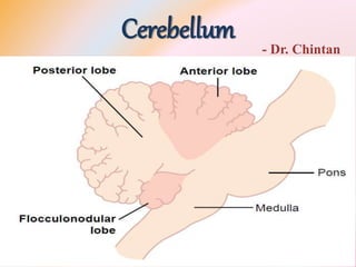
  • 8. Anatomical Functional Areas• Anatomically, the cerebellum is divided into three lobes by two deep fissures: • (1) the anterior lobe, (2) the posterior lobe, and (3) the flocculonodular lobe. • The flocculonodular lobe is the oldest of all portions of the cerebellum; • it developed along with (and functions with) the vestibular system in controlling body equilibrium
  • 9.
  • 10. Anatomical Functional Areas• the center of the cerebellum has a narrow band called the vermis – cerebellar control functions for muscle movements of the axial body, neck, shoulders, and hips are located. • To each side of the vermis is a large, laterally protruding cerebellar hemisphere, and each of these hemispheres is divided into an intermediate zone and a lateral zone. • The intermediate zone of the hemisphere is concerned with controlling muscle contractions in the distal portions of the upper and lower limbs - specially the hands and fingers and feet and toes.
  • 11.
  • 12. Anatomical Functional Areas• The lateral zone of the hemisphere operates at a much more distant level because this area joins with the cerebral cortex in the overall planning of sequential motor movements. • Without this lateral zone, most separate motor activities of the body lose their appropriate timing and sequencing and therefore become incoordinate
  • 13. Topographical Representation • axial portions of the body lie in the vermis part of the cerebellum, whereas the limbs and facial regions lie in the intermediate zones • These topographical representations receive afferent nerve signals from all the respective parts of the body as well as from corresponding topographical motor areas in the cerebral cortex and brain stem. • They send motor signals back to the same respective topographical areas of the cerebral motor cortex, as well as to topographical areas of the red nucleus and reticular formation in the brain stem.
  • 14.
  • 15. Topographical Representation • large lateral portions of the cerebellar hemispheres do not have topographical representations of the body. • They receive their input signals from the cerebral cortex, especially from the premotor areas of the frontal cortex and from the somatosensory and other sensory association areas of the parietal cortex. • this connectivity helps in planning and coordinating the body’s rapid sequential muscular activities that occur one after another within fractions of a second.
  • 16.
  • 17.
  • 18. Input Pathways to the Cerebellum • Afferent Pathways from Other Parts of the Brain • the corticopontocerebellar pathway, which originates in the cerebral motor and premotor cortices and also in the cerebral somatosensory cortex. • It passes by way of the pontine nuclei and pontocerebellar tracts mainly to the lateral divisions of the cerebellar hemispheres on the opposite side of the brain from the cerebral areas
  • 19. Input Pathways to the Cerebellum• (1) olivocerebellar tract, which passes from the inferior olive to all parts of the cerebellum and is excited in the olive by fibers from the cerebral motor cortex, basal ganglia, widespread areas of the reticular formation, and spinal cord; • (2) vestibulocerebellar fibers, some of which originate in the vestibular apparatus and others from the brain stem vestibular nuclei — almost all of these terminate in the flocculonodular lobe and fastigial nucleus of the cerebellum; • (3) reticulocerebellar fibers, which originate in different portions of the brain stem reticular formation and terminate in the midline cerebellar areas (mainly in the vermis).
  • 20.
  • 21. Input Pathways to the Cerebellum• Afferent Pathways from the Periphery • the dorsal spinocerebellar tract and the ventral spinocerebellar tract. • The dorsal tract enters the cerebellum through the inferior cerebellar peduncle and terminates in the vermis and intermediate zones of the cerebellum on the same side as its origin. • The ventral tract enters the cerebellum through the superior cerebellar peduncle, but it terminates in both sides of the cerebellum.
  • 22.
  • 23. Input Pathways to the Cerebellum• The signals transmitted in the dorsal spinocerebellar tracts come from the muscle spindles and Golgi tendon organs, large tactile receptors of the skin, and joint receptors. • All these signals tell the cerebellum of the quick status of • (1) muscle contraction, • (2) degree of tension on the muscle tendons, • (3) positions and rates of movement of the parts of the body, and • (4) forces acting on the surfaces of the body.
  • 24. Input Pathways to the Cerebellum• the ventral spinocerebellar tracts receive less information from the peripheral receptors. • they are excited mainly by motor signals arriving in the anterior horns of the spinal cord from • (1) the brain through the corticospinal and rubrospinal tracts and • (2) the internal motor pattern generators in the cord. • This ventral fiber pathway tells the cerebellum which motor signals have arrived at the anterior horns - efference copy of the anterior horn motor drive.
  • 25. Input Pathways to the Cerebellum• The spinocerebellar pathways can transmit impulses at velocities up to 120 m/sec, which is the most rapid conduction in any pathway in the CNS. • This extremely rapid conduction is important for rapid judgment of the cerebellum of changes in peripheral muscle actions. • In addition, signals are also transmitted into the cerebellum from the body periphery through the spinal dorsal columns to the dorsal column nuclei of the medulla and then relayed to the cerebellum.
  • 26. Input Pathways to the Cerebellum• signals are also transmitted up the spinal cord through the spinoreticular pathway to the reticular formation of the brain stem and also through the spino-olivary pathway to the inferior olivary nucleus. • Then signals are relayed from both of these areas to the cerebellum. • Thus, the cerebellum continually collects information about the movements and positions of all parts of the body
  • 27. Output Signals from the Cerebellum• Deep Cerebellar Nuclei and the Efferent Pathways • Located deep in the cerebellar mass on each side are three deep cerebellar nuclei — • the dentate, • interposed (interpositus – globose-globosus, emboliform) • fastigial • All the deep cerebellar nuclei receive signals from two sources: • (1) the cerebellar cortex and • (2) the deep sensory afferent tracts to the cerebellum.
  • 28. Output Signals from the Cerebellum• Each time an input signal arrives in the cerebellum, it divides and goes in two directions: • (1) directly to one of the cerebellar deep nuclei and • (2) to a corresponding area of the cerebellar cortex overlying the deep nucleus. • Then, the cerebellar cortex relays an inhibitory output signal to the deep nucleus. • Thus, all input signals that enter the cerebellum eventually end in the deep nuclei in the form of initial excitatory signals followed a fraction of a second later by inhibitory signals. • From the deep nuclei, output signals leave the cerebellum and are distributed to other parts of the brain.
  • 29. Output Signals from the Cerebellum• 1. A pathway that originates in the midline structures of the cerebellum (the vermis) and then passes through the fastigial nuclei into the medullary and pontile regions of the brain stem. • This circuit functions in close association with the equilibrium apparatus and brain stem vestibular nuclei to control equilibrium, • and also in association with the reticular formation of the brain stem to control the postural attitudes of the body
  • 30. Neuronal Connections of the Vestibular Apparatus • Most of the vestibular nerve fibers terminate in the brain stem in the vestibular nuclei, which are located at the junction of the medulla and the pons. • Some fibers pass directly to the brain stem reticular nuclei without synapsing and also to the cerebellar fastigial, uvular, and flocculonodular lobe nuclei. • The flocculonodular lobes of the cerebellum are especially concerned with dynamic equilibrium signals from the semicircular ducts
  • 31.
  • 32. Flocculonodular lobes • severe injury to either the lobes or the ducts causes loss of dynamic equilibrium during rapid changes in direction of motion • but does not seriously disturb equilibrium under static conditions. • the uvula of the cerebellum plays a similar important role in static equilibrium.
  • 33. Output Signals from the Cerebellum• 2. A pathway that originates in • (1) the intermediate zone of the cerebellar hemisphere to • (2) the interposed nucleus to • (3) the ventrolateral and ventroanterior nuclei of the thalamus to • (4) the cerebral cortex to • (5) several midline structures of the thalamus to • (6) the basal ganglia and • (7) the red nucleus and reticular formation of the upper portion of the brain stem. • This complex circuit helps to coordinate mainly the reciprocal contractions of agonist and antagonist muscles in the peripheral portions of the limbs - hands, fingers, and thumbs.
  • 34. Output Signals from the Cerebellum• 3. A pathway that begins in the cerebellar cortex of the lateral zone of the cerebellar hemisphere and • then passes to the dentate nucleus, • then to the ventrolateral and ventroanterior nuclei of the thalamus, and, • finally, to the cerebral cortex. • This pathway plays an important role in helping coordinate sequential motor activities initiated by the cerebral cortex.
  • 35.
  • 36. Functional Unit — Purkinje Cell and Deep Nuclear Cell • The cerebellum has about 30 million nearly identical functional units - a single, very large Purkinje cell (30 million of which are in the cerebellar cortex) and on a corresponding deep nuclear cell. • The three major layers of the cerebellar cortex are: • the molecular layer, • Purkinje cell layer, and • granule cell layer. • Beneath these cortical layers, in the center of the cerebellar mass, are the deep cerebellar nuclei that send output signals to other parts of the nervous system.
  • 37. Neuronal Circuit of the Functional Unit • The output from the functional unit is from a deep nuclear cell. • This cell is continually under both excitatory and inhibitory influences. • The excitatory influences arise from direct connections with afferent fibers that enter the cerebellum from the brain or the periphery. • The inhibitory influence arises entirely from the Purkinje cell in the cortex of the cerebellum. • The afferent inputs to the cerebellum are mainly of two types, one called the climbing fiber type and the other called the mossy fiber type.
  • 38.
  • 39. Neuronal Circuit of the Functional Unit • The climbing fibers originate from the inferior olives of the medulla. • There is 1 climbing fiber for about 5 to 10 Purkinje cells. • After sending branches to several deep nuclear cells, the climbing fiber continues all the way to the outer layers of the cerebellar cortex, where it makes about 300 synapses with the soma and dendrites of each Purkinje cell. • a single impulse in climbing fiber will always cause a single, prolonged (up to 1 second), peculiar type of action potential in each Purkinje cell with which it connects, beginning with a strong spike and followed by a trail of weakening secondary spikes - complex spike.
  • 40. Neuronal Circuit of the Functional Unit • The mossy fibers are from the higher brain, brain stem, and spinal cord. • These fibers also send collaterals to excite the deep nuclear cells. • Then they proceed to the granule cell layer of the cortex, where they too synapse with hundreds to thousands of granule cells • granule cells send small axons to the molecular layer. • Here the axons divide into two branches that extend in parallel direction
  • 41. Neuronal Circuit of the Functional Unit• There are many millions of these parallel nerve fibers because there are 500 to 1000 granule cells for every 1 Purkinje cell. • In this molecular layer, the dendrites of the Purkinje cells project and 80,000 to 200,000 of the parallel fibers synapse with each Purkinje cell. • The mossy fibers’ synaptic connections are weak, so that large numbers of mossy fibers must be stimulated simultaneously to excite the Purkinje cell. • Activation usually takes the form of a much weaker short duration Purkinje cell action potential called a simple spike
  • 42. Neuronal Circuit of the Functional Unit • Purkinje cells and deep nuclear cells - both of them fire continuously; • the Purkinje cell fires at about 50 to 100 action potentials per second, • the deep nuclear cells at much higher rates. • the output activity of both these cells can be modulated upward or downward.
  • 43. Neuronal Circuit of the Functional Unit • direct stimulation of the deep nuclear cells by both the climbing and the mossy fibers excites them. • signals arriving from the Purkinje cells inhibit them. • Normally, the balance between these two effects is slightly in favor of excitation • So output from the deep nuclear cell remains relatively constant at a moderate level of continuous stimulation.
  • 44. Neuronal Circuit of the Functional Unit• In execution of a rapid motor movement, the initiating signal from the cerebral motor cortex or brain stem at first greatly increases deep nuclear cell excitation. • Then, another few milliseconds later, feedback inhibitory signals from the Purkinje cell circuit arrive. • In this way, there is first a rapid excitatory signal sent by the deep nuclear cells into the motor output pathway to enhance the motor movement, • but this is followed within another small fraction of a second by an inhibitory signal.
  • 45. Neuronal Circuit of the Functional Unit • This inhibitory signal resembles a “delay-line” negative feedback signal of the type that is effective in providing damping. • when the motor system is excited, a negative feedback signal occurs after a short delay to stop the muscle movement from overshooting its mark. • basket cells and stellate cells - inhibitory cells - located in the molecular layer of the cerebellar cortex, lying among and stimulated by the small parallel fibers. • These cells send their axons at right angles across the parallel fibers and cause lateral inhibition of adjacent Purkinje cells
  • 46.
  • 47. Turn-On/Turn-Off and Turn-Off/Turn- On• The typical function of the cerebellum is to help provide rapid turn-on signals for the agonist muscles and simultaneous reciprocal turn-off signals for the antagonist muscles at the onset of a movement. • Then on approaching termination of the movement, the cerebellum is mainly responsible for timing and executing the turn-off signals to the agonists and turn-on signals to the antagonists • contraction at the onset of movement begins with signals from the cerebral cortex - pass through brain stem and cord pathways to the agonist muscle • At the same time, parallel signals are sent by way of the pontile mossy fibers into the cerebellum
  • 48. Turn-On/Turn-Off and Turn-Off/Turn- On• One branch of each mossy fiber goes directly to deep nuclear cells - instantly sends an excitatory signal back into the cerebral corticospinal motor system • So the turn-on signal becomes more powerful because it becomes the sum of both the cortical and the cerebellar signals • all mossy fibers have a second branch that transmits signals by way of the granule cells to the cerebellar cortex and by way of “parallel” fibers, to the Purkinje cells.
  • 49. Turn-On/Turn-Off and Turn-Off/Turn- On• The Purkinje cells in turn inhibit the deep nuclear cells • helps to turn off the movement after a short time. • Throughout the spinal cord there are reciprocal agonist/ antagonist circuits for virtually every movement that the cord can initiate • plus • inhibitory cells play roles in the initial inhibition of the antagonist muscles at onset of a movement and subsequent excitation at the end of a movement.
  • 50. The Purkinje Cells “Learn” • when a person first performs a new motor act, • the degree of motor enhancement by the cerebellum at the onset of contraction, • the degree of inhibition at the end of contraction, and • the timing of these • Are almost always incorrect for accurate performance of the movement. • But after the act has been performed many times, • the individual events become progressively more accurate, • sometimes requiring only a few movements before the desired result is achieved
  • 51. The Purkinje Cells “Learn” • Mechanism - Sensitivity levels of cerebellar circuits progressively adapt during the training process. • the sensitivity of the Purkinje cells to respond to the granule cell excitation becomes altered. • this sensitivity change is brought about by signals from the climbing fibers entering the cerebellum from the inferior olivary complex • the climbing fibers excites purkinje cells
  • 52. The Purkinje Cells “Learn” • When a person performs a new movement for the first time, feedback signals from the muscle and joint proprioceptors will usually denote to the cerebellum how much the actual movement fails to match the intended movement. • And the climbing fiber signals alter long-term sensitivity of the Purkinje cells. • Over a period of time, this change in sensitivity, along with other possible “learning” functions of the cerebellum, make the timing and other aspects of cerebellar control of movements perfect. • When this has been achieved, the climbing fibers no longer need to send “error” signals to the cerebellum to cause further change.
  • 53. Function of the Cerebellum • 1. The vestibulocerebellum - small flocculonodular lobes - provides neural circuits for most of the body’s equilibrium movements. • 2. The spinocerebellum - vermis of the posterior and anterior cerebellum plus the adjacent intermediate zones on both sides of the vermis. • It provides the circuitry for coordinating mainly movements of the distal portions of the limbs, especially the hands and fingers.
  • 54. Function of the Cerebellum • 3. The cerebrocerebellum - lateral zones of the cerebellar hemispheres, lateral to the intermediate zones. • It receives input from the cerebral motor cortex and adjacent premotor and somatosensory cortices of the cerebrum. • It transmits its output information in the upward direction back to the brain, • functioning in a feedback manner with the cerebral cortical sensorimotor system • to plan sequential voluntary body and limb movements, • planning these as much as tenths of a second in advance of the actual movements.
  • 55. Vestibulocerebellum • loss of the vestibulocerebellum causes extreme disturbance of equilibrium and postural movements. • in people with vestibulocerebellar dysfunction, equilibrium is far more disturbed during performance of rapid motions than during stasis, • especially so when these movements involve changes in direction of movement and stimulate the semicircular ducts. • vestibulocerebellum is especially important in controlling balance between agonist and antagonist muscle contractions of the spine, hips, and shoulders during rapid changes in body positions as required by the vestibular apparatus
  • 56. Vestibulocerebellum • the signals from the periphery tell the brain how rapidly and in which directions the body parts are moving. • It is then the function of the vestibulocerebellum to calculate in advance from these rates and directions where the different parts will be during the next few milliseconds. • information from both the body periphery and the vestibular apparatus is used to provide anticipatory correction of postural motor signals necessary for maintaining equilibrium during extremely rapid motion, • Including rapidly changing directions of motion.
  • 57. Spinocerebellum • intermediate zone of each cerebellar hemisphere receives two types of information when a movement is performed: • (1) information from the cerebral motor cortex and from the midbrain red nucleus, telling the cerebellum the prearranged sequential plan of movement for the next few fractions of a second, and • (2) feedback information from the peripheral parts of the body, especially from the distal proprioceptors of the limbs, telling the cerebellum what actual movements result.
  • 58. Spinocerebellum • After the intermediate zone of the cerebellum has compared the planned movements with the actual movements, the deep nuclear cells of the interposed nucleus send corrective output signals • (1) back to the cerebral motor cortex through relay nuclei in the thalamus and • (2) to the magnocellular portion (the lower portion) of the red nucleus that gives rise to the rubrospinal tract. • The rubrospinal tract in turn joins the corticospinal tract in innervating the lateral most motor neurons in the anterior horns of the spinal cord gray matter, • the neurons that control the distal parts of the limbs, particularly the hands and fingers.
  • 59. Spinocerebellum • smooth, coordinate movements of the agonist and antagonist muscles of the distal limbs for performing acute purposeful patterned movements • compare the “intentions” of the higher levels of the motor control system, as transmitted to the intermediate cerebellar zone through the corticopontocerebellar tract, • with the “performance” by the respective parts of the body, as transmitted back to the cerebellum from the periphery • if the signals do not compare favorably, the inferior olivary- Purkinje cell system along with other cerebellar learning mechanisms corrects the motions until they perform the desired function.
  • 60.
  • 61. Spinocerebellum • Almost all movements of the body are “pendular” - Because of momentum, all pendular movements have a tendency to overshoot • If overshooting does occur in a person whose cerebellum has been destroyed, • the conscious centers of the cerebrum eventually recognize this and initiate a movement in the reverse direction attempting to bring the arm to its planned position. • But the arm, by virtue of its momentum, overshoots once more in the opposite direction, and appropriate corrective signals must again be instituted. • Thus, the arm oscillates back and forth past its planned point for several cycles before it finally fixes on its mark. • action tremor or intention tremor.
  • 62. Spinocerebellum • But, if the cerebellum is intact, appropriate learned, subconscious signals stop the movement precisely at the intended point, thereby preventing the overshoot as well as the tremor. • This is the basic characteristic of a damping system • Most rapid movements of the body - movements of the fingers in typing, occur so rapidly that it is not possible to receive feedback information either from the periphery to the cerebellum or from the cerebellum back to the motor cortex before the movements are over. • These movements are called ballistic movements, meaning that the entire movement is preplanned and set into motion to go a specific distance and then to stop.
  • 63. Spinocerebellum • Another important example is the saccadic movements of the eyes, in which the eyes jump from one position to the next when reading or when looking at successive points along a road as a person is moving in a car. • the changes that occur in these ballistic movements when the cerebellum is removed. • (1) The movements are slow to develop and do not have the extra onset rush that the cerebellum usually provides, • (2) the force developed is weak, and • (3) the movements are slow to turn off, usually allowing the movement to go well beyond the proposed mark
  • 64. Spinocerebellum • So, in the absence of the cerebellar circuit, • the motor cortex has to think extra hard to turn ballistic movements on and • again has to think hard and take extra time to turn the movement off. • Thus, the automatism of ballistic movements is lost. • circuitry of the cerebellum is organized to perform this biphasic, first excitatory and then delayed inhibitory function that is required for preplanned rapid ballistic movements.
  • 65. Cerebrocerebellum - planning • In human beings, the lateral zones of the two cerebellar hemispheres are highly developed and greatly enlarged. • human abilities to plan and perform difficult sequential patterns of movement, especially with the hands and fingers, and to speak. • the “plan” of sequential movements actually begins in the sensory and premotor areas of the cerebral cortex, and • from there the plan is transmitted to the lateral zones of the cerebellar hemispheres.
  • 66. Cerebrocerebellum - planning • many neurons in the cerebellar dentate nuclei display the activity pattern for the sequential movement that is yet to come while the present movement is still occurring. • Lateral cerebellar zones appear to be involved not with what movement is happening at a given moment but with what will be happening during the next sequential movement a fraction of a second or perhaps even seconds later. • ability to progress smoothly from one movement to the next in orderly sequence.
  • 67. Cerebrocerebellum - timing • To provide appropriate timing for each subsequent movement. • In the absence of lateral cerebellar zones, one loses the subconscious ability to predict ahead of time how far the different parts of the body will move in a given time • person becomes unable to determine when the next sequential movement needs to begin. • As a result, the subsequent movement may begin too early or too late. • lesions in the lateral zones of the cerebellum cause complex movements (such as those required for writing, running or talking) to become incoordinate
  • 68. Cerebrocerebellum - timing • helps to “time” events other than movements of the body • the rates of progression of both auditory and visual phenomena can be predicted by the brain, but both of these require cerebellar participation. • A person can predict from the changing visual scene how rapidly he or she is approaching an object. • effects of removing the large lateral portions of the cerebellum in monkeys • Such a monkey is unable to predict when it will reach the wall.
  • 69. Applied • Dysmetria and Ataxia • in the absence of the cerebellum, the subconscious motor control system cannot predict how far movements will go - movements overshoot their proposed mark; • then the conscious portion of the brain overcompensates in the opposite direction for the subsequent compensatory movement. This effect is called dysmetria, • And it results in uncoordinated movements called ataxia. • Dysmetria and ataxia can also result from lesions in the spinocerebellar tracts because feedback information from the moving parts of the body to the cerebellum is essential for cerebellar timing of movement termination.
  • 70. Applied • Past pointing - a person moves the hand or some other moving part of the body beyond the point of intention. • normally the cerebellum initiates most of the motor signal that turns off a movement after it is begun; • Dysdiadochokinesia - When the motor control system fails to predict where the different parts of the body will be at a given time, it “loses” perception of the parts during rapid motor movements • the subsequent movement may begin much too early or much too late, so that no orderly “progression of movement” can occur • a series of delayed attempted & disorderly movements occurs
  • 71. Applied • Dysarthria - failure of progression occurs is in talking • because the formation of words depends on rapid and orderly sequence of individual muscle movements in the larynx, mouth, and respiratory system. • Lack of coordination among these and inability to adjust in advance either the intensity of sound or duration of each successive sound causes disorderly vocalization, • with some syllables loud, some weak, some held for long intervals, some held for short intervals, and resultant speech is often incomprehensible.
  • 72. Applied • Intention Tremor • When a person who has lost the cerebellum performs a voluntary act, • the movements tend to oscillate, • especially when they approach the planned mark, • first overshooting the mark and then vibrating back and forth several times before settling on the mark. • intention tremor or action tremor - results from cerebellar overshooting and failure of the cerebellar system to “damp” the motor movements.
  • 73. Applied • Cerebellar nystagmus is tremor of the eyeballs that occurs usually when one attempts to fix the eyes on a scene to one side of the head. • This type of fixation results in rapid, tremulous movements of the eyes rather than steady fixation, and it is manifestation of failure of damping by the cerebellum. • It occurs especially when the flocculonodular lobes of the cerebellum are damaged • also associated with loss of equilibrium
  • 74. Applied • Hypotonia • Loss of the deep cerebellar nuclei - the dentate and interposed nuclei, causes decreased tone of the peripheral body musculature on the side of the cerebellar lesion. • The hypotonia results from loss of cerebellar facilitation of the motor cortex and brain stem motor nuclei by tonic signals from the deep cerebellar nuclei.