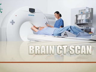
SlideShare une entreprise Scribd logo
1  sur  74
Anatomy &
Physiology
BRAIN
• is one of the largest and
most complex organ in the
human body.
• The adult human brain
weighs on average about 3
lbs. (1.5 kg)with a volume
of around 1130 cubic
centimeters (cm3) in
women and 1260 cm3 in
men
• Our brain gives us awareness of
ourselves and of our environment,
processing a constant stream of
sensory data.

• It controls our muscle movements,
the secretions of our glands, and
even our breathing and internal
temperature.
• Every creative thought, feeling, and
plan is developed by our brain.
• The brain’s neurons record the
memory of every event in our lives.
IT HAS THREE MAIN PARTS:

•Cerebrum
• the largest part of the mature brain.
• It consists of two large masses, called
cerebral hemispheres, which are
almost mirror images of each other.
• The cerebrum is concerned with
higher brain functions, interpreting
sensory impulses and initiating
muscle movements.
• It stores information and uses it to
process reasoning.
• It also functions in determining
intelligence and personality.
• The surface of the cerebrum has a
folded appearance called the cortex.
• The cortex contains about 70% of
the 100 billion nerve cells.
• The nerve cell bodies color the
cortex grey-brown giving it its name
– gray matter.
• Beneath the cortex are long
connecting fibers between neurons,
called axons, which make up the
white matter.
•Cerebellum
• a rounded structure located
behind the brain stem, to which
it is linked by thick nerve tracts.
• It accounts for about 11% of
the whole brain weight and
appears similar to the
cerebrum.
• It is concerned mainly with the
maintenance of posture and
balance and the coordination of
movement.
•Brain Stem
• Houses the midbrain
(mesencephalon), pons (part of
the metencephalon), and medulla
oblongata (myelencephalon).
• This is the posterior area of the
brain that attaches to the spinal
cord.
• It's here, at the brain stem, where
information is sent back and forth
between the cerebrum or
cerebellum and the body.
• Cranial nerves 3-12 are located
here as well as significant
processing centers.
THE BRAIN IS ALSO DIVIDED INTO
SEVERAL LOBES:

Frontal lobe
• Personality, behavior,
emotions,
• Judgment, planning,
problem solving
• Speech: speaking and
writing (Broca’s area)
• Body movement (motor
strip)

• Intelligence, concentration,
self awareness

Parietal lobe
• Interprets language, words
• Sense of touch, pain,
temperature (sensory strip)
• Interprets signals from
vision, hearing, motor,
sensory and memory
• Spatial and visual perception
Occipital lobe
• Interprets vision (color,
light, movement)

Temporal lobe
• Understanding language
(Wernicke’s area)
• Memory
• Hearing
• Sequencing and organization
DEEP STRUCTURES
• Hypothalamus - is located in the
floor of the third ventricle and is the
master control of the autonomic
system. It plays a role in controlling
behaviors such as hunger, thirst,
sleep, and sexual response. It also
regulates body temperature, blood
pressure, emotions, and secretion of
hormones.
• Pineal gland - is located behind the third
ventricle. It helps regulate the body’s
internal clock and circadian rhythms by
secreting melatonin. It has some role in
sexual development.

• Thalamus - serves as a relay station for
almost all information that comes and goes
to the cortex. It plays a role in pain
sensation, attention, alertness and memory.
• Basal ganglia - includes the caudate,
putamen and globus pallidus. These nuclei
work with the cerebellum to coordinate
fine motions, such as fingertip
movements.
• Limbic system - is the center of our
emotions, learning, and memory. Included
in this system are the cingulate gyri,
hypothalamus, amygdala (emotional
reactions) and hippocampus (memory).
SKULL
• The purpose of the bony skull is to protect the
brain from injury.
• The skull is formed from 8 bones that fuse together
along suture lines.
• These bones include the frontal, parietal (2),
temporal (2), sphenoid, occipital and ethmoid.
The face is formed from 14 paired bones
including the maxilla , zygoma, nasal, palatine,
lacrimal, inferior nasal conchae, mandible, and
vomer.
CT scanning of the head is typically used to detect:
• bleeding, brain injury and skull fractures in patients with
head injuries.
• bleeding caused by a ruptured or leaking aneurysm in a
patient with a sudden severe headache.
• a blood clot or bleeding within the brain shortly after a
patient exhibits symptoms of a stroke.
• a stroke, especially with a new technique called Perfusion CT.
• brain tumors.
• enlarged brain cavities (ventricles) in patients with
hydrocephalus.
• diseases or malformations of the skull.
CT scanning is also performed to:
• evaluate the extent of bone and soft tissue damage in patients with
facial trauma, and planning surgical reconstruction.
• diagnose diseases of the temporal bone on the side of the skull, which
may be causing hearing problems.
• determine whether inflammation or other changes are present in the
paranasal sinuses.
• plan radiation therapy for cancer of the brain or other tissues.
• guide the passage of a needle used to obtain a tissue sample (biopsy)
from the brain.
• assess aneurysms or arteriovenous malformations through a technique
called CT angiography.
• CT scanning has no absolute contraindications.
• But due to the relatively high radiation dose
involved in CT scans, it is important to avoid
scanning patients who are pregnant. Radiation
exposure to a fetus can cause developmental
problems. Thus, CT should only be performed for
pregnant patients in critical situations and only
after discussion of the potential risks.
• Patients who have an allergy to the IV contrast
media (IVCM) used in CT scans.
• Renal impairment may also prohibit your patient
from having IVCM.
Before the CT scan, tell your doctor if you:
• Are or might be pregnant.
• Are allergic to any medicines, including iodine dyes.
• Have a heart condition, such as heart failure.
• Have diabetes or take metformin (Glucophage) for your
diabetes. You may have to adjust your medicine for a day
before and after the test.
• Have had kidney problems.
• Have asthma.
• Have a medical device, such as a pacemaker or an insulin
pump.

• Become very nervous in small spaces. You need to lie still
inside the CT scanner, so you may need a medicine (sedative)
to help you relax.
• You should wear comfortable, loose-fitting
clothing to your exam. You may be given a
gown to wear during the procedure.

• Metal objects, including jewelry, eyeglasses,
dentures and hairpins, may affect the CT
images and should be left at home or
removed prior to your exam. You may also be
asked to remove hearing aids and removable
dental work. Women will be asked to remove
bras containing metal underwire. You may be
asked to remove any piercings, if possible.
• If your procedure involves the use of
contrast dye, you will be asked to sign a
consent form that gives permission to do the
procedure. Read the form carefully and ask
questions if something is not clear.
• Generally, there is no fasting requirement
prior to a CT scan, unless a contrast dye is to
be used. Your doctor will give you special
instructions ahead of time if contrast is to be
used and if you will need to withhold food
and drink.
• Procedure with contrast:
• Kidney function test (BUN and Creatinine)
must be done 72 hrs. before the procedure.
• NPO (4 hours) before the procedure.
• You may drink clear liquids up until the
time of your scan. Clear liquids include
water, black coffee or tea, apple juice, clear
soda or clear broth.
• Secure a consent form.
CONTRAST MEDIA
• (Iopamiro/Ultravist)
>300 mg I/mL and 370 mg I/mL
• You will lie on a scan table that slides into a large, circular
opening of the scanning machine. Your head may be
immobilized to prevent movement during the procedure.
• The technologist will be in another room where the scanner
controls are located. However, you will be in constant sight of
the technologist through a window. Speakers inside the
scanner will enable the technologist to communicate with
and hear you. You may have a call button so that you can let
the technologist know if you have any problems during the
procedure. The technologist will be watching you at all times
and will be in constant communication.
• As the scanner begins to rotate around you, X-rays will pass
through the body for short amounts of time. You will hear
clicking sounds, which are normal.
• The X-rays absorbed by the body's tissues will be
detected by the scanner and transmitted to the
computer. The computer will transform the
information into an image to be interpreted by the
radiologist.
• It will be very important for you to remain very still
during the procedure. You may be asked to hold
your breath at various times during the procedure.
• If contrast dye is used for your procedure, you will
be removed from the scanner after the first set of
scans has been completed. A second set of scans will
be taken after the contrast dye has been
administered.
• If contrast dye is used for your procedure, you may
feel some effects when the dye is injected into the
IV line. These effects include a flushing sensation, a
salty or metallic taste in the mouth, a brief
headache, or nausea and/or vomiting. These effects
usually last for a few moments.
• You should notify the technologist if you feel any
breathing difficulties, sweating, numbness, or heart
palpitations.
• When the procedure has been completed, you will
be removed from the scanner.
• If an IV line was inserted for
contrast administration, the line
will be removed.
• You may be asked to wait for a
short period of time while the
radiologist examines the scans to
make sure they are clear.
• If contrast dye was used during your procedure,
you may be monitored for a period of time for
any side effects or reactions to the contrast dye,
such as itching, swelling, rash, or difficulty
breathing. Notify the radiologist or your doctor
if you experience any of these symptoms.
• If you notice any pain, redness, and/or swelling
at the IV site after you return home following
your procedure, you should notify your doctor
as this could indicate an infection or other type
of reaction.
• Otherwise, there is no special type of care required
after a CT scan of the brain. You may resume your
usual diet and activities unless your doctor advises
you differently.
• Your doctor may give you additional or alternate
instructions after the procedure, depending on
your particular situation.
• If you are a diabetic who takes any medication that
contains metformin, you must have a blood test to
check your kidney function before you can start
taking metformin again. Call your doctor for the
results of the blood test and for instructions about
resuming metformin.
ROUTINE ADULT HEAD (BRAIN)

PATIENT POSITIONING:
• • Patient should be supine, head first into the gantry, with the head in the
head-holder whenever possible.
• • Center the table height such that the external auditory meatus (EAM) is at
the center of the gantry.
• • To reduce or avoid ocular lens exposure, the scan angle should be parallel
to a line created by the supraorbital ridge and the inner table of the
posterior margin of the foramen magnum.
• This may be accomplished by either tilting the patient’s chin toward the
chest (“tucked” position) or tilting the gantry.
• While there may be some situations where this is not possible due to
scanner or patient positioning limitations, it is considered good practice to
perform one or both of these maneuvers whenever possible.
• SCAN RANGE: Top of C1 lamina through top of calvarium.
CONTRAST:
• Oral: None.
• Injected: Some indications require injection of intravenous or
intrathecal contrast media during imaging
• of the brain.
• Intravenous contrast administration should be performed as
directed by the supervising radiologist
• using appropriate injection protocols and in accordance with
the ACR Practice Guideline for the Use of Intravascular
Contrast Media. A typical amount would be 100 cc at 300
mg/cc strength, injected at 1 cc/sec. A delay of 4 minutes
between contrast injection and the start of scanning is
typical.
AXIAL VERSUS HELICAL SCAN MODE (both are
provided in the following sample protocols):
• There are advantages and disadvantages to using
either axial or helical scans for routine head CT
exams. The decision as to whether to use axial or
helical should be influenced by the specific patient
indication, scanner capabilities, and image quality
requirements.

• Users of this document should consider the
information in the following table and consult with
both the manufacturer and a
• medical physicist to assist in determining which
mode to use.
SCOUT: LATERAL
LANDMARK: OML
SLICE PLANE: CORONAL & AXIAL
I.V. CONTRAST: 100-140 ML
BREATH HOLD: NONE
DFOV
SLICE THICKNESS: 1-1.5 mm
12
FILMING: BONE & SOFT TISSUE

PITUITARY AND SELLA TURCICA
PATHOLOGY
EPIDURAL HEMATOMA
• An epidural hematoma is
usually associated with a
skull fracture. It often
occurs when an impact
fractures the calvarium.
The fractured bone
lacerates a dural artery or
a venous sinus. The blood
from the ruptured vessel
collects between the skull
and dura.

Biconvex (lenticellular) epidural
hematoma (arrowheads),
deep to the parietal skull fracture
(arrow).
SUBARACHNOID
HEMORRHAGE
• A subarachnoid hemorrhage occurs
with injury of small arteries or veins on
the surface of the brain. The ruptured
vessel bleeds into the space between
the pia and arachnoid matter. The most
common cause of subarachnoid
hemorrhage is trauma. In the absence of
significant trauma, the most common
cause of subarachnoid hemorrhage is
the rupture of a cerebral aneurysm.

High density blood (arrowheads)
fills the sulci over the
right cerebral convexity in this
subarachnoid hemorrhage.
SUBDURAL HEMATOMA
• is a type of hematoma,
usually associated
with traumatic brain injury.
• Blood gathers with the
outermost meninges layer,
between the dura mater,
which adheres to the skull,
and the arachnoid mater,
which envelops the brain.

High density, crescent shaped
hematoma (arrowheads)
overlying the right cerebral
hemisphere. Note the shift of the
normally midline septum
pellucidum due to the mass effect
arrow.
HEMORRHAGIC
STROKE
• are due to rupture of a
cerebral blood vessel
that causes bleeding
into or around the brain

Hemorrhage in the cerebellum (arrow).
HYDROCEPHALUS
• Hydrocephalus, a problem with the ratio of
production of CSF to its re-absorbtion, is
most frequent in children.
INTRACRANIAL
TUMOR
• Intracranial tumors
generally present
with a focal
neurological deficit,
seizure, or
headache.

Multiple enhancing masses located at
the grey-white junction zones.
MENINGIOMA
• Meningiomas are the
most common extraaxial neoplasm of the
brain. Middle-aged
women are most
frequently affected.
Twenty percent of
meningiomas calcify.
On CT, meningiomas
are usually isointense
to gray matter.

Axial, post contrast CT demonstrating
broad based enhancing extra-axial mass.
CT IMAGES
A. Orbit
B. Sphenoid Sinus
C. Temporal Lobe
D. External Auditory Canal
E. Mastoid Air Cells
F. Cerebellar Hemisphere
A. Frontal Lobe
B. Frontal Bone (Superior Surface of Orbital Part)
C. Dorsum Sellae
D. Basilar Artery
E. Temporal Lobe
F. Mastoid Air Cells
G. Cerebellar Hemisphere
A. Frontal Lobe
B. Sylvian Fissure
C. Temporal Lobe
D. Suprasellar Cistern
E. Midbrain
F. Fourth Ventricle
G. Cerebellar Hemisphere
A. Falx Cerebri
B. Frontal Lobe
C. Anterior Horn of Lateral Ventricle
D. Third Ventricle
E. Quadrigeminal Plate Cistern
F. Cerebellum
THE END
Re p o r t e d b y :
J o a n C. Va r g a s

Contenu connexe

Tendances

Contrast media in CT
Contrast media in CTContrast media in CT
Contrast media in CT
Kyle Rousseau
 
Digital subtraction angiography
Digital subtraction angiographyDigital subtraction angiography
Digital subtraction angiography
subhayanmandal
 

Tendances (20)

Ncct and cect brain and orbit
Ncct and cect brain and orbitNcct and cect brain and orbit
Ncct and cect brain and orbit
 
MCU- Micturating cysto-urethrogram
MCU- Micturating cysto-urethrogramMCU- Micturating cysto-urethrogram
MCU- Micturating cysto-urethrogram
 
Ct head protocols
Ct head protocolsCt head protocols
Ct head protocols
 
Contrast media in CT
Contrast media in CTContrast media in CT
Contrast media in CT
 
Cerebral angiography technique
Cerebral angiography techniqueCerebral angiography technique
Cerebral angiography technique
 
Digital subtraction angiography
Digital subtraction angiographyDigital subtraction angiography
Digital subtraction angiography
 
CT angiography
CT angiographyCT angiography
CT angiography
 
CT Procedure OF Abdomen & Pelvis
CT Procedure OF Abdomen & Pelvis CT Procedure OF Abdomen & Pelvis
CT Procedure OF Abdomen & Pelvis
 
MRI Brain
MRI BrainMRI Brain
MRI Brain
 
Ct thorax
Ct thoraxCt thorax
Ct thorax
 
Ct Basics
Ct BasicsCt Basics
Ct Basics
 
Skull radiography
Skull radiography Skull radiography
Skull radiography
 
CT numbers, window width and window level
CT numbers, window width and window levelCT numbers, window width and window level
CT numbers, window width and window level
 
Contrast media used with ct
Contrast media used with ctContrast media used with ct
Contrast media used with ct
 
CT Angiography presentation
CT Angiography presentation CT Angiography presentation
CT Angiography presentation
 
MR spectroscopy
MR spectroscopyMR spectroscopy
MR spectroscopy
 
Approach to head ct
Approach to head ctApproach to head ct
Approach to head ct
 
Barium meal follow through
Barium meal follow throughBarium meal follow through
Barium meal follow through
 
Loopogram
LoopogramLoopogram
Loopogram
 
INTRAVENOUS UROGRAM (IVU)
INTRAVENOUS UROGRAM (IVU)INTRAVENOUS UROGRAM (IVU)
INTRAVENOUS UROGRAM (IVU)
 

En vedette (6)

Ct brain presentation
Ct brain presentationCt brain presentation
Ct brain presentation
 
How to read a Head CT, CT Brain
How to read a Head CT, CT BrainHow to read a Head CT, CT Brain
How to read a Head CT, CT Brain
 
CT Scan - Basics
CT Scan - BasicsCT Scan - Basics
CT Scan - Basics
 
Ct brain basics and anatomy
Ct brain basics and anatomyCt brain basics and anatomy
Ct brain basics and anatomy
 
BASICS of CT Head
BASICS of CT HeadBASICS of CT Head
BASICS of CT Head
 
Computer Tomography (CT Scan)
Computer Tomography (CT Scan)Computer Tomography (CT Scan)
Computer Tomography (CT Scan)
 

Similaire à BRAIN CT SCAN

Neurologic Examination for PC II.pptx
Neurologic Examination for PC II.pptxNeurologic Examination for PC II.pptx
Neurologic Examination for PC II.pptx
L3miD1
 
Craniosacral Inservice
Craniosacral InserviceCraniosacral Inservice
Craniosacral Inservice
Sarah Guarino
 

Similaire à BRAIN CT SCAN (20)

Diagnostic test in neurological disorder and it's nursing management
Diagnostic test in neurological disorder and it's nursing managementDiagnostic test in neurological disorder and it's nursing management
Diagnostic test in neurological disorder and it's nursing management
 
Diagnostic test in neurological disorder
Diagnostic test in neurological disorderDiagnostic test in neurological disorder
Diagnostic test in neurological disorder
 
Epilepsy
EpilepsyEpilepsy
Epilepsy
 
CNS DIAGNOSTIC TESTS
CNS DIAGNOSTIC TESTSCNS DIAGNOSTIC TESTS
CNS DIAGNOSTIC TESTS
 
MRI BRAIN(AIIMS DELHI).pptx
MRI BRAIN(AIIMS DELHI).pptxMRI BRAIN(AIIMS DELHI).pptx
MRI BRAIN(AIIMS DELHI).pptx
 
MRI Contrast And Plain.pptx
MRI Contrast And Plain.pptxMRI Contrast And Plain.pptx
MRI Contrast And Plain.pptx
 
00183 (2).ppt
00183 (2).ppt00183 (2).ppt
00183 (2).ppt
 
Week 2 to 3
Week 2 to 3Week 2 to 3
Week 2 to 3
 
Traumatic brain injury
Traumatic brain injuryTraumatic brain injury
Traumatic brain injury
 
Epilepsy
EpilepsyEpilepsy
Epilepsy
 
NERVOUS SYSTEM BY MS. TANVI P. VAGHELA..
NERVOUS SYSTEM BY MS. TANVI P. VAGHELA..NERVOUS SYSTEM BY MS. TANVI P. VAGHELA..
NERVOUS SYSTEM BY MS. TANVI P. VAGHELA..
 
12 - Basic Medical Instruments Basic Medical Instruments
12 - Basic Medical Instruments Basic Medical Instruments12 - Basic Medical Instruments Basic Medical Instruments
12 - Basic Medical Instruments Basic Medical Instruments
 
Myelogram pdf
Myelogram pdfMyelogram pdf
Myelogram pdf
 
Magnetic Resonance Imaging (MRI)
Magnetic Resonance Imaging (MRI)Magnetic Resonance Imaging (MRI)
Magnetic Resonance Imaging (MRI)
 
Neurologic Examination for PC II.pptx
Neurologic Examination for PC II.pptxNeurologic Examination for PC II.pptx
Neurologic Examination for PC II.pptx
 
Coplats dee 2010
Coplats dee 2010Coplats dee 2010
Coplats dee 2010
 
Patient preparation for MRI CT and Nuclear Medicine
Patient preparation for MRI CT and Nuclear Medicine Patient preparation for MRI CT and Nuclear Medicine
Patient preparation for MRI CT and Nuclear Medicine
 
Supratentorial masses excision -anaesthetic implication
Supratentorial masses excision -anaesthetic implication Supratentorial masses excision -anaesthetic implication
Supratentorial masses excision -anaesthetic implication
 
Craniotomy.pptx
Craniotomy.pptxCraniotomy.pptx
Craniotomy.pptx
 
Craniosacral Inservice
Craniosacral InserviceCraniosacral Inservice
Craniosacral Inservice
 

Dernier

Dehradun Call Girls Service {8854095900} ❤️VVIP ROCKY Call Girl in Dehradun U...
Dehradun Call Girls Service {8854095900} ❤️VVIP ROCKY Call Girl in Dehradun U...Dehradun Call Girls Service {8854095900} ❤️VVIP ROCKY Call Girl in Dehradun U...
Dehradun Call Girls Service {8854095900} ❤️VVIP ROCKY Call Girl in Dehradun U...
Sheetaleventcompany
 
Call Girls in Gagan Vihar (delhi) call me [🔝 9953056974 🔝] escort service 24X7
Call Girls in Gagan Vihar (delhi) call me [🔝  9953056974 🔝] escort service 24X7Call Girls in Gagan Vihar (delhi) call me [🔝  9953056974 🔝] escort service 24X7
Call Girls in Gagan Vihar (delhi) call me [🔝 9953056974 🔝] escort service 24X7
9953056974 Low Rate Call Girls In Saket, Delhi NCR
 

Dernier (20)

Call Girls Ahmedabad Just Call 9630942363 Top Class Call Girl Service Available
Call Girls Ahmedabad Just Call 9630942363 Top Class Call Girl Service AvailableCall Girls Ahmedabad Just Call 9630942363 Top Class Call Girl Service Available
Call Girls Ahmedabad Just Call 9630942363 Top Class Call Girl Service Available
 
Models Call Girls In Hyderabad 9630942363 Hyderabad Call Girl & Hyderabad Esc...
Models Call Girls In Hyderabad 9630942363 Hyderabad Call Girl & Hyderabad Esc...Models Call Girls In Hyderabad 9630942363 Hyderabad Call Girl & Hyderabad Esc...
Models Call Girls In Hyderabad 9630942363 Hyderabad Call Girl & Hyderabad Esc...
 
Coimbatore Call Girls in Coimbatore 7427069034 genuine Escort Service Girl 10...
Coimbatore Call Girls in Coimbatore 7427069034 genuine Escort Service Girl 10...Coimbatore Call Girls in Coimbatore 7427069034 genuine Escort Service Girl 10...
Coimbatore Call Girls in Coimbatore 7427069034 genuine Escort Service Girl 10...
 
Jogeshwari ! Call Girls Service Mumbai - 450+ Call Girl Cash Payment 90042684...
Jogeshwari ! Call Girls Service Mumbai - 450+ Call Girl Cash Payment 90042684...Jogeshwari ! Call Girls Service Mumbai - 450+ Call Girl Cash Payment 90042684...
Jogeshwari ! Call Girls Service Mumbai - 450+ Call Girl Cash Payment 90042684...
 
Kollam call girls Mallu aunty service 7877702510
Kollam call girls Mallu aunty service 7877702510Kollam call girls Mallu aunty service 7877702510
Kollam call girls Mallu aunty service 7877702510
 
Dehradun Call Girls Service {8854095900} ❤️VVIP ROCKY Call Girl in Dehradun U...
Dehradun Call Girls Service {8854095900} ❤️VVIP ROCKY Call Girl in Dehradun U...Dehradun Call Girls Service {8854095900} ❤️VVIP ROCKY Call Girl in Dehradun U...
Dehradun Call Girls Service {8854095900} ❤️VVIP ROCKY Call Girl in Dehradun U...
 
Call Girls in Gagan Vihar (delhi) call me [🔝 9953056974 🔝] escort service 24X7
Call Girls in Gagan Vihar (delhi) call me [🔝  9953056974 🔝] escort service 24X7Call Girls in Gagan Vihar (delhi) call me [🔝  9953056974 🔝] escort service 24X7
Call Girls in Gagan Vihar (delhi) call me [🔝 9953056974 🔝] escort service 24X7
 
Call Girls Hyderabad Just Call 8250077686 Top Class Call Girl Service Available
Call Girls Hyderabad Just Call 8250077686 Top Class Call Girl Service AvailableCall Girls Hyderabad Just Call 8250077686 Top Class Call Girl Service Available
Call Girls Hyderabad Just Call 8250077686 Top Class Call Girl Service Available
 
Call Girls Rishikesh Just Call 8250077686 Top Class Call Girl Service Available
Call Girls Rishikesh Just Call 8250077686 Top Class Call Girl Service AvailableCall Girls Rishikesh Just Call 8250077686 Top Class Call Girl Service Available
Call Girls Rishikesh Just Call 8250077686 Top Class Call Girl Service Available
 
Low Rate Call Girls Bangalore {9179660964} ❤️VVIP NISHA Call Girls in Bangalo...
Low Rate Call Girls Bangalore {9179660964} ❤️VVIP NISHA Call Girls in Bangalo...Low Rate Call Girls Bangalore {9179660964} ❤️VVIP NISHA Call Girls in Bangalo...
Low Rate Call Girls Bangalore {9179660964} ❤️VVIP NISHA Call Girls in Bangalo...
 
Independent Call Girls Service Mohali Sector 116 | 6367187148 | Call Girl Ser...
Independent Call Girls Service Mohali Sector 116 | 6367187148 | Call Girl Ser...Independent Call Girls Service Mohali Sector 116 | 6367187148 | Call Girl Ser...
Independent Call Girls Service Mohali Sector 116 | 6367187148 | Call Girl Ser...
 
Top Rated Pune Call Girls (DIPAL) ⟟ 8250077686 ⟟ Call Me For Genuine Sex Serv...
Top Rated Pune Call Girls (DIPAL) ⟟ 8250077686 ⟟ Call Me For Genuine Sex Serv...Top Rated Pune Call Girls (DIPAL) ⟟ 8250077686 ⟟ Call Me For Genuine Sex Serv...
Top Rated Pune Call Girls (DIPAL) ⟟ 8250077686 ⟟ Call Me For Genuine Sex Serv...
 
Premium Bangalore Call Girls Jigani Dail 6378878445 Escort Service For Hot Ma...
Premium Bangalore Call Girls Jigani Dail 6378878445 Escort Service For Hot Ma...Premium Bangalore Call Girls Jigani Dail 6378878445 Escort Service For Hot Ma...
Premium Bangalore Call Girls Jigani Dail 6378878445 Escort Service For Hot Ma...
 
Andheri East ) Call Girls in Mumbai Phone No 9004268417 Elite Escort Service ...
Andheri East ) Call Girls in Mumbai Phone No 9004268417 Elite Escort Service ...Andheri East ) Call Girls in Mumbai Phone No 9004268417 Elite Escort Service ...
Andheri East ) Call Girls in Mumbai Phone No 9004268417 Elite Escort Service ...
 
Top Rated Hyderabad Call Girls Chintal ⟟ 9332606886 ⟟ Call Me For Genuine Se...
Top Rated  Hyderabad Call Girls Chintal ⟟ 9332606886 ⟟ Call Me For Genuine Se...Top Rated  Hyderabad Call Girls Chintal ⟟ 9332606886 ⟟ Call Me For Genuine Se...
Top Rated Hyderabad Call Girls Chintal ⟟ 9332606886 ⟟ Call Me For Genuine Se...
 
Call Girls Mysore Just Call 8250077686 Top Class Call Girl Service Available
Call Girls Mysore Just Call 8250077686 Top Class Call Girl Service AvailableCall Girls Mysore Just Call 8250077686 Top Class Call Girl Service Available
Call Girls Mysore Just Call 8250077686 Top Class Call Girl Service Available
 
Most Beautiful Call Girl in Bangalore Contact on Whatsapp
Most Beautiful Call Girl in Bangalore Contact on WhatsappMost Beautiful Call Girl in Bangalore Contact on Whatsapp
Most Beautiful Call Girl in Bangalore Contact on Whatsapp
 
Russian Call Girls Service Jaipur {8445551418} ❤️PALLAVI VIP Jaipur Call Gir...
Russian Call Girls Service  Jaipur {8445551418} ❤️PALLAVI VIP Jaipur Call Gir...Russian Call Girls Service  Jaipur {8445551418} ❤️PALLAVI VIP Jaipur Call Gir...
Russian Call Girls Service Jaipur {8445551418} ❤️PALLAVI VIP Jaipur Call Gir...
 
Independent Call Girls In Jaipur { 8445551418 } ✔ ANIKA MEHTA ✔ Get High Prof...
Independent Call Girls In Jaipur { 8445551418 } ✔ ANIKA MEHTA ✔ Get High Prof...Independent Call Girls In Jaipur { 8445551418 } ✔ ANIKA MEHTA ✔ Get High Prof...
Independent Call Girls In Jaipur { 8445551418 } ✔ ANIKA MEHTA ✔ Get High Prof...
 
(Low Rate RASHMI ) Rate Of Call Girls Jaipur ❣ 8445551418 ❣ Elite Models & Ce...
(Low Rate RASHMI ) Rate Of Call Girls Jaipur ❣ 8445551418 ❣ Elite Models & Ce...(Low Rate RASHMI ) Rate Of Call Girls Jaipur ❣ 8445551418 ❣ Elite Models & Ce...
(Low Rate RASHMI ) Rate Of Call Girls Jaipur ❣ 8445551418 ❣ Elite Models & Ce...
 

BRAIN CT SCAN

  • 1.
  • 3. BRAIN • is one of the largest and most complex organ in the human body. • The adult human brain weighs on average about 3 lbs. (1.5 kg)with a volume of around 1130 cubic centimeters (cm3) in women and 1260 cm3 in men
  • 4. • Our brain gives us awareness of ourselves and of our environment, processing a constant stream of sensory data. • It controls our muscle movements, the secretions of our glands, and even our breathing and internal temperature. • Every creative thought, feeling, and plan is developed by our brain. • The brain’s neurons record the memory of every event in our lives.
  • 5. IT HAS THREE MAIN PARTS: •Cerebrum • the largest part of the mature brain. • It consists of two large masses, called cerebral hemispheres, which are almost mirror images of each other. • The cerebrum is concerned with higher brain functions, interpreting sensory impulses and initiating muscle movements. • It stores information and uses it to process reasoning. • It also functions in determining intelligence and personality.
  • 6. • The surface of the cerebrum has a folded appearance called the cortex. • The cortex contains about 70% of the 100 billion nerve cells. • The nerve cell bodies color the cortex grey-brown giving it its name – gray matter. • Beneath the cortex are long connecting fibers between neurons, called axons, which make up the white matter.
  • 7. •Cerebellum • a rounded structure located behind the brain stem, to which it is linked by thick nerve tracts. • It accounts for about 11% of the whole brain weight and appears similar to the cerebrum. • It is concerned mainly with the maintenance of posture and balance and the coordination of movement.
  • 8. •Brain Stem • Houses the midbrain (mesencephalon), pons (part of the metencephalon), and medulla oblongata (myelencephalon). • This is the posterior area of the brain that attaches to the spinal cord. • It's here, at the brain stem, where information is sent back and forth between the cerebrum or cerebellum and the body. • Cranial nerves 3-12 are located here as well as significant processing centers.
  • 9. THE BRAIN IS ALSO DIVIDED INTO SEVERAL LOBES: Frontal lobe • Personality, behavior, emotions, • Judgment, planning, problem solving • Speech: speaking and writing (Broca’s area) • Body movement (motor strip) • Intelligence, concentration, self awareness Parietal lobe • Interprets language, words • Sense of touch, pain, temperature (sensory strip) • Interprets signals from vision, hearing, motor, sensory and memory • Spatial and visual perception
  • 10. Occipital lobe • Interprets vision (color, light, movement) Temporal lobe • Understanding language (Wernicke’s area) • Memory • Hearing • Sequencing and organization
  • 11.
  • 12. DEEP STRUCTURES • Hypothalamus - is located in the floor of the third ventricle and is the master control of the autonomic system. It plays a role in controlling behaviors such as hunger, thirst, sleep, and sexual response. It also regulates body temperature, blood pressure, emotions, and secretion of hormones.
  • 13. • Pineal gland - is located behind the third ventricle. It helps regulate the body’s internal clock and circadian rhythms by secreting melatonin. It has some role in sexual development. • Thalamus - serves as a relay station for almost all information that comes and goes to the cortex. It plays a role in pain sensation, attention, alertness and memory.
  • 14. • Basal ganglia - includes the caudate, putamen and globus pallidus. These nuclei work with the cerebellum to coordinate fine motions, such as fingertip movements. • Limbic system - is the center of our emotions, learning, and memory. Included in this system are the cingulate gyri, hypothalamus, amygdala (emotional reactions) and hippocampus (memory).
  • 15.
  • 16. SKULL • The purpose of the bony skull is to protect the brain from injury. • The skull is formed from 8 bones that fuse together along suture lines. • These bones include the frontal, parietal (2), temporal (2), sphenoid, occipital and ethmoid. The face is formed from 14 paired bones including the maxilla , zygoma, nasal, palatine, lacrimal, inferior nasal conchae, mandible, and vomer.
  • 17.
  • 18.
  • 19.
  • 20. CT scanning of the head is typically used to detect: • bleeding, brain injury and skull fractures in patients with head injuries. • bleeding caused by a ruptured or leaking aneurysm in a patient with a sudden severe headache. • a blood clot or bleeding within the brain shortly after a patient exhibits symptoms of a stroke. • a stroke, especially with a new technique called Perfusion CT. • brain tumors. • enlarged brain cavities (ventricles) in patients with hydrocephalus. • diseases or malformations of the skull.
  • 21. CT scanning is also performed to: • evaluate the extent of bone and soft tissue damage in patients with facial trauma, and planning surgical reconstruction. • diagnose diseases of the temporal bone on the side of the skull, which may be causing hearing problems. • determine whether inflammation or other changes are present in the paranasal sinuses. • plan radiation therapy for cancer of the brain or other tissues. • guide the passage of a needle used to obtain a tissue sample (biopsy) from the brain. • assess aneurysms or arteriovenous malformations through a technique called CT angiography.
  • 22.
  • 23. • CT scanning has no absolute contraindications. • But due to the relatively high radiation dose involved in CT scans, it is important to avoid scanning patients who are pregnant. Radiation exposure to a fetus can cause developmental problems. Thus, CT should only be performed for pregnant patients in critical situations and only after discussion of the potential risks. • Patients who have an allergy to the IV contrast media (IVCM) used in CT scans. • Renal impairment may also prohibit your patient from having IVCM.
  • 24.
  • 25. Before the CT scan, tell your doctor if you: • Are or might be pregnant. • Are allergic to any medicines, including iodine dyes. • Have a heart condition, such as heart failure. • Have diabetes or take metformin (Glucophage) for your diabetes. You may have to adjust your medicine for a day before and after the test. • Have had kidney problems. • Have asthma. • Have a medical device, such as a pacemaker or an insulin pump. • Become very nervous in small spaces. You need to lie still inside the CT scanner, so you may need a medicine (sedative) to help you relax.
  • 26. • You should wear comfortable, loose-fitting clothing to your exam. You may be given a gown to wear during the procedure. • Metal objects, including jewelry, eyeglasses, dentures and hairpins, may affect the CT images and should be left at home or removed prior to your exam. You may also be asked to remove hearing aids and removable dental work. Women will be asked to remove bras containing metal underwire. You may be asked to remove any piercings, if possible.
  • 27. • If your procedure involves the use of contrast dye, you will be asked to sign a consent form that gives permission to do the procedure. Read the form carefully and ask questions if something is not clear. • Generally, there is no fasting requirement prior to a CT scan, unless a contrast dye is to be used. Your doctor will give you special instructions ahead of time if contrast is to be used and if you will need to withhold food and drink.
  • 28. • Procedure with contrast: • Kidney function test (BUN and Creatinine) must be done 72 hrs. before the procedure. • NPO (4 hours) before the procedure. • You may drink clear liquids up until the time of your scan. Clear liquids include water, black coffee or tea, apple juice, clear soda or clear broth. • Secure a consent form.
  • 29.
  • 30.
  • 31.
  • 32.
  • 34.
  • 35. • You will lie on a scan table that slides into a large, circular opening of the scanning machine. Your head may be immobilized to prevent movement during the procedure. • The technologist will be in another room where the scanner controls are located. However, you will be in constant sight of the technologist through a window. Speakers inside the scanner will enable the technologist to communicate with and hear you. You may have a call button so that you can let the technologist know if you have any problems during the procedure. The technologist will be watching you at all times and will be in constant communication. • As the scanner begins to rotate around you, X-rays will pass through the body for short amounts of time. You will hear clicking sounds, which are normal.
  • 36. • The X-rays absorbed by the body's tissues will be detected by the scanner and transmitted to the computer. The computer will transform the information into an image to be interpreted by the radiologist. • It will be very important for you to remain very still during the procedure. You may be asked to hold your breath at various times during the procedure. • If contrast dye is used for your procedure, you will be removed from the scanner after the first set of scans has been completed. A second set of scans will be taken after the contrast dye has been administered.
  • 37. • If contrast dye is used for your procedure, you may feel some effects when the dye is injected into the IV line. These effects include a flushing sensation, a salty or metallic taste in the mouth, a brief headache, or nausea and/or vomiting. These effects usually last for a few moments. • You should notify the technologist if you feel any breathing difficulties, sweating, numbness, or heart palpitations. • When the procedure has been completed, you will be removed from the scanner.
  • 38. • If an IV line was inserted for contrast administration, the line will be removed. • You may be asked to wait for a short period of time while the radiologist examines the scans to make sure they are clear.
  • 39.
  • 40. • If contrast dye was used during your procedure, you may be monitored for a period of time for any side effects or reactions to the contrast dye, such as itching, swelling, rash, or difficulty breathing. Notify the radiologist or your doctor if you experience any of these symptoms. • If you notice any pain, redness, and/or swelling at the IV site after you return home following your procedure, you should notify your doctor as this could indicate an infection or other type of reaction.
  • 41. • Otherwise, there is no special type of care required after a CT scan of the brain. You may resume your usual diet and activities unless your doctor advises you differently. • Your doctor may give you additional or alternate instructions after the procedure, depending on your particular situation. • If you are a diabetic who takes any medication that contains metformin, you must have a blood test to check your kidney function before you can start taking metformin again. Call your doctor for the results of the blood test and for instructions about resuming metformin.
  • 42.
  • 43. ROUTINE ADULT HEAD (BRAIN) PATIENT POSITIONING: • • Patient should be supine, head first into the gantry, with the head in the head-holder whenever possible. • • Center the table height such that the external auditory meatus (EAM) is at the center of the gantry. • • To reduce or avoid ocular lens exposure, the scan angle should be parallel to a line created by the supraorbital ridge and the inner table of the posterior margin of the foramen magnum. • This may be accomplished by either tilting the patient’s chin toward the chest (“tucked” position) or tilting the gantry. • While there may be some situations where this is not possible due to scanner or patient positioning limitations, it is considered good practice to perform one or both of these maneuvers whenever possible. • SCAN RANGE: Top of C1 lamina through top of calvarium.
  • 44. CONTRAST: • Oral: None. • Injected: Some indications require injection of intravenous or intrathecal contrast media during imaging • of the brain. • Intravenous contrast administration should be performed as directed by the supervising radiologist • using appropriate injection protocols and in accordance with the ACR Practice Guideline for the Use of Intravascular Contrast Media. A typical amount would be 100 cc at 300 mg/cc strength, injected at 1 cc/sec. A delay of 4 minutes between contrast injection and the start of scanning is typical.
  • 45. AXIAL VERSUS HELICAL SCAN MODE (both are provided in the following sample protocols): • There are advantages and disadvantages to using either axial or helical scans for routine head CT exams. The decision as to whether to use axial or helical should be influenced by the specific patient indication, scanner capabilities, and image quality requirements. • Users of this document should consider the information in the following table and consult with both the manufacturer and a • medical physicist to assist in determining which mode to use.
  • 46.
  • 47.
  • 48.
  • 49.
  • 50.
  • 51.
  • 52.
  • 53.
  • 54.
  • 55.
  • 56.
  • 57. SCOUT: LATERAL LANDMARK: OML SLICE PLANE: CORONAL & AXIAL I.V. CONTRAST: 100-140 ML BREATH HOLD: NONE DFOV SLICE THICKNESS: 1-1.5 mm 12 FILMING: BONE & SOFT TISSUE PITUITARY AND SELLA TURCICA
  • 59. EPIDURAL HEMATOMA • An epidural hematoma is usually associated with a skull fracture. It often occurs when an impact fractures the calvarium. The fractured bone lacerates a dural artery or a venous sinus. The blood from the ruptured vessel collects between the skull and dura. Biconvex (lenticellular) epidural hematoma (arrowheads), deep to the parietal skull fracture (arrow).
  • 60. SUBARACHNOID HEMORRHAGE • A subarachnoid hemorrhage occurs with injury of small arteries or veins on the surface of the brain. The ruptured vessel bleeds into the space between the pia and arachnoid matter. The most common cause of subarachnoid hemorrhage is trauma. In the absence of significant trauma, the most common cause of subarachnoid hemorrhage is the rupture of a cerebral aneurysm. High density blood (arrowheads) fills the sulci over the right cerebral convexity in this subarachnoid hemorrhage.
  • 61. SUBDURAL HEMATOMA • is a type of hematoma, usually associated with traumatic brain injury. • Blood gathers with the outermost meninges layer, between the dura mater, which adheres to the skull, and the arachnoid mater, which envelops the brain. High density, crescent shaped hematoma (arrowheads) overlying the right cerebral hemisphere. Note the shift of the normally midline septum pellucidum due to the mass effect arrow.
  • 62. HEMORRHAGIC STROKE • are due to rupture of a cerebral blood vessel that causes bleeding into or around the brain Hemorrhage in the cerebellum (arrow).
  • 63. HYDROCEPHALUS • Hydrocephalus, a problem with the ratio of production of CSF to its re-absorbtion, is most frequent in children.
  • 64. INTRACRANIAL TUMOR • Intracranial tumors generally present with a focal neurological deficit, seizure, or headache. Multiple enhancing masses located at the grey-white junction zones.
  • 65. MENINGIOMA • Meningiomas are the most common extraaxial neoplasm of the brain. Middle-aged women are most frequently affected. Twenty percent of meningiomas calcify. On CT, meningiomas are usually isointense to gray matter. Axial, post contrast CT demonstrating broad based enhancing extra-axial mass.
  • 67. A. Orbit B. Sphenoid Sinus C. Temporal Lobe D. External Auditory Canal E. Mastoid Air Cells F. Cerebellar Hemisphere
  • 68. A. Frontal Lobe B. Frontal Bone (Superior Surface of Orbital Part) C. Dorsum Sellae D. Basilar Artery E. Temporal Lobe F. Mastoid Air Cells G. Cerebellar Hemisphere
  • 69. A. Frontal Lobe B. Sylvian Fissure C. Temporal Lobe D. Suprasellar Cistern E. Midbrain F. Fourth Ventricle G. Cerebellar Hemisphere
  • 70. A. Falx Cerebri B. Frontal Lobe C. Anterior Horn of Lateral Ventricle D. Third Ventricle E. Quadrigeminal Plate Cistern F. Cerebellum
  • 71.
  • 72.
  • 73.
  • 74. THE END Re p o r t e d b y : J o a n C. Va r g a s

Notes de l'éditeur

  1. Picture and text with reflection(Basic)To reproduce the picture effects on this slide, do the following:On the Home tab, in the Slides group, click Layout and then click Blank. On the Insert tab, in the Illustrations group, click Picture. In the Insert Picture dialog box, select a picture, and then click Insert. Under Picture Tools, on the Format tab, in the bottom right corner of the Size group, click the Size and Position dialog box launcher. In the Size and Position dialog box, on the Size tab, resize or crop the picture as needed so that under Size and rotate, the Height box is set to 3.17” and the Width box is set to 10”. Resize the picture under Size and rotate by entering values into the Height and Width boxes. Crop the picture under Crop from by entering values into the Left, Right, Top, and Bottom boxes. Select the picture. On the Home tab, in the Drawing group, click Arrange, point to Align, and then do the following:Click Align to Slide. Click Align Top.Under Picture Tools, on the Format tab, in the Picture Styles group, click Picture Effects, point to Reflections, and then under Reflection Variations click Half Reflection, touching (first row, second option from the left). On the Insert tab, in the Text group, click Text Box, and then on the slide, drag to draw the text box.Enter text in the text box, select the text, and then on the Home tab, in the Font group, select Impact from the Font list and then enter 42 in the Font Size box.On the Home tab, in the Paragraph group, click Align Text Right to align the text right in the text box.Select the text box. Under Drawing Tools, on the Format tab, in the WordArt Styles group, click Text Effects, point to Reflection, and then under Reflection Variations click Half Reflection, touching (first row, second option from the left). Under Drawing Tools, on the Format tab, in the bottom right corner of the WordArt Styles group, click the Format Text Effects dialog box launcher. In the Format Text Effects dialog box, click Text Fill in the left pane, select Solid fill in the Text Fill pane, and then do the following:Click the button next to Color, and then under Theme Colors, click White, Background 1 (first row, first option from the left). In the Transparency box, enter 12%.On the slide, drag the text box onto the picture to position as needed. To reproduce the background on this slide, do the following: Right-click the slide background area, and then click Format Background. In the Format Background dialog box, click Fill in the left pane, select Gradient fill in the Fill pane, and then do the following:In the Type list, select Radial.Click the button next to Direction, and then click From Center (third option from the left).In the Angle box, enter 0⁰.Under Gradient stops, click Add or Remove until two stops appear in the drop-down list.Also under Gradient stops, customize the gradient stops that you added as follows:Select Stop 1 from the list, and then do the following:In the Stop position box, enter 10%.Click the button next to Color, and then under Theme Colors click White, Background 1, Darker 5% (second row, first option from the left).Select Stop 2 from the list, and then do the following: In the Stop position box, enter 99%.Click the button next to Color, and then under Theme Colors click White, Background 1, Darker 35% (fifth row, first option from the left).