SlideShare une entreprise Scribd logo
1  sur  82
Temporomandibular Joint
-Surgical Anatomy and
Approaches
INDIAN DENTAL ACADEMY
Leader in continuing dental education
www.indiandentalacademy.com

www.indiandentalacademy.com
TEMPOROMANDIBULAR JOINT
Unique features
•
•

Covered with fibrocartilage

•

•

Simultaneous movements

Presence of teeth

Bicondylar, ginglymoarthroidal, compound, complex,
secondary, synovial joint.

www.indiandentalacademy.com
Evolution

Agnatha

Gnathostomes

Osteichthyes

Amphibians
www.indiandentalacademy.com
Reptiles

Mammals

Mammals like reptiles
www.indiandentalacademy.com
Prenatal development

www.indiandentalacademy.com
Post natal development
Condyle


Mediolateral width
• 9.6mm at birth
• 12.4mm at deciduous point
• 15mm in permanent dentition



Anteroposterior
• Faster than mediolateral growth
• 6.5mm - from eruption to completion
of deciduous teeth.
• 7.3mm - adult size
www.indiandentalacademy.com
Glenoid fossa


3 times more deeper in adult than infant.



Cartilage slowly replaced by fibrous tissue with age.

Articular eminence :


Rudimentary at birth



Growth increases after eruption of permanent incisors

www.indiandentalacademy.com
Age changes in Mandible

www.indiandentalacademy.com
ANATOMY &
BIOMECHANICS OF THE
TEMPOROMANDIBULAR
JOINT
www.indiandentalacademy.com
TMJ

BONY COMPONENTS

SOFT-TISSUE
COMPONENTS
1. Articular disk
2. Joint capsule
3. Ligaments

1. Glenoid fossa
2. Condylar head
3. Articular eminence



MUSCLES
1.
Muscles of mastication
2.
Muscles attached to the joint
3.
Muscles of facial expression
4.
Muscles of the neck

www.indiandentalacademy.com
BONY COMPONENTS

www.indiandentalacademy.com
CONDYLAR HEAD


Oval – mediolaterally – ‘Rugby ball’



15-20 mm long (M-L); 8-10 mm wide (A-P); 8-120 mm thick



Medial pole > lateral pole



Posterior surface > anterior surface



Articulating surface – Fibrous tissue



140o with line connecting EAM on both sides



Axes – meet anterior to foramen magnum

www.indiandentalacademy.com
ARTICULAR EMINENCE
•

Sigmoid shape, Anterior & posterior slopes

•

Saddle – shaped in coronal section – concave
mediolaterally – path of condyle

•

With disc, guides mandibular movement during
jaw opening

•

Has 3 layers
−

Fibrocartilagenous layer (gradually
diminishes with age but persists)

−

Undifferentiated connective tissue

−

Fibrous connective tissue
www.indiandentalacademy.com
JOINT CAPSULE / CAPSULAR
LIGAMENT


Fibrous, non-elastic membrane
surrounding the TMJ

Functions:


Seals joint space



Provides passive stability



Active stability - proprioceptive
nerve-endings in capsule
www.indiandentalacademy.com
Articular Disc

www.indiandentalacademy.com
Attachments of articular disk –

1. Anteriorly – Joint capsule,
Lateral pterygoid muscle fibres –
‘Sphenomeniscus’ fibres
- stabilize disk during mastication &
deglutition
2. Posteriorly disc attached Retrodiscal tissue
www.indiandentalacademy.com
Discal ligaments

www.indiandentalacademy.com
Retrodiscal tissue


loose connective tissue



Between bilaminar
zone of disc



SRL – Meniscotemporal frenum



IRL – Meniscomandibular frenum



Rich blood supply &
nerve supply,
Compressible
www.indiandentalacademy.com
SYNOVIAL MEMBRANE


Lines inner surface of capsule – villi



Functions:

1.

Medium for metabolic exchange to avascular articulating
surfaces

2.

Lubricant – minimizes friction

www.indiandentalacademy.com


Lubrication by 2 mechanisms –

1.

BOUNDARY LUBRICATION
- primary mechanism
- moving joint
- synovial fluid forced from one area of cavity to
another

2.

WEEPING LUBRICATION:
- Compressed but not moving joint
- synovial fluid forced in & out of articular surfaces
by compression
- prolonged loading will exhaust fluid
- mechanism of www.indiandentalacademy.com
metabolic exchange
LIGAMENTS


Non-elastic collagenous structures - restricts and limits
movements a joint



Maintains – joint spaces, without causing tissue damage



True ligaments:
1. COLLATERAL / DISCAL LIGAMENTS
2. CAPSULAR LIGAMENT
3. TEMPOROMANDIBULAR / LATERAL LIGAMENT



Accessory ligaments:
1. SPHENOMANDIBULAR LIGAMENT
www.indiandentalacademy.com
2. STYLOMANDIBULAR LIGAMENT
COLLATERAL / DISCAL
LIGAMENT
Functions:
1.

Restricts movement of disc
away from condyle

2.

Hinge movement between
condyle & disc

3.

Disc moves passively with
condyle
www.indiandentalacademy.com
TEMPOROMANDIBULAR /
LATERAL LIGAMENT


FUNCTIONAL LIGAMENT



Fan-shaped reinforcement of
lateral wall of capsule



2 parts

1.

Outer oblique – outer surface of
condylar neck
resists excessive dropping of
condyle
limits extent of mouth opening

www.indiandentalacademy.com


Horizontal part –
lateral pole of condyle & lateral margin of disk
•
•
•

limits posterior movement of condyle & disc
protects RDT from trauma
protects lateral pterygoid from over lengthening or
extension

Functions:

Prevents lateral (same side) & medial (contralateral) dislocation

www.indiandentalacademy.com
Accessory Ligaments


Sphenomandibular L
igament – no role
• Remnants of Meckel’ s cartilage
• Important landmark during surgery



Stylomandibular L
igament – limits excessive
protrusive movements



Retinacular ligament

www.indiandentalacademy.com
MUSCLES INVOLVED
IN JAW-MOVEMENTS

www.indiandentalacademy.com
Classification:
1.

Jaw-closing group –
1. Temporalis
2. Masseter
3. Medial pterygoid

2.

Jaw-opening group –
1. Lateral Pterygoid
2. Suprahyoid muscles
3. Infrahyoid muscles
www.indiandentalacademy.com


TEMPORALIS



Three parts
• Anterior part – almost vertical –
elevation
• Middle part – oblique – elevate
& retrude
• Posterior portion – almost
horizontal - retrusion & joint
loading shared with pterygo
massetric sling

www.indiandentalacademy.com


MASSETER
• Origin
 superficial –
• Ant 2/3rd of zygomatic
arch
 Middle layer• ant 2/3rd of deep surface
and post 1/3rd of lower
border of Z arch
 Deep layer• Deep surface of Z arch
• Insertion
 Angle of mandible and ramus
 Lower part of lat surface of
ramus
 Middle & deep fibers – middle
and upper part of ramus

www.indiandentalacademy.com
MEDIAL PTERYGOID:
OriginSuferficial – tuberosity of maxilla and
adjoining bone
deep – medial surface of lat pterygoid
plate
Insertion
roughened medial surface of angle of
mandible
Functions
Elevation
 Protrusion
 Unilateral – Mediotrusive
 With masseter – muscular sling to
support angle of mandible

www.indiandentalacademy.com


LATERAL PTERYGOID:



Origin• Upper head- Crest of greater wing
of sphenoid.
• Lower head- lat surface of lateral
pterygoid plate.



Insertion• Pterygoid fovea
• Ant margin of articular disc &
capsule

www.indiandentalacademy.com
SUPRAHYOID MUSCLES:
Digastrics
Mylohyoid
 Stylohyoid

FUNCTIONS
Jaw opening & swallowing
 Pull mandible downward & hyoid
backward
www.indiandentalacademy.com
BIOMECHANICS OF
TMJ

www.indiandentalacademy.com
At Rest


Occlusion - physiological rest position



Tonus of elevators – maintain constant contact



Intra articular pressure

www.indiandentalacademy.com
1. INFERIOR JOINT CAVITY




Tightly bound – discal ligaments
Condyle + disc
Rotational / Hinge

2. SUPERIOR JOINT CAVITY


Disc not tightly attached to fossa



Translatory / sliding movements

www.indiandentalacademy.com
Jaw Movements

www.indiandentalacademy.com
TMJ Relations
Superficial relations


Skin, superficial fascia and branches of the facial
nerve



Auriculo-temporal nerve



Superficial temporal artery



Glenoid lobe of the parotid gland

Superior relations


Temporal lobe of brain



Tympanic cavity



Chorda tympani and anterior ligament to malleus
www.indiandentalacademy.com
Inferior relations


Parotid gland



Lower head of the lateral pterygoid.



Venous channels.



Branches from the pterygoid venous plexus

Anterior relations


The lateral pterygoid.



The masseteric and deep
temporal nerves

www.indiandentalacademy.com
Posterior relations


Auriculo-temporal nerve



Superficial temporal artery.



Parotid gland



Styloid process

Medial relations


squamo-tympanic fissure, chorda tympani nerve



spine of the sphenoid, sphenomandibular ligament.



middle meningeal artery, carotid sheath.



auriculo-temporal nerve, mandibular nerve.



middle, inner ear, auditor tube.
www.indiandentalacademy.com
Distances of important structures medial to TMJ.
Structures
from
zygomatic
arch

Mean
mediolateral

Mean
anteroposte
rior

Middle
meningeal
artery

31mm

2.4mm

Carotid artery

37mm

-6.5mm

Internal
jugular
vein

38.3mm

-8.7mm

Mandibular
nerve (from
GF)

18.7mm

9.2mm

Nojan et al [OOO 1999; 88: 674-8].
www.indiandentalacademy.com
IMPORTANT STRUCTURES
Auriculotemporal nerve


Runs from deep to superficial
layers as it reaches
preauricular region.



Inevitable damage –
preauricular approach.

Superficial temporal artery


Deep to parotid.



Posterior to neck of condyle
and crosses zygomatic process



Runs in superficial fascia
www.indiandentalacademy.com
Maxillary artery


Beneath – condylar neck.



Immediate posteromedial relation.



Subperiosteal guard.



Endangered in condylotomy and
resection of bony ankylosis.

www.indiandentalacademy.com
Facial nerve

www.indiandentalacademy.com
Mandibular and cervical branch

www.indiandentalacademy.com
Approaches



Many approaches have been proposed.
Can be grouped as follows
•
•
•
•
•
•
•
•

Pre-auricular
Endaural
Post auricular
Submandibular
Intra-oral
Closed condylotomy
Rhytidectomy incision
Horizontal incision along the lower border of the malar
arch
• Through soft tissue lacerations or scars.
www.indiandentalacademy.com


Ideal approach characteristics.
• Be based on sound anatomical principles. Have clear
anatomical landmarks.
• Be designed to give protection to both the facial and the
auriculo-temporal nerves, and to the external auditory
canal.
• Provide a relatively bloodless field.
• Provide excellent visibility of the lesional site without flap
tension.
• Be rapidly and confidently executed.
• Be uncomplicated in its repair.
• Give a good cosmetic result with minimal functional
sequelae.
www.indiandentalacademy.com
Pre-auricular


Started by Risdon in 1934

www.indiandentalacademy.com
•Popularized by Blair (1936) –
inverted L shape.
•Dingman used Blairs
modification - obtuse angulated
vertical incision.
Vertical component –
anterior to tragus.
Superior leg – obliquely
anterior to pinna.
www.indiandentalacademy.com


1979 extensive study by Alkayat and Bramley – the first
modified preauricular incision

www.indiandentalacademy.com
Indications


When maximum exposure is required.



When lateral and anterior exposure is desired.

Advantage:


There is minimal bleeding and less sensory loss.
• Spares the main branches of vessels and nerves.



Fascial planes are easily identified.



There is excellent visibility.



The potential complications of muscle herniation and
fibrosis are avoided.
• The muscle is never exposed.

www.indiandentalacademy.com
Disadvantages:
 Scarring present.
 Threat of damage to facial nerve
branches.
 Sensory loss over post-auricular skin.
 Frey syndrome.
 Damage to superficial temporal artery.

www.indiandentalacademy.com
Endaural approach







Introduced by Shanbaugh – middle ear surgeries.
Lemperts – use for TMJ.
Different from Dingman that it involved external auditory
meatus to a greater depth.
Davidson modification – superior preauricular component.

www.indiandentalacademy.com
Surgical approach
I-part



•

Anterior endaural incision in superior meatal
wall (depth-bony cartilagenous junction).

•

Then outward incision for 3-5mm at conchal
cartilage.
II-part



•

Extends from superior extent endaural incision
directly upwards to a point about halfway
between meatus and upper edge of the auricle.
III-part



•

Continuous superiorly in the inter
cartilagenous cleft and becomes the facial
www.indiandentalacademy.com
Indications:


When lateral and posterior exposure is required.



To avoid scarring.

Advantages:


Excellent lateral and posterior exposure.



Scar exposure is less.

Disadvantage:


Limited anterior visibility.



Demands greater skills.



Tragal cartilage degeneration.

www.indiandentalacademy.com
Post-auricular approach


Introduced by Bockenheimer (1920)



Modified by Axhausen.

www.indiandentalacademy.com
Indications:
 When lateral and posterior exposure is required.
 Normal scar formation in the patient's history.
 Healthy ear apparatus and absence of aural
sepsis.
 Normal width of the external auditory canal.
 Absence of infection or inflammation of the
joint structures.
 General health of the patient does not restrict
length of operating period.

www.indiandentalacademy.com
Advantages:
 Excellent accessibility especially
posterior and lateral.
 Reduction in facial nerve damage.
 No excessive bleeding.
Disadvantages:
 Limited anterior accessibility.
 Perforation of cartilaginous external
auditory meatus.
 External auditory canal
stenosis.
 Infections.

www.indiandentalacademy.com
Risdon’s approach (Submandibular)


Incision about finger breadth
below angle of mandible
parallel to lower border.



Lies between cervical
branches of facial nerve,
lower boundary of bony
EAM at least 3cm inferior.
www.indiandentalacademy.com
Indications


Usually for subcondylar procedure



Severe bony ankylosis



Direct condylar fracture fixation



Costochondral grafting

Advantages:


Less chances of facial nerve damage

Disadvantages:


Inadequate accessibility



Increased reflection and traction of tissue



Temporary parasthesia may be present

www.indiandentalacademy.com
Retromandibular approach


Developed by E.C. Hinds and W.J. Girotin (1967)

www.indiandentalacademy.com
Indications
 For condylar neck fractures
 Condylotomy
 Vertical ramus osteotomies
Advantages:
 Less chances of damage to facial nerve
Disadvantages:
 Reduced accessibility
 Parasthesia of facial nerve
 Damage to retromandibular vessels
www.indiandentalacademy.com
Rhitidectomy approach


A variant of retromandibular approach.

www.indiandentalacademy.com
Indications


Esthetic is a concerned and extensive exposure
is required.

Advantages


Less conspicuous facial carve



Good exposure

Disadvantage


Added time required
www.indiandentalacademy.com
Bicoronal flap


Incision following hair line about
4cm behind it.



Depth – till subgleal loose tissue



Inferior extent – continue as
preauricular



Blunt dissection to reflect the flap till
2cm above the infraorbital rim and
superior temporal line.



Pericranium is incised about 3-4cm
superior to orbital rim,



Incision of Alkayat and Bramley
www.indiandentalacademy.com
continued
www.indiandentalacademy.com
Indication
 Bilateral exposure
 Extensive exposure required
Advantages
 Good exposure
 Easy to get the facial phase
 Reduced risk of damage to facial nerve branches
 Hidden scar
Disadvantages
 Bleeding in initial phase
 Extensive dissection required
 Not esthetic in completely bald patients
www.indiandentalacademy.com
Intra-oral approach


Vertical incision in the retromolar region along the ascending
ramus.



Expose the entire medial surface of the ramus protecting the
lingual nerve and inferior dental bundle with a retractor.



The condylar notch is visualize.



Elevation of temporal attachment might be necessary.



Winstanely’s used a long, vertical incision from the tip of the
coronoid process to the depth of the buccal sulcus.



Sear….. advocates lateral and medial exposure for
condylectomy.
www.indiandentalacademy.com
Indications
 Oblique subcondylar osteotomy
 Open condylotomy (asymmetry)
Advantages
 No risk to facial and auricular temporal nerves
 No scar
Disadvantages
 Limited accessibility
 Risk of damage to lingual nerve, Inferior
alveolar bundle and maxillary artery.

www.indiandentalacademy.com
Arthroscopic approach




Arthroscopy of human TMJ
was first described Ohnishi
(1975).
3 primary approaches
•
•
•



Lateral posterior (most safe)
Lateral anterior
Endaural

Landmarks
• Condyle, zygomatic arch,
superficial temporal artery
and posterior aspect of
mandible.

www.indiandentalacademy.com


2 points are marked on tragocanthal line
• 10mm and 15mm anterior to tragus



18 or 19 gauge needle is passed in the upper joint cavity from point A
through posterior approach inclining 30° anterior and superior
direction.



For the inferior joint cavity needle and cannula are passed at the same
point and directed inferiorly and posteriorly at 45° rather than
anteroinferiorly

www.indiandentalacademy.com
Indications
 Joint arthritis
 For diagnostic purpose
 Hyperextensibility
Advantages
 Closed procedure
 No scar
Disadvantages
 Risk of damage to the encountering structures
 Massive bleeding
 AV fistula formation
 Intracranial entry

www.indiandentalacademy.com
Conclusion

www.indiandentalacademy.com
References

www.indiandentalacademy.com
Thank you…
www.indiandentalacademy.com

Contenu connexe

Tendances

6 maxillary osteotomies
6  maxillary osteotomies6  maxillary osteotomies
6 maxillary osteotomies
vasanramkumar
 
TMJ PPT By Dr.Nasser
TMJ PPT  By Dr.NasserTMJ PPT  By Dr.Nasser
TMJ PPT By Dr.Nasser
Gamal Nasser
 
Surgical anatomy of maxillary sinus – note on (2)
Surgical anatomy of maxillary sinus – note on (2)Surgical anatomy of maxillary sinus – note on (2)
Surgical anatomy of maxillary sinus – note on (2)
DrDona Bhattacharya
 

Tendances (20)

Temporomandibular joint development and applied aspects
Temporomandibular joint development and applied aspectsTemporomandibular joint development and applied aspects
Temporomandibular joint development and applied aspects
 
6 maxillary osteotomies
6  maxillary osteotomies6  maxillary osteotomies
6 maxillary osteotomies
 
Paediatric Facial Fractures
Paediatric Facial FracturesPaediatric Facial Fractures
Paediatric Facial Fractures
 
Condylar fractures
Condylar fracturesCondylar fractures
Condylar fractures
 
Surgical anatomy of mandible
Surgical anatomy of mandibleSurgical anatomy of mandible
Surgical anatomy of mandible
 
Condylar #
Condylar #Condylar #
Condylar #
 
TMJ Ankylosis & It's Management
TMJ Ankylosis & It's ManagementTMJ Ankylosis & It's Management
TMJ Ankylosis & It's Management
 
Intra-oral Extra-Mucosal Fixation of Atrophic Mandible
Intra-oral Extra-Mucosal Fixation of Atrophic MandibleIntra-oral Extra-Mucosal Fixation of Atrophic Mandible
Intra-oral Extra-Mucosal Fixation of Atrophic Mandible
 
Development,anatomy and applied anatomy of Maxilla
Development,anatomy and applied anatomy of MaxillaDevelopment,anatomy and applied anatomy of Maxilla
Development,anatomy and applied anatomy of Maxilla
 
6.pre prosthetic surgery(54) Dr. RAHUL TIWARI
6.pre prosthetic surgery(54) Dr. RAHUL TIWARI6.pre prosthetic surgery(54) Dr. RAHUL TIWARI
6.pre prosthetic surgery(54) Dr. RAHUL TIWARI
 
Masticatory space infection
Masticatory space infectionMasticatory space infection
Masticatory space infection
 
Maxillary sinus sinus
Maxillary sinus sinus Maxillary sinus sinus
Maxillary sinus sinus
 
TMJ PPT By Dr.Nasser
TMJ PPT  By Dr.NasserTMJ PPT  By Dr.Nasser
TMJ PPT By Dr.Nasser
 
Submandibular and retromandibular approach
Submandibular and retromandibular approachSubmandibular and retromandibular approach
Submandibular and retromandibular approach
 
Maxillofacial space infections
Maxillofacial space infectionsMaxillofacial space infections
Maxillofacial space infections
 
Surgical anatomy of maxillary sinus – note on (2)
Surgical anatomy of maxillary sinus – note on (2)Surgical anatomy of maxillary sinus – note on (2)
Surgical anatomy of maxillary sinus – note on (2)
 
Genioplasty
GenioplastyGenioplasty
Genioplasty
 
Le fort i maxillary osteotomy
Le fort i maxillary osteotomyLe fort i maxillary osteotomy
Le fort i maxillary osteotomy
 
Middle third fractures
Middle third fracturesMiddle third fractures
Middle third fractures
 
Maxillary Orthognathic surgery
Maxillary Orthognathic surgeryMaxillary Orthognathic surgery
Maxillary Orthognathic surgery
 

En vedette

surgical anatomy of TMJ
surgical anatomy of TMJsurgical anatomy of TMJ
surgical anatomy of TMJ
Dhaval Trivedi
 
Temporo mandibular joint
Temporo mandibular jointTemporo mandibular joint
Temporo mandibular joint
Martin Bush
 

En vedette (10)

Surgical approaches to tmj
Surgical approaches to tmjSurgical approaches to tmj
Surgical approaches to tmj
 
surgical anatomy of TMJ
surgical anatomy of TMJsurgical anatomy of TMJ
surgical anatomy of TMJ
 
A VEIW ON TEMPOROMANDIBULAR JOINT
A VEIW ON TEMPOROMANDIBULAR JOINTA VEIW ON TEMPOROMANDIBULAR JOINT
A VEIW ON TEMPOROMANDIBULAR JOINT
 
Temporo mandibular joint
Temporo mandibular jointTemporo mandibular joint
Temporo mandibular joint
 
Surgical approaches of TMJ /certified fixed orthodontic courses by Indian d...
Surgical approaches of TMJ   /certified fixed orthodontic courses by Indian d...Surgical approaches of TMJ   /certified fixed orthodontic courses by Indian d...
Surgical approaches of TMJ /certified fixed orthodontic courses by Indian d...
 
Tmj & ankylosis ppt
Tmj & ankylosis pptTmj & ankylosis ppt
Tmj & ankylosis ppt
 
Surgical anatomy of the temporomandibular joint and surgical (nx power lite) ...
Surgical anatomy of the temporomandibular joint and surgical (nx power lite) ...Surgical anatomy of the temporomandibular joint and surgical (nx power lite) ...
Surgical anatomy of the temporomandibular joint and surgical (nx power lite) ...
 
anatomy of TMJ
anatomy of TMJanatomy of TMJ
anatomy of TMJ
 
Tmj anatomy
Tmj anatomyTmj anatomy
Tmj anatomy
 
Tmj.ppt
Tmj.pptTmj.ppt
Tmj.ppt
 

Similaire à Tmj surgical anatomy and approaches (nx power lite) /certified fixed orthodontic courses by Indian dental academy

Similaire à Tmj surgical anatomy and approaches (nx power lite) /certified fixed orthodontic courses by Indian dental academy (20)

Temporomandibular joint /certified fixed orthodontic courses by Indian denta...
Temporomandibular joint  /certified fixed orthodontic courses by Indian denta...Temporomandibular joint  /certified fixed orthodontic courses by Indian denta...
Temporomandibular joint /certified fixed orthodontic courses by Indian denta...
 
Temperomandibular joint /certified fixed orthodontic courses by Indian dent...
Temperomandibular  joint  /certified fixed orthodontic courses by Indian dent...Temperomandibular  joint  /certified fixed orthodontic courses by Indian dent...
Temperomandibular joint /certified fixed orthodontic courses by Indian dent...
 
1 anatomy & physiology of tmj
1 anatomy & physiology of tmj1 anatomy & physiology of tmj
1 anatomy & physiology of tmj
 
Osteology of maxilla and mandible swt/ dental implant courses
Osteology of maxilla and mandible swt/ dental implant coursesOsteology of maxilla and mandible swt/ dental implant courses
Osteology of maxilla and mandible swt/ dental implant courses
 
Osteology of maxilla and mandible / oral surgery courses  
Osteology of maxilla and mandible / oral surgery courses  Osteology of maxilla and mandible / oral surgery courses  
Osteology of maxilla and mandible / oral surgery courses  
 
temporomandibular joint
temporomandibular jointtemporomandibular joint
temporomandibular joint
 
Temporomandibular Joint
Temporomandibular JointTemporomandibular Joint
Temporomandibular Joint
 
Tmj prostho
Tmj prosthoTmj prostho
Tmj prostho
 
Muscles of mastication
Muscles of masticationMuscles of mastication
Muscles of mastication
 
Muscles of mastication/ orthodontics courses in india
Muscles of mastication/ orthodontics courses in indiaMuscles of mastication/ orthodontics courses in india
Muscles of mastication/ orthodontics courses in india
 
Tmj
TmjTmj
Tmj
 
Mandi movements exam/ dental crown & bridge courses
Mandi movements exam/ dental crown & bridge coursesMandi movements exam/ dental crown & bridge courses
Mandi movements exam/ dental crown & bridge courses
 
Muscles of masstication s2
Muscles of masstication s2Muscles of masstication s2
Muscles of masstication s2
 
Temporomandibular Joint (Anatomy)
Temporomandibular Joint (Anatomy) Temporomandibular Joint (Anatomy)
Temporomandibular Joint (Anatomy)
 
Muscles of mastication.ppt/certified fixed orthodontic courses by Indian dent...
Muscles of mastication.ppt/certified fixed orthodontic courses by Indian dent...Muscles of mastication.ppt/certified fixed orthodontic courses by Indian dent...
Muscles of mastication.ppt/certified fixed orthodontic courses by Indian dent...
 
Anatomy of supportin structures/ oral surgery courses  
Anatomy of supportin structures/ oral surgery courses  Anatomy of supportin structures/ oral surgery courses  
Anatomy of supportin structures/ oral surgery courses  
 
Tmj (4)
Tmj (4)Tmj (4)
Tmj (4)
 
temporomandibular joint.pptx
temporomandibular joint.pptxtemporomandibular joint.pptx
temporomandibular joint.pptx
 
Muscles of mastication 3
Muscles of mastication 3Muscles of mastication 3
Muscles of mastication 3
 
Muscles of mastication /certified fixed orthodontic courses by Indian dental ...
Muscles of mastication /certified fixed orthodontic courses by Indian dental ...Muscles of mastication /certified fixed orthodontic courses by Indian dental ...
Muscles of mastication /certified fixed orthodontic courses by Indian dental ...
 

Plus de Indian dental academy

Plus de Indian dental academy (20)

Indian Dentist - relocate to united kingdom
Indian Dentist - relocate to united kingdomIndian Dentist - relocate to united kingdom
Indian Dentist - relocate to united kingdom
 
1ST, 2ND AND 3RD ORDER BENDS IN STANDARD EDGEWISE APPLIANCE SYSTEM /Fixed ort...
1ST, 2ND AND 3RD ORDER BENDS IN STANDARD EDGEWISE APPLIANCE SYSTEM /Fixed ort...1ST, 2ND AND 3RD ORDER BENDS IN STANDARD EDGEWISE APPLIANCE SYSTEM /Fixed ort...
1ST, 2ND AND 3RD ORDER BENDS IN STANDARD EDGEWISE APPLIANCE SYSTEM /Fixed ort...
 
Invisalign -invisible aligners course in india
Invisalign -invisible aligners course in india Invisalign -invisible aligners course in india
Invisalign -invisible aligners course in india
 
Invisible aligners for your orthodontics pratice
Invisible aligners for your orthodontics praticeInvisible aligners for your orthodontics pratice
Invisible aligners for your orthodontics pratice
 
online fixed orthodontics course
online fixed orthodontics courseonline fixed orthodontics course
online fixed orthodontics course
 
online orthodontics course
online orthodontics courseonline orthodontics course
online orthodontics course
 
Development of muscles of mastication / dental implant courses
Development of muscles of mastication / dental implant coursesDevelopment of muscles of mastication / dental implant courses
Development of muscles of mastication / dental implant courses
 
Corticosteriods uses in dentistry/ oral surgery courses  
Corticosteriods uses in dentistry/ oral surgery courses  Corticosteriods uses in dentistry/ oral surgery courses  
Corticosteriods uses in dentistry/ oral surgery courses  
 
Cytotoxicity of silicone materials used in maxillofacial prosthesis / dental ...
Cytotoxicity of silicone materials used in maxillofacial prosthesis / dental ...Cytotoxicity of silicone materials used in maxillofacial prosthesis / dental ...
Cytotoxicity of silicone materials used in maxillofacial prosthesis / dental ...
 
Diagnosis and treatment planning in completely endntulous arches/dental courses
Diagnosis and treatment planning in completely endntulous arches/dental coursesDiagnosis and treatment planning in completely endntulous arches/dental courses
Diagnosis and treatment planning in completely endntulous arches/dental courses
 
Properties of Denture base materials /rotary endodontic courses
Properties of Denture base materials /rotary endodontic coursesProperties of Denture base materials /rotary endodontic courses
Properties of Denture base materials /rotary endodontic courses
 
Use of modified tooth forms in complete denture occlusion / dental implant...
Use of modified  tooth forms  in  complete denture occlusion / dental implant...Use of modified  tooth forms  in  complete denture occlusion / dental implant...
Use of modified tooth forms in complete denture occlusion / dental implant...
 
Dental luting cements / oral surgery courses  
Dental   luting cements / oral surgery courses  Dental   luting cements / oral surgery courses  
Dental luting cements / oral surgery courses  
 
Dental casting alloys/ oral surgery courses  
Dental casting alloys/ oral surgery courses  Dental casting alloys/ oral surgery courses  
Dental casting alloys/ oral surgery courses  
 
Dental casting investment materials/endodontic courses
Dental casting investment materials/endodontic coursesDental casting investment materials/endodontic courses
Dental casting investment materials/endodontic courses
 
Dental casting waxes/ oral surgery courses  
Dental casting waxes/ oral surgery courses  Dental casting waxes/ oral surgery courses  
Dental casting waxes/ oral surgery courses  
 
Dental ceramics/prosthodontic courses
Dental ceramics/prosthodontic coursesDental ceramics/prosthodontic courses
Dental ceramics/prosthodontic courses
 
Dental implant/ oral surgery courses  
Dental implant/ oral surgery courses  Dental implant/ oral surgery courses  
Dental implant/ oral surgery courses  
 
Dental perspective/cosmetic dentistry courses
Dental perspective/cosmetic dentistry coursesDental perspective/cosmetic dentistry courses
Dental perspective/cosmetic dentistry courses
 
Dental tissues and their replacements/ oral surgery courses  
Dental tissues and their replacements/ oral surgery courses  Dental tissues and their replacements/ oral surgery courses  
Dental tissues and their replacements/ oral surgery courses  
 

Dernier

Russian Escort Service in Delhi 11k Hotel Foreigner Russian Call Girls in Delhi
Russian Escort Service in Delhi 11k Hotel Foreigner Russian Call Girls in DelhiRussian Escort Service in Delhi 11k Hotel Foreigner Russian Call Girls in Delhi
Russian Escort Service in Delhi 11k Hotel Foreigner Russian Call Girls in Delhi
kauryashika82
 
Activity 01 - Artificial Culture (1).pdf
Activity 01 - Artificial Culture (1).pdfActivity 01 - Artificial Culture (1).pdf
Activity 01 - Artificial Culture (1).pdf
ciinovamais
 
Ecosystem Interactions Class Discussion Presentation in Blue Green Lined Styl...
Ecosystem Interactions Class Discussion Presentation in Blue Green Lined Styl...Ecosystem Interactions Class Discussion Presentation in Blue Green Lined Styl...
Ecosystem Interactions Class Discussion Presentation in Blue Green Lined Styl...
fonyou31
 

Dernier (20)

Russian Escort Service in Delhi 11k Hotel Foreigner Russian Call Girls in Delhi
Russian Escort Service in Delhi 11k Hotel Foreigner Russian Call Girls in DelhiRussian Escort Service in Delhi 11k Hotel Foreigner Russian Call Girls in Delhi
Russian Escort Service in Delhi 11k Hotel Foreigner Russian Call Girls in Delhi
 
Advanced Views - Calendar View in Odoo 17
Advanced Views - Calendar View in Odoo 17Advanced Views - Calendar View in Odoo 17
Advanced Views - Calendar View in Odoo 17
 
Unit-IV- Pharma. Marketing Channels.pptx
Unit-IV- Pharma. Marketing Channels.pptxUnit-IV- Pharma. Marketing Channels.pptx
Unit-IV- Pharma. Marketing Channels.pptx
 
Mattingly "AI & Prompt Design: The Basics of Prompt Design"
Mattingly "AI & Prompt Design: The Basics of Prompt Design"Mattingly "AI & Prompt Design: The Basics of Prompt Design"
Mattingly "AI & Prompt Design: The Basics of Prompt Design"
 
Sports & Fitness Value Added Course FY..
Sports & Fitness Value Added Course FY..Sports & Fitness Value Added Course FY..
Sports & Fitness Value Added Course FY..
 
Activity 01 - Artificial Culture (1).pdf
Activity 01 - Artificial Culture (1).pdfActivity 01 - Artificial Culture (1).pdf
Activity 01 - Artificial Culture (1).pdf
 
Call Girls in Dwarka Mor Delhi Contact Us 9654467111
Call Girls in Dwarka Mor Delhi Contact Us 9654467111Call Girls in Dwarka Mor Delhi Contact Us 9654467111
Call Girls in Dwarka Mor Delhi Contact Us 9654467111
 
Advance Mobile Application Development class 07
Advance Mobile Application Development class 07Advance Mobile Application Development class 07
Advance Mobile Application Development class 07
 
Ecosystem Interactions Class Discussion Presentation in Blue Green Lined Styl...
Ecosystem Interactions Class Discussion Presentation in Blue Green Lined Styl...Ecosystem Interactions Class Discussion Presentation in Blue Green Lined Styl...
Ecosystem Interactions Class Discussion Presentation in Blue Green Lined Styl...
 
social pharmacy d-pharm 1st year by Pragati K. Mahajan
social pharmacy d-pharm 1st year by Pragati K. Mahajansocial pharmacy d-pharm 1st year by Pragati K. Mahajan
social pharmacy d-pharm 1st year by Pragati K. Mahajan
 
Arihant handbook biology for class 11 .pdf
Arihant handbook biology for class 11 .pdfArihant handbook biology for class 11 .pdf
Arihant handbook biology for class 11 .pdf
 
Interactive Powerpoint_How to Master effective communication
Interactive Powerpoint_How to Master effective communicationInteractive Powerpoint_How to Master effective communication
Interactive Powerpoint_How to Master effective communication
 
Explore beautiful and ugly buildings. Mathematics helps us create beautiful d...
Explore beautiful and ugly buildings. Mathematics helps us create beautiful d...Explore beautiful and ugly buildings. Mathematics helps us create beautiful d...
Explore beautiful and ugly buildings. Mathematics helps us create beautiful d...
 
Grant Readiness 101 TechSoup and Remy Consulting
Grant Readiness 101 TechSoup and Remy ConsultingGrant Readiness 101 TechSoup and Remy Consulting
Grant Readiness 101 TechSoup and Remy Consulting
 
General AI for Medical Educators April 2024
General AI for Medical Educators April 2024General AI for Medical Educators April 2024
General AI for Medical Educators April 2024
 
Z Score,T Score, Percential Rank and Box Plot Graph
Z Score,T Score, Percential Rank and Box Plot GraphZ Score,T Score, Percential Rank and Box Plot Graph
Z Score,T Score, Percential Rank and Box Plot Graph
 
Disha NEET Physics Guide for classes 11 and 12.pdf
Disha NEET Physics Guide for classes 11 and 12.pdfDisha NEET Physics Guide for classes 11 and 12.pdf
Disha NEET Physics Guide for classes 11 and 12.pdf
 
Software Engineering Methodologies (overview)
Software Engineering Methodologies (overview)Software Engineering Methodologies (overview)
Software Engineering Methodologies (overview)
 
fourth grading exam for kindergarten in writing
fourth grading exam for kindergarten in writingfourth grading exam for kindergarten in writing
fourth grading exam for kindergarten in writing
 
Measures of Dispersion and Variability: Range, QD, AD and SD
Measures of Dispersion and Variability: Range, QD, AD and SDMeasures of Dispersion and Variability: Range, QD, AD and SD
Measures of Dispersion and Variability: Range, QD, AD and SD
 

Tmj surgical anatomy and approaches (nx power lite) /certified fixed orthodontic courses by Indian dental academy

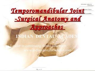
  • 1. Temporomandibular Joint -Surgical Anatomy and Approaches INDIAN DENTAL ACADEMY Leader in continuing dental education www.indiandentalacademy.com www.indiandentalacademy.com
  • 2. TEMPOROMANDIBULAR JOINT Unique features • • Covered with fibrocartilage • • Simultaneous movements Presence of teeth Bicondylar, ginglymoarthroidal, compound, complex, secondary, synovial joint. www.indiandentalacademy.com
  • 6. Post natal development Condyle  Mediolateral width • 9.6mm at birth • 12.4mm at deciduous point • 15mm in permanent dentition  Anteroposterior • Faster than mediolateral growth • 6.5mm - from eruption to completion of deciduous teeth. • 7.3mm - adult size www.indiandentalacademy.com
  • 7. Glenoid fossa  3 times more deeper in adult than infant.  Cartilage slowly replaced by fibrous tissue with age. Articular eminence :  Rudimentary at birth  Growth increases after eruption of permanent incisors www.indiandentalacademy.com
  • 8. Age changes in Mandible www.indiandentalacademy.com
  • 9. ANATOMY & BIOMECHANICS OF THE TEMPOROMANDIBULAR JOINT www.indiandentalacademy.com
  • 10. TMJ BONY COMPONENTS SOFT-TISSUE COMPONENTS 1. Articular disk 2. Joint capsule 3. Ligaments 1. Glenoid fossa 2. Condylar head 3. Articular eminence  MUSCLES 1. Muscles of mastication 2. Muscles attached to the joint 3. Muscles of facial expression 4. Muscles of the neck www.indiandentalacademy.com
  • 12. CONDYLAR HEAD  Oval – mediolaterally – ‘Rugby ball’  15-20 mm long (M-L); 8-10 mm wide (A-P); 8-120 mm thick  Medial pole > lateral pole  Posterior surface > anterior surface  Articulating surface – Fibrous tissue  140o with line connecting EAM on both sides  Axes – meet anterior to foramen magnum www.indiandentalacademy.com
  • 13. ARTICULAR EMINENCE • Sigmoid shape, Anterior & posterior slopes • Saddle – shaped in coronal section – concave mediolaterally – path of condyle • With disc, guides mandibular movement during jaw opening • Has 3 layers − Fibrocartilagenous layer (gradually diminishes with age but persists) − Undifferentiated connective tissue − Fibrous connective tissue www.indiandentalacademy.com
  • 14. JOINT CAPSULE / CAPSULAR LIGAMENT  Fibrous, non-elastic membrane surrounding the TMJ Functions:  Seals joint space  Provides passive stability  Active stability - proprioceptive nerve-endings in capsule www.indiandentalacademy.com
  • 16. Attachments of articular disk – 1. Anteriorly – Joint capsule, Lateral pterygoid muscle fibres – ‘Sphenomeniscus’ fibres - stabilize disk during mastication & deglutition 2. Posteriorly disc attached Retrodiscal tissue www.indiandentalacademy.com
  • 18. Retrodiscal tissue  loose connective tissue  Between bilaminar zone of disc  SRL – Meniscotemporal frenum  IRL – Meniscomandibular frenum  Rich blood supply & nerve supply, Compressible www.indiandentalacademy.com
  • 19. SYNOVIAL MEMBRANE  Lines inner surface of capsule – villi  Functions: 1. Medium for metabolic exchange to avascular articulating surfaces 2. Lubricant – minimizes friction www.indiandentalacademy.com
  • 20.  Lubrication by 2 mechanisms – 1. BOUNDARY LUBRICATION - primary mechanism - moving joint - synovial fluid forced from one area of cavity to another 2. WEEPING LUBRICATION: - Compressed but not moving joint - synovial fluid forced in & out of articular surfaces by compression - prolonged loading will exhaust fluid - mechanism of www.indiandentalacademy.com metabolic exchange
  • 21. LIGAMENTS  Non-elastic collagenous structures - restricts and limits movements a joint  Maintains – joint spaces, without causing tissue damage  True ligaments: 1. COLLATERAL / DISCAL LIGAMENTS 2. CAPSULAR LIGAMENT 3. TEMPOROMANDIBULAR / LATERAL LIGAMENT  Accessory ligaments: 1. SPHENOMANDIBULAR LIGAMENT www.indiandentalacademy.com 2. STYLOMANDIBULAR LIGAMENT
  • 22. COLLATERAL / DISCAL LIGAMENT Functions: 1. Restricts movement of disc away from condyle 2. Hinge movement between condyle & disc 3. Disc moves passively with condyle www.indiandentalacademy.com
  • 23. TEMPOROMANDIBULAR / LATERAL LIGAMENT  FUNCTIONAL LIGAMENT  Fan-shaped reinforcement of lateral wall of capsule  2 parts 1. Outer oblique – outer surface of condylar neck resists excessive dropping of condyle limits extent of mouth opening www.indiandentalacademy.com
  • 24.  Horizontal part – lateral pole of condyle & lateral margin of disk • • • limits posterior movement of condyle & disc protects RDT from trauma protects lateral pterygoid from over lengthening or extension Functions:  Prevents lateral (same side) & medial (contralateral) dislocation www.indiandentalacademy.com
  • 25. Accessory Ligaments  Sphenomandibular L igament – no role • Remnants of Meckel’ s cartilage • Important landmark during surgery  Stylomandibular L igament – limits excessive protrusive movements  Retinacular ligament www.indiandentalacademy.com
  • 27. Classification: 1. Jaw-closing group – 1. Temporalis 2. Masseter 3. Medial pterygoid 2. Jaw-opening group – 1. Lateral Pterygoid 2. Suprahyoid muscles 3. Infrahyoid muscles www.indiandentalacademy.com
  • 28.  TEMPORALIS  Three parts • Anterior part – almost vertical – elevation • Middle part – oblique – elevate & retrude • Posterior portion – almost horizontal - retrusion & joint loading shared with pterygo massetric sling www.indiandentalacademy.com
  • 29.  MASSETER • Origin  superficial – • Ant 2/3rd of zygomatic arch  Middle layer• ant 2/3rd of deep surface and post 1/3rd of lower border of Z arch  Deep layer• Deep surface of Z arch • Insertion  Angle of mandible and ramus  Lower part of lat surface of ramus  Middle & deep fibers – middle and upper part of ramus www.indiandentalacademy.com
  • 30. MEDIAL PTERYGOID: OriginSuferficial – tuberosity of maxilla and adjoining bone deep – medial surface of lat pterygoid plate Insertion roughened medial surface of angle of mandible Functions Elevation  Protrusion  Unilateral – Mediotrusive  With masseter – muscular sling to support angle of mandible www.indiandentalacademy.com
  • 31.  LATERAL PTERYGOID:  Origin• Upper head- Crest of greater wing of sphenoid. • Lower head- lat surface of lateral pterygoid plate.  Insertion• Pterygoid fovea • Ant margin of articular disc & capsule www.indiandentalacademy.com
  • 32. SUPRAHYOID MUSCLES: Digastrics Mylohyoid  Stylohyoid FUNCTIONS Jaw opening & swallowing  Pull mandible downward & hyoid backward www.indiandentalacademy.com
  • 34. At Rest  Occlusion - physiological rest position  Tonus of elevators – maintain constant contact  Intra articular pressure www.indiandentalacademy.com
  • 35. 1. INFERIOR JOINT CAVITY    Tightly bound – discal ligaments Condyle + disc Rotational / Hinge 2. SUPERIOR JOINT CAVITY  Disc not tightly attached to fossa  Translatory / sliding movements www.indiandentalacademy.com
  • 38. TMJ Relations Superficial relations  Skin, superficial fascia and branches of the facial nerve  Auriculo-temporal nerve  Superficial temporal artery  Glenoid lobe of the parotid gland Superior relations  Temporal lobe of brain  Tympanic cavity  Chorda tympani and anterior ligament to malleus www.indiandentalacademy.com
  • 39. Inferior relations  Parotid gland  Lower head of the lateral pterygoid.  Venous channels.  Branches from the pterygoid venous plexus Anterior relations  The lateral pterygoid.  The masseteric and deep temporal nerves www.indiandentalacademy.com
  • 40. Posterior relations  Auriculo-temporal nerve  Superficial temporal artery.  Parotid gland  Styloid process Medial relations  squamo-tympanic fissure, chorda tympani nerve  spine of the sphenoid, sphenomandibular ligament.  middle meningeal artery, carotid sheath.  auriculo-temporal nerve, mandibular nerve.  middle, inner ear, auditor tube. www.indiandentalacademy.com
  • 41. Distances of important structures medial to TMJ. Structures from zygomatic arch Mean mediolateral Mean anteroposte rior Middle meningeal artery 31mm 2.4mm Carotid artery 37mm -6.5mm Internal jugular vein 38.3mm -8.7mm Mandibular nerve (from GF) 18.7mm 9.2mm Nojan et al [OOO 1999; 88: 674-8]. www.indiandentalacademy.com
  • 42. IMPORTANT STRUCTURES Auriculotemporal nerve  Runs from deep to superficial layers as it reaches preauricular region.  Inevitable damage – preauricular approach. Superficial temporal artery  Deep to parotid.  Posterior to neck of condyle and crosses zygomatic process  Runs in superficial fascia www.indiandentalacademy.com
  • 43. Maxillary artery  Beneath – condylar neck.  Immediate posteromedial relation.  Subperiosteal guard.  Endangered in condylotomy and resection of bony ankylosis. www.indiandentalacademy.com
  • 46. Mandibular and cervical branch www.indiandentalacademy.com
  • 47. Approaches   Many approaches have been proposed. Can be grouped as follows • • • • • • • • Pre-auricular Endaural Post auricular Submandibular Intra-oral Closed condylotomy Rhytidectomy incision Horizontal incision along the lower border of the malar arch • Through soft tissue lacerations or scars. www.indiandentalacademy.com
  • 48.  Ideal approach characteristics. • Be based on sound anatomical principles. Have clear anatomical landmarks. • Be designed to give protection to both the facial and the auriculo-temporal nerves, and to the external auditory canal. • Provide a relatively bloodless field. • Provide excellent visibility of the lesional site without flap tension. • Be rapidly and confidently executed. • Be uncomplicated in its repair. • Give a good cosmetic result with minimal functional sequelae. www.indiandentalacademy.com
  • 50. Pre-auricular  Started by Risdon in 1934 www.indiandentalacademy.com
  • 52. •Popularized by Blair (1936) – inverted L shape. •Dingman used Blairs modification - obtuse angulated vertical incision. Vertical component – anterior to tragus. Superior leg – obliquely anterior to pinna. www.indiandentalacademy.com
  • 53.  1979 extensive study by Alkayat and Bramley – the first modified preauricular incision www.indiandentalacademy.com
  • 55. Indications  When maximum exposure is required.  When lateral and anterior exposure is desired. Advantage:  There is minimal bleeding and less sensory loss. • Spares the main branches of vessels and nerves.  Fascial planes are easily identified.  There is excellent visibility.  The potential complications of muscle herniation and fibrosis are avoided. • The muscle is never exposed. www.indiandentalacademy.com
  • 56. Disadvantages:  Scarring present.  Threat of damage to facial nerve branches.  Sensory loss over post-auricular skin.  Frey syndrome.  Damage to superficial temporal artery. www.indiandentalacademy.com
  • 57. Endaural approach     Introduced by Shanbaugh – middle ear surgeries. Lemperts – use for TMJ. Different from Dingman that it involved external auditory meatus to a greater depth. Davidson modification – superior preauricular component. www.indiandentalacademy.com
  • 58. Surgical approach I-part  • Anterior endaural incision in superior meatal wall (depth-bony cartilagenous junction). • Then outward incision for 3-5mm at conchal cartilage. II-part  • Extends from superior extent endaural incision directly upwards to a point about halfway between meatus and upper edge of the auricle. III-part  • Continuous superiorly in the inter cartilagenous cleft and becomes the facial www.indiandentalacademy.com
  • 59. Indications:  When lateral and posterior exposure is required.  To avoid scarring. Advantages:  Excellent lateral and posterior exposure.  Scar exposure is less. Disadvantage:  Limited anterior visibility.  Demands greater skills.  Tragal cartilage degeneration. www.indiandentalacademy.com
  • 60. Post-auricular approach  Introduced by Bockenheimer (1920)  Modified by Axhausen. www.indiandentalacademy.com
  • 62. Indications:  When lateral and posterior exposure is required.  Normal scar formation in the patient's history.  Healthy ear apparatus and absence of aural sepsis.  Normal width of the external auditory canal.  Absence of infection or inflammation of the joint structures.  General health of the patient does not restrict length of operating period. www.indiandentalacademy.com
  • 63. Advantages:  Excellent accessibility especially posterior and lateral.  Reduction in facial nerve damage.  No excessive bleeding. Disadvantages:  Limited anterior accessibility.  Perforation of cartilaginous external auditory meatus.  External auditory canal stenosis.  Infections. www.indiandentalacademy.com
  • 64. Risdon’s approach (Submandibular)  Incision about finger breadth below angle of mandible parallel to lower border.  Lies between cervical branches of facial nerve, lower boundary of bony EAM at least 3cm inferior. www.indiandentalacademy.com
  • 66. Indications  Usually for subcondylar procedure  Severe bony ankylosis  Direct condylar fracture fixation  Costochondral grafting Advantages:  Less chances of facial nerve damage Disadvantages:  Inadequate accessibility  Increased reflection and traction of tissue  Temporary parasthesia may be present www.indiandentalacademy.com
  • 67. Retromandibular approach  Developed by E.C. Hinds and W.J. Girotin (1967) www.indiandentalacademy.com
  • 69. Indications  For condylar neck fractures  Condylotomy  Vertical ramus osteotomies Advantages:  Less chances of damage to facial nerve Disadvantages:  Reduced accessibility  Parasthesia of facial nerve  Damage to retromandibular vessels www.indiandentalacademy.com
  • 70. Rhitidectomy approach  A variant of retromandibular approach. www.indiandentalacademy.com
  • 71. Indications  Esthetic is a concerned and extensive exposure is required. Advantages  Less conspicuous facial carve  Good exposure Disadvantage  Added time required www.indiandentalacademy.com
  • 72. Bicoronal flap  Incision following hair line about 4cm behind it.  Depth – till subgleal loose tissue  Inferior extent – continue as preauricular  Blunt dissection to reflect the flap till 2cm above the infraorbital rim and superior temporal line.  Pericranium is incised about 3-4cm superior to orbital rim,  Incision of Alkayat and Bramley www.indiandentalacademy.com continued
  • 74. Indication  Bilateral exposure  Extensive exposure required Advantages  Good exposure  Easy to get the facial phase  Reduced risk of damage to facial nerve branches  Hidden scar Disadvantages  Bleeding in initial phase  Extensive dissection required  Not esthetic in completely bald patients www.indiandentalacademy.com
  • 75. Intra-oral approach  Vertical incision in the retromolar region along the ascending ramus.  Expose the entire medial surface of the ramus protecting the lingual nerve and inferior dental bundle with a retractor.  The condylar notch is visualize.  Elevation of temporal attachment might be necessary.  Winstanely’s used a long, vertical incision from the tip of the coronoid process to the depth of the buccal sulcus.  Sear….. advocates lateral and medial exposure for condylectomy. www.indiandentalacademy.com
  • 76. Indications  Oblique subcondylar osteotomy  Open condylotomy (asymmetry) Advantages  No risk to facial and auricular temporal nerves  No scar Disadvantages  Limited accessibility  Risk of damage to lingual nerve, Inferior alveolar bundle and maxillary artery. www.indiandentalacademy.com
  • 77. Arthroscopic approach   Arthroscopy of human TMJ was first described Ohnishi (1975). 3 primary approaches • • •  Lateral posterior (most safe) Lateral anterior Endaural Landmarks • Condyle, zygomatic arch, superficial temporal artery and posterior aspect of mandible. www.indiandentalacademy.com
  • 78.  2 points are marked on tragocanthal line • 10mm and 15mm anterior to tragus  18 or 19 gauge needle is passed in the upper joint cavity from point A through posterior approach inclining 30° anterior and superior direction.  For the inferior joint cavity needle and cannula are passed at the same point and directed inferiorly and posteriorly at 45° rather than anteroinferiorly www.indiandentalacademy.com
  • 79. Indications  Joint arthritis  For diagnostic purpose  Hyperextensibility Advantages  Closed procedure  No scar Disadvantages  Risk of damage to the encountering structures  Massive bleeding  AV fistula formation  Intracranial entry www.indiandentalacademy.com