Acute coronary syndrome NSTEMI
•
78 j'aime•18,531 vues

This was another didactics during our rotation in Internal Medicine--Out-patient Department. Source: Harrison's Textbook of Internal Medicine 18th ed.

Recommandé
Contenu connexe
Tendances
Tendances (20)
En vedette
En vedette (20)
Similaire à Acute coronary syndrome NSTEMI
Similaire à Acute coronary syndrome NSTEMI (20)
Dernier
Dernier (20)
Acute coronary syndrome NSTEMI
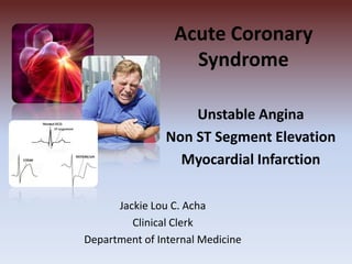
- 1. Acute Coronary Syndrome Unstable Angina Non ST Segment Elevation Myocardial Infarction Jackie Lou C. Acha Clinical Clerk Department of Internal Medicine
- 2. Objectives • Definition of terms • Pathophysiology • Clinical Presentation • Physical Examination findings • Diagnosis • Management
- 3. Definition of Terms • Ischemic Heart Disease – Inadequate supply of blood and oxygen to a portion of the Myocardium => IMBALANCE between Myocardial Oxygen Supply and Demand
- 4. Definition of Terms • Patients with IHD fall into Two Large Groups 1. Patients with CAD -commonly present with STABLE ANGINA 2. Patients with Acute Coronary Syndromes - Unstable Angina and Non-ST-Segment Elevation MI (NSTEMI) - Acute Myocardial Infarction (MI) with ST-Elevation (STEMI)
- 5. • Stable Angina – Episodic clinical syndrome – Due to transient Myocardial Ischemia – Angina usually crescendo-decrescendo in nature – Caused by exertion or emotion, relieved by rest – Typically lasts 2-5 mins – Can radiate to either shoulder and both arms Definition of Terms
- 6. Definition of Terms • Unstable Angina – myocardial ischemia with no evidence of heart muscle death (myocardial necrosis) – angina pectoris or equivalent ischemic discomfort with at least one of three features: (1) it occurs at rest (or with minimal exertion), usually lasting >10 minutes; (2) it is severe and of new onset (i.e., within the prior 4–6 weeks); and/or (3) it occurs with a crescendo pattern (i.e., distinctly more severe, prolonged, or frequent than previously)
- 7. Stable Angina Unstable Angina Episodic Severe and of New onset Crescendo - Decrescendo Crescendo pattern Occurs on exertion, relieved by rest Occurs at rest Lasts 2-5 mins Lasts > 10 min Stable vs. Unstable Angina
- 8. Definition of Terms • NSTEMI – Clinical features of Unstable Angina + evidence of Myocardial Necrosis as reflected by Elevated Cardiac Enzymes
- 10. Pathophysiology • Reduction in oxygen supply and/or an increase in myocardial oxygen demand • Four Pathophysiologic Processes 1. Plaque Rupture or Erosion with Superimposed Nonocclusive Thrombus (most common) 2. Dynamic Obstruction (Coronary Spasm as in Prinzmetal’s Variant Angina) 3. Progressive Mechanical Obstruction 4. Secondary UA related to Increased Myocardial O2 demand and/or Decreased Supply ( e.g. Tachycardia, Anemia)
- 11. Pathophysiology More than 90% of patients with IHD have atherosclerosis of one or more of the epicardial coronary arteries progressive narrowing of the lumen leading to stenosis (“fixed” obstructions) or to acute plaque disruption with thrombosis, both of which compromise blood flow A fixed lesion obstructing 75% or greater of the lumen is generally required to cause symptomatic ischemia precipitated by exercise (most often manifested as chest pain, known as angina) Obstruction of 90% of the lumen can lead to inadequate coronary blood flow even at rest
- 12. Pathophysiology of MI Within minutes the thrombus can evolve to completely occlude the coronary lumen of the coronary vessel Vasospasm (platelet aggregation and mediator release) Other mediators activate the extrinsic pathway of coagulation Release potent secondary aggregators(thromboxane A2, adenosine diphosphate, and serotonin) Platelets adhere, aggregate, become activated A sudden disruption of an atheromatous plaque
- 14. Commonly affected Coronary Arteries in MI - left anterior descending artery - left circumflex artery - right coronary artery 5% have stenosis of the left main coronary artery 15% have three-vessel CAD 30% have two-vessel disease 40% have single-vessel disease
- 15. Clinical Presentation • Clinical Hallmark: CHEST PAIN- substernal region or sometimes epigastrium, radiates to neck, left shoulder, and left arm; severe enough to be considered painful Anginal equivalents: Dyspnea, Epigastric Discomfort
- 16. Physical Examination – Unremarkable – If there is large area of MI or Large NSTEMI, • Diaphoresis • Pale cool skin • Sinus tachycardia • 3rd and 4th heart sound • Basilar rales • hypotension
- 17. DIAGNOSIS • Four major diagnostic tools used in the Diagnosis of UA/NSTEMI in ED 1. History 2. ECG 3. Cardiac markers 4. Stress Testing
- 18. DIAGNOSIS • ECG – ST Segment Depression – Transient ST-segment Elevation – T-wave Inversion • Cardiac Biomarkers – CK-MB – Troponin
- 19. Copyright ©2007 American College of Cardiology Foundation. Restrictions may apply. Anderson, J. L. et al. J Am Coll Cardiol 2007;50:e1-e157 Timing of Release of Various Biomarkers After Acute Myocardial Infarction
- 20. Killip Classification Killip Class Description Mortality Rate Class I no signs of pulmonary or venous congestion 0–5% Class II moderate heart failure as evidenced by rales at the lung bases, S3 gallop, tachypnea, or signs of failure of the right side of the heart, including venous and hepatic congestion 10–20% Class III severe heart failure, pulmonary edema 35–45% Class IV shock with systolic pressure <90 mmHg and evidence of peripheral vasoconstriction, peripheral cyanosis, mental confusion, and oliguria 85–95%
- 21. Risk Stratification TIMI Risk Score for UA/NSTEMI
- 22. Algorithm for risk stratification and treatment of patients with suspected coronary artery disease
- 23. Management A. Medical Treatment - Bed rest with continuous ECG monitoring for ST-Segment Deviation and Cardiac rhythm - ambulation is permitted if patient shows no recurrence of ischemia and does not develop a biomarker necrosis for 12-24 hours
- 24. Medical Therapy Drug Category Clinical Condition When to Avoida Dosage Nitrates Administer sublingually, and, if symptoms persist, intravenously Hypotension Patient receiving sildenafil or other PDE-5 inhibitor Topical, oral, or buccal nitrates are acceptable alternatives for patients without ongoing or refractory symptoms 5–10 g/min by continuous infusion titrated up to 75–100 g/min until relief of symptoms or limiting side effects (headache or hypotension with a systolic blood pressure <90 mmHg or more than 30% below starting mean arterial pressure levels if significant hypertension is present)
- 25. Medical Therapy Drug Category Clinical Condition When to Avoida Dosage Beta blockersb Unstable angina PR interval (ECG) >0.24 s 2° or 3° atrioventricular block Heart rate <60 beats/min Systolic pressure <90 mmHg Shock Left ventricular failure Severe reactive airway disease Metoprolol 25–50 mg by mouth every 6 h If needed, and no heart failure, 5-mg increments by slow (over 1–2 min) IV administration
- 26. Drug Category Clinical Condition When to Avoida Dosage Calcium channel blockers Patients whose symptoms are not relieved by adequate doses of nitrates and beta blockers, or in patients unable to tolerate adequate doses of one or both of these agents, or in patients with variant angina Pulmonary edema Evidence of left ventricular dysfunction (for diltiazem or verapamil) Dependent on specific agent Morphine sulfate Patients whose symptoms are not relieved after three serial sublingual nitroglycerin tablets or whose symptoms recur with adequate anti-ischemic therapy Hypotension Respiratory depression Confusion Obtundation 2–5 mg IV dose May be repeated every 5– 30 min as needed to relieve symptoms and maintain patient comfort
- 27. • Anti-Ischemic Treatment – Bed rest – Nitrates – Beta Blockers • Anti-Thrombotic Therapy – Aspirin – Clopidogrel • Anticoagulation Therapy – Unfractionated Heparin – LMWH Enoxaparin
- 28. Oral Antiplatelet Therapy Aspirin Initial dose of 162–325 mg nonenteric formulation followed by 75–162 mg/d of an enteric or a nonenteric formulation Clopidogrel Loading dose of 300-600 mg followed by 75 mg/d Prasugrel Pre-PCI: Loading dose 60 mg followed by 10 mg/d Intravenous Antiplatelet Therapy Abciximab 0.25 mg/kg bolus followed by infusion of 0.125 g/kg per min (maximum 10 g/min) for 12 to 24 h Eptifibatide 180 g/kg bolus followed by infusion of 2.0 g/kg per min for 72 to 96 h Tirofiban 0.4 g/kg per min for 30 min followed by infusion of 0.1 g/kg per min for 48 to 96 h
- 29. Heparins* Unfractionated Heparin (UFH) Bolus 60–70 U/kg (maximum 5000 U) IV followed by infusion of 12–15 U/kg per h (initial maximum 1000 U/h) titrated to a PTT 50–70 s Enoxaparin 1 mg/kg SC every 12 h; the first dose may be preceded by a 30-mg IV bolus; renal adjustment to 1 mg/kg once daily if creatine Cl < 30 cc/min Fondaparinux 2.5 mg SC qd Bivalirudin Initial bolus intravenous bolus of 0.1 mg/kg and an infusion of 0.25 mg/kg per hour. Before PCI, an additional intravenous bolus of 0.5 mg/kg was administered, and the infusion was increased to 1.75 mg/kg per hour.
- 30. Invasive Strategy • Class I recommendations for Use of an Early Invasive Strategy – Recurrent angina at rest/ low level activity despite medications – Elevated TnT or TnI – New ST-segment Depression – Recurrent angina/ Ischemia with CHF symptoms, rales, MR – Positive Stress Test – EF <0.40 – Decreased BP – Sustained VT – PCI <6 moths, prior CABG
- 31. Long Term Management • Risk factor modification • Long Term Treatment – Beta blockers (anti-ischemic; reduce triggers for MI) – Statins (long term plaque stabilization) – ACE Inhibitors (long term plaque stabilization) – Anti platelet therapy (Aspirin + Clopidogrel for at least 9-12 months)
- 32. THANK YOU!
Notes de l'éditeur
- More than 90% of patients with IHD have atherosclerosis of one or more of the epicardial coronary arteries. The clinical manifestations of coronary atherosclerosis are generally due to progressive narrowing of the lumen leading to stenosis (“fixed” obstructions) or to acute plaque disruption with thrombosis, both of which compromise blood flow. A fixed lesion obstructing 75% or greater of the lumen is generally required to cause symptomatic ischemia precipitated by exercise (most often manifested as chest pain, known as angina); with this degree of obstruction, compensatory coronary arterial vasodilation is no longer sufficient to meet even moderate increases in myocardial demand. Obstruction of 90% of the lumen can lead to inadequate coronary blood flow even at rest. The progressive myocardial ischemia induced by slowly developing occlusions may stimulate the formation of collateral vessels over time, which can protect against myocardial ischemia and infarction and mitigate the effects of high-grade stenoses
- Following disruption of a vulnerable plaque, patients experience ischemic discomfort resulting from a reduction of flow through the affected epicardial coronary artery. The flow reduction may be caused by a completely occlusive thrombus (right) or subtotally occlusive thrombus (left). Patients with ischemic discomfort may present with or without ST-segment elevation. Of patients with ST-segment elevation, the majority (wide red arrow) ultimately develop a Q wave on the ECG (QwMI), while a minority (thin red arrow) do not develop Q wave and, in older literature, were said to have sustained a non-Q-wave MI (NQMI). Patients who present without ST-segment elevation are suffering from either unstable angina or a non-ST-segment elevation MI (NSTEMI) (wide green arrows), a distinction that is ultimately made on the presence or absence of a serum cardiac marker such as CKMB or a cardiac troponin detected in the blood. The majority of patients presenting with NSTEMI do not develop a Q wave on the ECG; a minority develop a QwMI (thin green arrow)
- a simple but comprehensive clinical risk stratification score to identify increasing risk of death, myocardial infarction, or urgent revascularization to day 14